
SIMULIIDAE, Newman, 1834
|
publication ID |
https://doi.org/10.1111/j.1096-3642.2007.00314.x |
|
DOI |
https://doi.org/10.5281/zenodo.10545234 |
|
persistent identifier |
https://treatment.plazi.org/id/0E458F02-FF99-B247-FBF8-F94BFE88FD89 |
|
treatment provided by |
Felipe |
|
scientific name |
SIMULIIDAE |
| status |
|
SIMULIIDAE View in CoL View at ENA
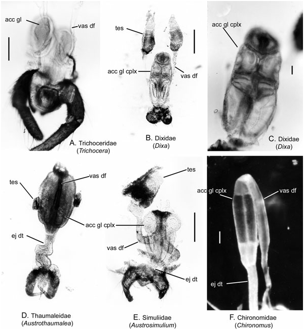
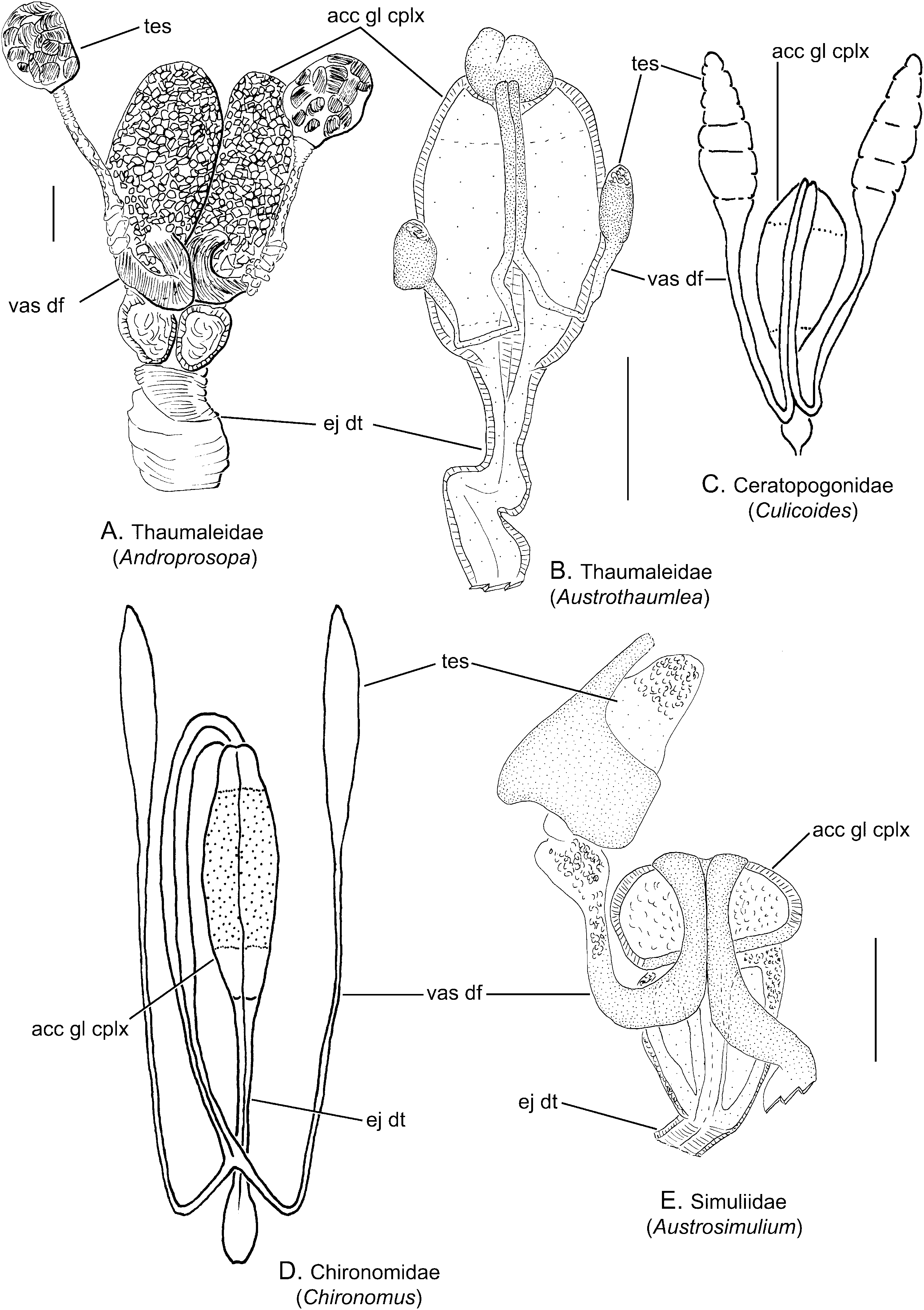
Description ( Figs 5E View Figure 5 , 7E View Figure 7 )
Testis: Each testis is ovoid, pear-shaped or an elongate tapered organ, clothed in pigment granules or a fat body ( Crosskey, 1990). The internal details are outlined by Raminani & Crupp (1978) and Rubtsov (1989).
Epididymis: Not differentiated.
Vas deferens: The vasa deferentia are short, a little longer than the testes. The ducts curve anteriorly to lie juxtaposed on the dorsal surface of the accessory gland complex. Each duct is rather broad, stout and enters the anterior chamber separately ( Jobling & Lewis, 1987). Spermatozoa normally fill the distal portion of the vasa deferentia ( Raminani & Crupp, 1978). The ducts are also often covered in pigmented fat cells.
Accessory gland and seminal vesicle: Each side of the accessory gland complex is two-chambered. The anterior chamber is oval and granular secretory material has been observed inside ( Raminani & Crupp, 1978). The walls of this chamber are rich in glandular cells ( Jobling & Lewis, 1987; Rubtsov, 1989: fig. 10A, fpsd). The second pair of chambers is elongate, approximately twice as long as the anterior chamber and more than three times as long as wide ( Fig. 7E View Figure 7 ). Strong musculature encircles the anterior glands ( Rubtsov, 1989: fig. 10A, epsd).
Ejaculatory duct: A pair of ducts exits the constricted end of the second pair of chambers, but remains divided until near the base of the aedeagus. The duct is rather stout, containing circular muscle.
Ejaculatory apodeme, sperm pump, and aedeagus: The ejaculatory apodeme and sperm pump are absent in Simuliidae . The aedeagus is mostly membranous, although a dorsal plate is occasionally present in the dorsal wall of the aedeagus. The median sclerite is viewed as a dorsal extension of the ventral plate rather than derived from the aedeagus ( Adler, Currie & wood, 2004). The ventral plate (ventral aedeagal guide) articulates with the gonocoxite and serves to aid in opening the female genital chamber by lifting the anal lobes ( Wood, 1978).
Remarks: In Simuliidae , two chambers within the accessory gland complex are present ( Fig. 7E View Figure 7 ; Raminani & Crupp, 1978; Rubtsov, 1989; Crosskey, 1990), in contrast to the three chambers observed in other Chironomoidea .
Spermatophores have been confirmed in Simuliidae , with detailed descriptions of their shape and position during copulation ( Davies, 1965; Wenk, 1965). A spermatophore in Simuliidae is an organized structure containing two packets of spermatozoa ( Wood, 1978), which are partly separated by an internal ridge or groove ( Davies, 1965). The presence of two separate packets is considered to have resulted from spermatozoa from each of the two halves of the accessory gland complex coming together within the ejaculatory duct ( Wood, 1978).
No known copyright restrictions apply. See Agosti, D., Egloff, W., 2009. Taxonomic information exchange and copyright: the Plazi approach. BMC Research Notes 2009, 2:53 for further explanation.