
Anomalophylla tsangpoana, Ahrens, 2005
|
publication ID |
https://doi.org/ 10.11646/zootaxa.1076.1.1 |
|
publication LSID |
lsid:zoobank.org:pub:6B9A5402-EF49-446E-B261-3C0800A925E2 |
|
DOI |
https://doi.org/10.5281/zenodo.10533192 |
|
persistent identifier |
https://treatment.plazi.org/id/0F26A030-8256-2C2B-4307-FA9BFCD0F924 |
|
treatment provided by |
Felipe |
|
scientific name |
Anomalophylla tsangpoana |
| status |
sp. nov. |
Anomalophylla tsangpoana sp. n.
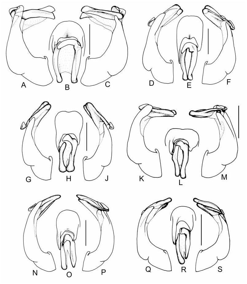
( Fig. 1G J View FIGURE 1 , 5 View FIGURE 5 )
Type material. Holotype: ♂ ”SE Tibet, 2.vi.1996 Nyaingentanglha Shan, Basamtso, 3500 m, V. Major leg.” ( MMBC via TICB) . Paratypes: 6 ♂♂ same data as holotype ( TICB, CA) , 70 ♂♂, 14 ♀♀ “CHINAE. Tibet; 7.vi.1997 Gyamda; 4200 m; 110 km E of Mila Pass ; A. Wrzecionko leg.” ( TICB, CA) .
Additional material examined. ‘ Hybrid’ population ( A. tristicula x A. tsangpoana ): Tibet: 37 specimens ”E Tibet, Bomi env. 29°52’N, 95°45’E 9.–10.VII.1997 mixed forest, ca. 3000 m M. Trýzna & O. Safránek lgt.” ( TICB) GoogleMaps , 2 specimens ”E Tibet, Bomi env. 3000 m 29°52’N, 95°45’E mixed forest, 9.–10.VII.1997 Jaroslav Turna leg.” ( TICB) GoogleMaps , 12 specimens ”China SE Tibet TomeBomi 3.7.1996 leg. V. Paulus ” ( CA, TICB) , 17 specimens ” China: E. Tibet, TomeTangmai , 2000 m 30 km W of Donjung 12.– 13.6.1997 leg. A. Wrzecionko ” ( CA) , 13 specimens “ChinaE. Tibet Lenang [Lunang] 3250 m 30 km W.[N] of Tome leg. Wrzecionko 15.6.2000 ” ( TICB) .
Doubtful determination (♀): 2 ♀♀ “ China, ETibet 2050–2400 m N of Brahmaputra great bend 30°00’–07’ / 94°52’–95°09’ 16.–20.7.92 L.+ R. Businsky lgt.” ( NHMW, CA) .
Holotype description. Length: 6.6 mm, length of elytra: 4.0 mm, width: 3.5 mm. Body oblong, black, elytra reddish brown with dark borders; dorsal surface dull with long, dense, erect setae; pilosity black, setae on elytra and sometimes those on pronotum posteriorly white. Head: Labroclypeus transverse, widest just apical to base; lateral margins strongly convex and convergent anteriorly toward base; anterior angles strongly rounded; lateral border and ocular canthus producing a distinct blunt angle; anterior and lateral margin strongly reflexed and anteriorly sinuate medially; surface almost flat medially and moderately shiny, with double punctation, coarse, dense punctures each bearing a long, erect seta mixed with fine glabrous setae. Frontoclypeal suture distinct, weakly curved and slightly elevated; smooth area anterior to eye as wide as long. Ocular canthus moderately long and slender, finely and densely punctate, densely setose. Frons with double punctation, coarse and moderately dense punctures each bearing a long, erect seta mixed with fine glabrous setae; basal punctation less dense. Eyes small, ratio of diameter / interocular width: 0.44. Antenna with ten antennomeres, black, some basal antennomeres brown; club with five equal in length antennomeres; club 2.5 times as long as the remaining antennomeres combined, reflexed. Prementum almost flat. Pronotum: widest at base; lateral margins in posterior half straight and weakly convergent anteriorly, in anterior third strongly convex and convergent; anterior angles weakly produced and moderately rounded; posterior angles blunt and moderately rounded, anterior margin medially weakly convex with distinct, fine marginal line; basal margin with a fine marginal line. Pronotal surface with moderately dense, double punctation; fine glabrous punctures mixed with large punctures bearing a long erect seta which is slightly directed anteriorly and weakly curved posteriorly. Anterior and lateral borders setose; basal margin of hypomeron not produced ventrally, not transversely sulcate anterior to base. Scutellum : moderately long; apex sharp with fine and sparse punctures; medially smooth; minute setae but no longer setae present in the punctures. Elytra: oblong, widest medially, striae indistinctly impressed and finely densely punctate; intervals flat, with fine, moderately dense punctures; punctures on all intervals with long and erect setae, which are anteriorly longer and denser; sutural interval with thick and long single seta. Epipleural edge fine, ending at the strongly convex external apical angle of elytra, epipleura densely setose, strongly curved in anterior third of elytra, apical border chitinous, without short microtrichomes. Venter: Ventral surface dull with fine, dense punctures; metasternum densely setose; setae partially appressed, partially erect. Metacoxa glabrous near the articulation of the leg; elsewhere with fine, long appressed setae. Abdominal sternites with an indistinct transverse row of coarse punctures bearing thick setae between fine, dense punctation; all sternites with fine, long setae; tegument of abdominal sternites (60x magnification) with fine polygonal mesh pattern formed by microtrichomes; penultimate sternite at midline with longitudinally impressed line. Mesosternum between mesocoxae narrow, narrower than mesofemur, with irregularly scattered, strong setae. Ratio of length of metepisternum / metacoxa: 1 / 1.5. Pygidium strongly convex, posteriorly moderately shiny; with fine, dense punctures bearing fine, long setae; without smooth midline. Legs: slender with shiny surface. Femora with two longitudinal rows of setae, finely densely punctate and setose; anterior edge of metafemur acute, lacking adjacent serrated line; posterior margin of metafemur weakly convex with a few fine setae medially; ventral posterior margin weakly widened in apical half; posterior margin smooth ventrally and dorsally,. Metatibia moderately slender and long, widest at apex; ratio width / length: 1 / 3.8; dorsal margin sharply carinate and smooth with two groups of spines, the basal group at one third, apical one at two thirds of metatibial length, basally with a few single spines in punctures; lateral face longitudinally convex with dense, fine punctures; ventral edge serrated, with five strong, long, equally spaced spines; medial face finely punctate, apex sharply truncate interiorly near tarsal articulation. Tarsomeres dorsally glabrous, coarsely punctate; ventrally with sparse, short setae; metatarsomeres dorsally without longitudinal impressions, ventrally with strongly serrated ridge adjacent to strong longitudinal carina, laterally without strong longitudinal carina; first metatarsomere slightly shorter than the following two tarsomeres combined and one third of its length longer than the upper tibial spur. Protibia short, bidentate. Protarsal claws symmetrical, basal tooth of inner protarsal claw evenly pointed. Aedeagus: Fig. 1G–J View FIGURE 1 .
Intraspecific variation. Length: 5.4–6.8 mm, length of elytra: 3.6–4.3 mm, width: 2.9– 3.3 mm. In some specimens dark lateral border of elytra broadly extended. Density of punctation on dorsal surface variable. ♀: Antennal club with three antennomeres and distinctly shorter than the remaining antennomeres combined; eyes equal in size to that of male.
Diagnosis. Anomalophylla tsangpoana is in genital shape and habitus similar to A. tristicula . It is differentiated from A. tristicula by the shorter basal lobe and the lacking sharp lateral margin of right paramere and more rounded apex of parameres.
Etymology. Named after the Tsangpo River in Tibet.
Remarks. Anomalophylla tsangpoana represents a distinct species that is geographically separated from A. tristicula . In the cladistic analysis (Ahrens accepted) the typical form of Anomalophylla tristicula and A. tristicula form B are sister taxa, while Anomalophylla tsangpoana is the sister taxon to this clade. Anomalophylla tristicula and A. tsangpoana occur together in a limited hybrid zone with specimens of A. tsangpoana ( Fig. 5 View FIGURE 5 , grey squares, see additional material examined above) showing in some features an intermediate character compared to those of A. tristicula , possibly indicating some limited interbreeding between the two species.

No known copyright restrictions apply. See Agosti, D., Egloff, W., 2009. Taxonomic information exchange and copyright: the Plazi approach. BMC Research Notes 2009, 2:53 for further explanation.
|
Kingdom |
|
|
Phylum |
|
|
Class |
|
|
Order |
|
|
Family |
|
|
Genus |