
Spinonychiurus natashae, Pomorski, Romuald J. & Kapruś, Igor J., 2015
|
publication ID |
https://doi.org/ 10.11646/zootaxa.3914.2.1 |
|
publication LSID |
lsid:zoobank.org:pub:84210442-8F95-440B-8D50-9EDD4C75BA8D |
|
DOI |
https://doi.org/10.5281/zenodo.6118368 |
|
persistent identifier |
https://treatment.plazi.org/id/10737675-FFD6-8F51-BDB0-23E461A2AF37 |
|
treatment provided by |
Plazi |
|
scientific name |
Spinonychiurus natashae |
| status |
sp. nov. |
Spinonychiurus natashae sp. nov.
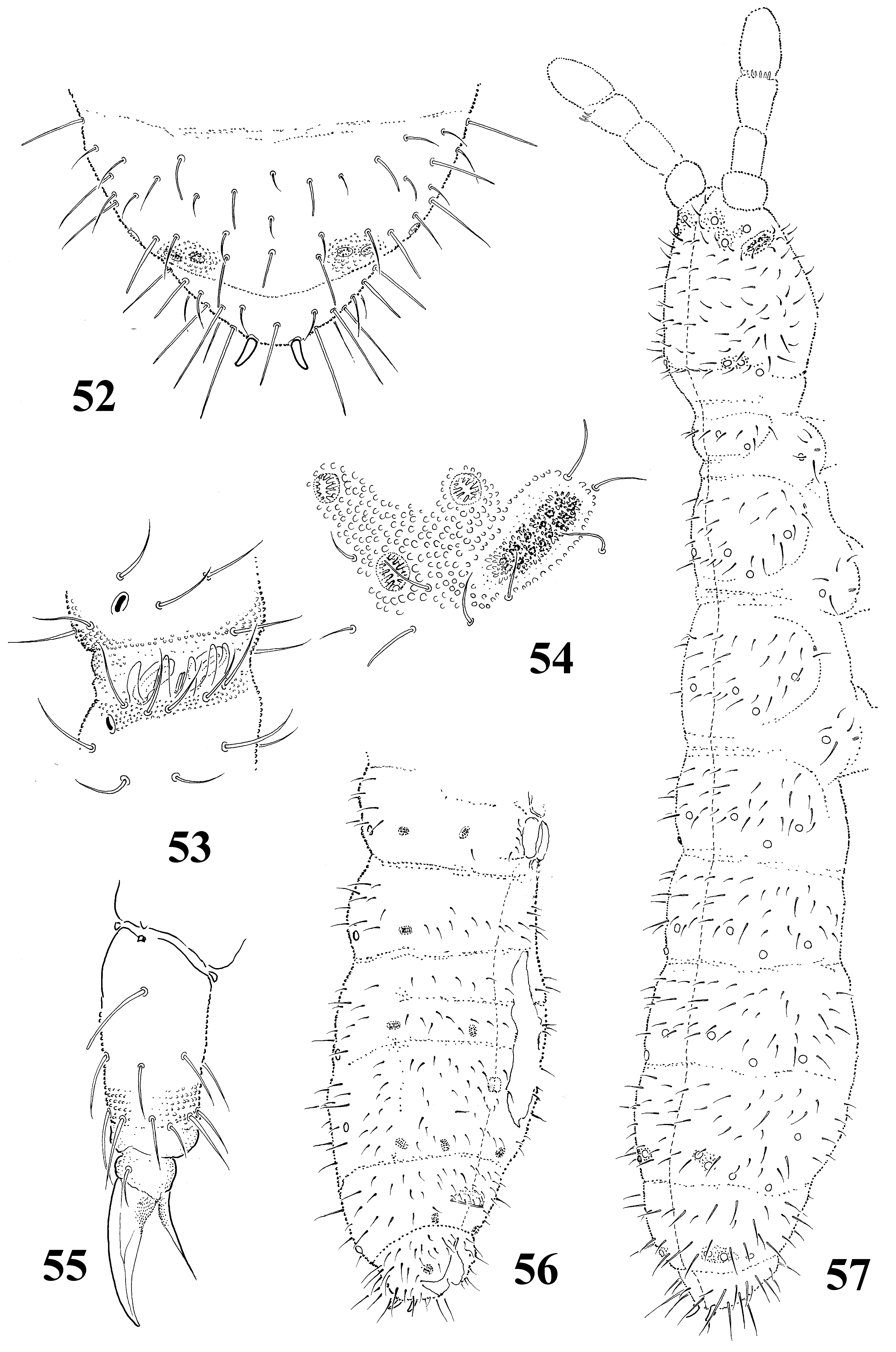
( Figs 52–57 View FIGURES 52 – 57 )
Type material. Holotype (female) and 5 paratypes (4 females, 1 juv., mounted on slide); spruce forest on steep slope in Chinturgen canyon; 22.viii.1991; Zaliyisky Alatau, near Issyk town, South East Kazakhstan, leg. N. A. Kuznetsova. (preserved in the collection the Department of Biodiversity and Evolutionary Taxonomy, Zoological Institute, Wrocław University, Wrocław)
Etymology. The species is cordially dedicated to Natasha A. Kuznetsova from Moscow Pedagogical State University, who personally collected specimens of the new species.
Description. Color white. Body length of females 0.7–0.8 mm. Body shape cylindrical with curved anal spines, distinctly shorter than inner edge of claw (in relation 0.75) ( Figs 52, 57 View FIGURES 52 – 57 ). Granulation of body surface fine and rather uniform, distinctly stronger around pseudocelli on the front of head, hind margin of head and subaxial pseudocelli on abdominal terga IV and V. Antennal bases not marked.
Antennae nearly as long as head or a little longer. Antennal segment IV with typical subapical organite and two poorly marked sensilla (dorsal-subapical and internal-subbasal). Microsensillum on antennal segment IV in lateroexternal position, in proximal whorl of chaetae ( Fig. 53 View FIGURES 52 – 57 ). Antennal III sensory organ with 5 guard chaetae, 5 papillae, 2 small sensory rods and 2 bent, smooth with longitudinal ribs sensory clubs, and microsensillum located slightly below antennal III sensory organ ( Fig. 53 View FIGURES 52 – 57 ).
Postantennal organ consists of 10–13 granulated vesicles ( Fig. 54 View FIGURES 52 – 57 ). Labial palp of AB type. Pseudocellar formula: dorsally 33/233/33353, ventrally 1/000/0000. Subcoxae1 of I–III legs with 1 pseudocellus each. Parapseudocellar formula: 2/000/212213 (each anal valve with parapseudocellus). Localization of parapseudocelli on abdominal sterna I–VI as in Fig. 56 View FIGURES 52 – 57 . Ventral anterior cephalic parapseudocelli located near labial palp. Subcoxae1 of I–III legs with 1 parapseudocellus each.
Dorsal chaetotaxy as in Fig. 57 View FIGURES 52 – 57 , nearly symmetrical, poorly differentiated into meso- and microchaetae. Thoracic terga II and III with microsensilla laterally. Body sensory chaetae s not differentiated. Thoracic tergum I with 6–7 + 6–7 chaetae. Abdominal tergum IV with p0 chaeta, because of asymmetry other axial chaetae difficult to recognize. Abdominal tergum V with a0 and p0 chaetae, and sometimes with m0 chaeta ( Figs 52, 57 View FIGURES 52 – 57 ). Abdominal tergum VI with 2 axial chaetae and 1+1 prespinal chaetae. Subcoxae1 of I–III legs with 4, 5, 5 chaetae respectively.
Thoracic sterna I–III with 0+0, 1+1, 1+1 chaetae respectively. Ventral tube with 7+7 chaetae, and 1+1 chaetae at base. Furca reduced to small area of fine granulation and three rows of manubrial chaetae behind its posterior edge ( Fig. 54 View FIGURES 52 – 57 ). Claws without teeth. Empodial appendage without basal lamella, shorter than inner edge of claw (ratio 0.8). Tibiotarsi I–III with 9 chaetae in distal whorl ( Fig. 55 View FIGURES 52 – 57 ). Males unknown.
Remarks. S. natashae sp. nov. belongs to the group of species with 9 chaetae in distal whorl of tibiotarsi and very similar to S. issykkulensis sp. nov. For differences see: “Diagnosis” of S. issykkulensis sp. nov.
No known copyright restrictions apply. See Agosti, D., Egloff, W., 2009. Taxonomic information exchange and copyright: the Plazi approach. BMC Research Notes 2009, 2:53 for further explanation.