
Spinonychiurus edinensis ( Bagnall, 1935 )
|
publication ID |
https://doi.org/ 10.11646/zootaxa.3914.2.1 |
|
publication LSID |
lsid:zoobank.org:pub:84210442-8F95-440B-8D50-9EDD4C75BA8D |
|
DOI |
https://doi.org/10.5281/zenodo.6118350 |
|
persistent identifier |
https://treatment.plazi.org/id/10737675-FFD9-8F5C-BDB0-27556661A88C |
|
treatment provided by |
Plazi |
|
scientific name |
Spinonychiurus edinensis ( Bagnall, 1935 ) |
| status |
|
Spinonychiurus edinensis ( Bagnall, 1935)
( Figs 1–6 View FIGURES 1 – 6 )
Onychiurus edinensis Bagnall, 1935: 117 .
Syn. Onychiurus spinosus Bagnall, 1949: 51 .
Type material. Five paratypes (females mounted on two slides); Great Britain, Scotland, Edinburgh district, Corstorphine, ii.1935, leg.? (preserved in the collection of the Museum National d’Histoire Naturelle, Paris).
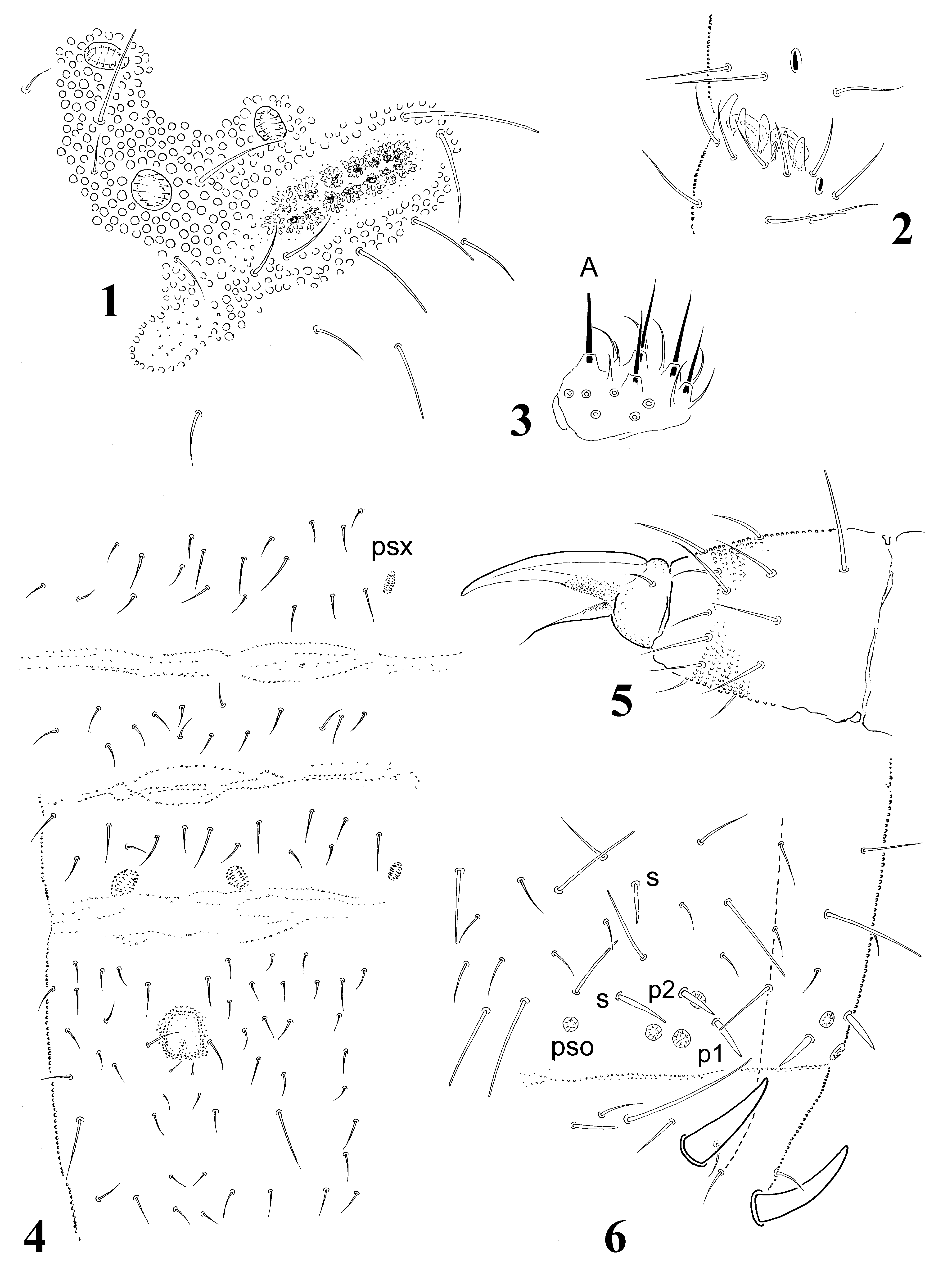
Redescription. Body length of females: 1.1–1.2 mm. Body shape cylindrical with relatively long and strong anal spines (ratio anal spine/inner edge of claw = 1.3) ( Fig. 6 View FIGURES 1 – 6 ). Granulation of body surface fine and uniform, areas of stronger granulation invisible. Antennal bases not marked.
Antennae nearly as long as head or slightly longer. Antennal segment IV with typical subapical organite. Microsensillum on antennal segment IV in latero-external position, above of first proximal whorl of chaetae ( Fig. 2 View FIGURES 1 – 6 ). Antennal III sensory organ with 5 guard chaetae, 5 papillae, 2 small sensory rods and 2 bent, smooth with longitudinal ribs sensory clubs, and microsensillum located slightly below antennal III sensory organ ( Fig. 2 View FIGURES 1 – 6 ). Postantennal organ consists of 14–17 granulated vesicles ( Fig. 1 View FIGURES 1 – 6 ). Labial palp of A type ( Fig. 3 View FIGURES 1 – 6 ).
Pseudocellar formula: dorsally 34/233/44454, ventrally 1/000/0000. Subcoxae1 of I–III legs with 1 pseudocellus each. Parapseudocellar formula:?/000/112? ( Fig. 4 View FIGURES 1 – 6 ). Parapseudocelli on subcoxae1 of I–III legs invisible.
Dorsal chaetotaxy with asymmetries (also in axial chaetotaxy), well differentiated into macro- meso- and microchaetae. Thoracic terga II and III with microsensilla laterally. Body sensory chaetae s dorsally cylindrical, poorly marked, distributed according to formula: 1/011/222121. Thoracic tergum I with 8+8 chaetae. Abdominal tergum IV with p0 chaeta; m0 chaeta difficult to be recognized because of asymmetries. Abdominal tergum V with a0 and p0 chaetae (m0 in two paratypes present), chaetae p1 and p2 as strong spines ( Fig. 6 View FIGURES 1 – 6 ). Abdominal tergum VI with 2 axial chaetae and 1+1 prespinal chaetae. Subcoxae1 of I–III legs with 3, 4, 4 chaetae respectively.
Thoracic sterna I–III with 0+0, 1+1, 1+1 chaetae respectively. Ventral tube with 7+7 chaetae, and 1+1 chaetae at base. Furca reduced to small area of fine granulation and three rows of manubrial chaetae behind its posterior edge ( Fig. 4 View FIGURES 1 – 6 ).
Claws without teeth ( Fig. 5 View FIGURES 1 – 6 ). Empodial appendage without basal lamella, shorter than inner edge of claw (ratio 0.7–0.75). Tibiotarsi I–III with 11 chaetae in distal whorl ( Fig. 5 View FIGURES 1 – 6 ). Anal spines 1.3 as long as inner edge of claw III. Males unknown.
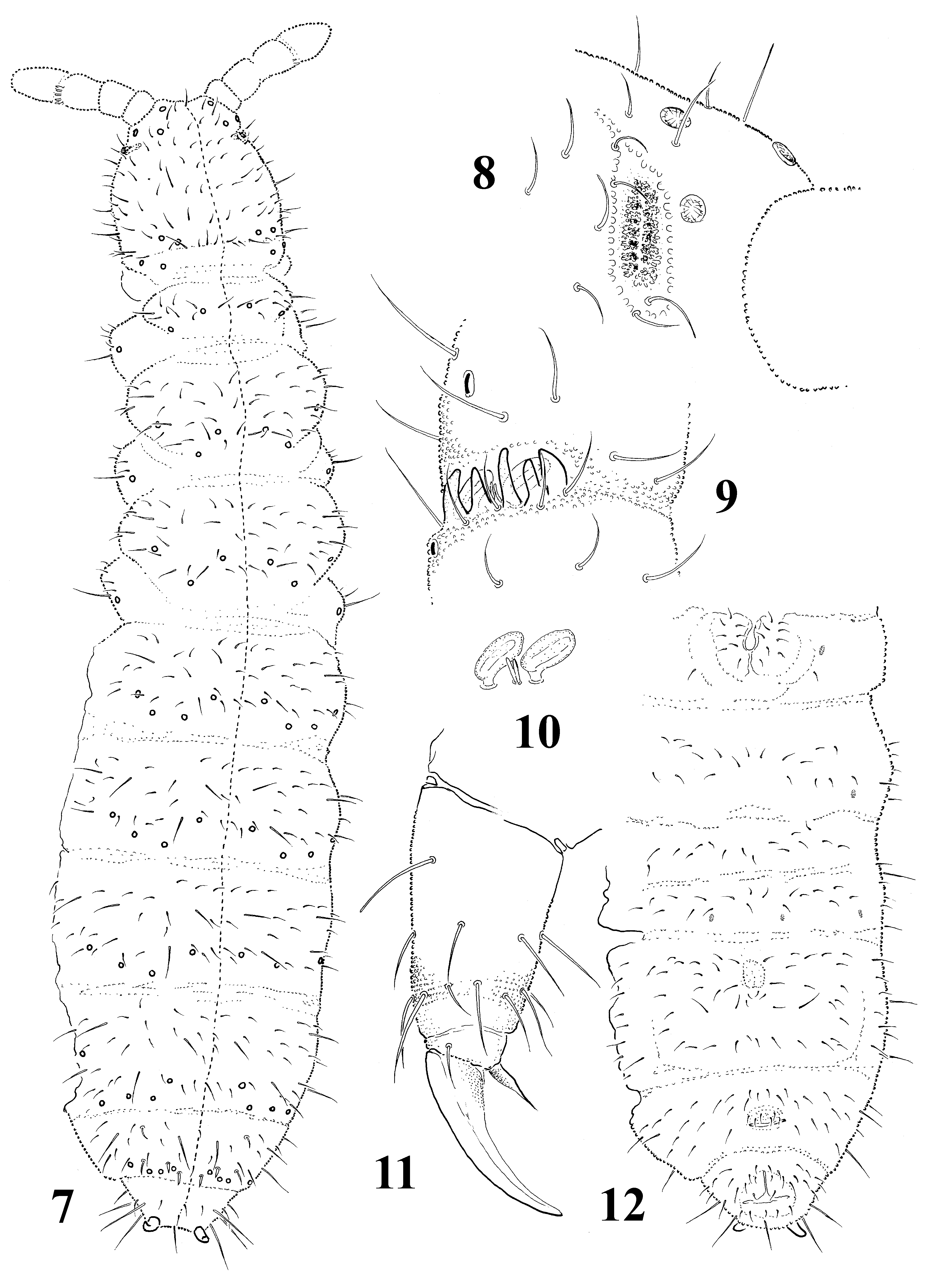
Remarks. The condition of paratypes of S. edinesis is rather poor. They are strongly shrunken, crumpled and therefore some characters are invisible. Examining of fresh material of the species is necessary. Redescription of S. edinesis should to explain the taxonomic status of S. spinularius , which was considered as its synonym ( Murphy, 1960). Comparison of characters of both species available to examining reveals differences in length of empodial appendage, length of anal spines and number chaetae on subcoxae 1. In S. edinensis empodial appendage is shorter than inner edge of claw in relation 0.7–0.75, length of anal spines reach 1.3 of inner edge of claw III and subcoxae 1 of I–III legs with 3, 4, 4 chaetae respectively while in S. spinularius the same ratios attained values 0.4–0.5 and 1.5–1.6 respectively and subcoxae 1 carry 5, 5, 5 chaetae (compare also Figs 5 View FIGURES 1 – 6 , 11 View FIGURES 7 – 12 ).
Onychiurus spinosus Bagnall 1949 was described from North Ireland and according to original description it differs from S. edinesis by shorter anal spines and smaller body size (0.6 mm). The type material of this species is not available and probably has been lost, but judging by small size of body the description could be made on the base of juveniles in which proportions of body parts are different than in adult’s specimens. In this light O. spinosus is hereby considered to be a junior synonym of S. edinensis .
No known copyright restrictions apply. See Agosti, D., Egloff, W., 2009. Taxonomic information exchange and copyright: the Plazi approach. BMC Research Notes 2009, 2:53 for further explanation.
|
Kingdom |
|
|
Phylum |
|
|
Class |
|
|
Order |
|
|
Family |
|
|
Genus |
Spinonychiurus edinensis ( Bagnall, 1935 )
| Pomorski, Romuald J. & Kapruś, Igor J. 2015 |
Onychiurus edinensis
| Bagnall 1935: 117 |