
Enicospilus diae Lima & Kumagai
|
publication ID |
https://doi.org/ 10.5281/zenodo.213294 |
|
DOI |
https://doi.org/10.5281/zenodo.6178607 |
|
persistent identifier |
https://treatment.plazi.org/id/110C879E-FFEB-FF84-65BF-03DAFD607143 |
|
treatment provided by |
Plazi |
|
scientific name |
Enicospilus diae Lima & Kumagai |
| status |
sp. nov. |
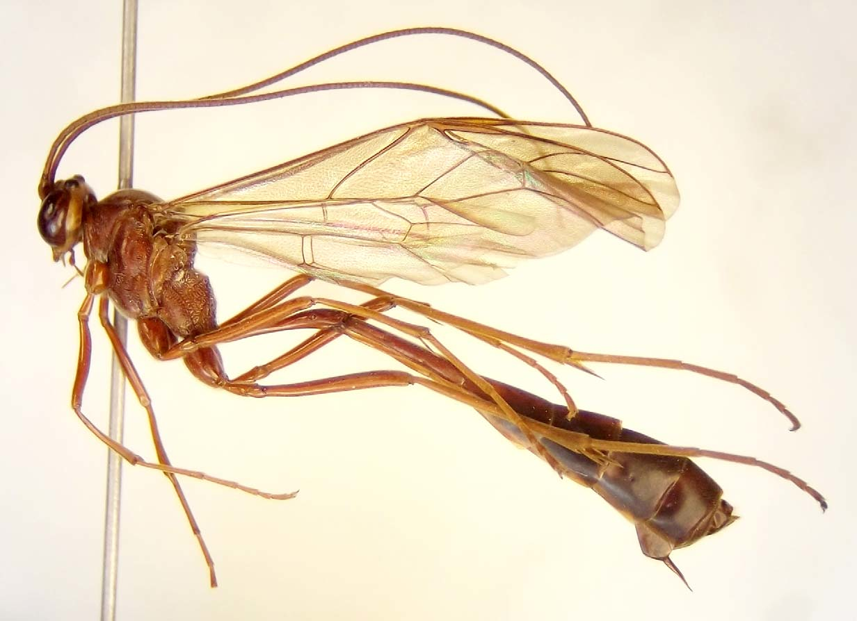
Enicospilus diae Lima & Kumagai sp. n.
( Figs 3‒4 View FIGURE 3 View FIGURE 4 )
Type locality. Brazil, Minas Gerais, Belo Horizonte. Campus of the Universidade Federal de Minas Gerais. Geographic coordinates UTM 23K 0607592/ 7802025, 842m.
Diagnosis. Enicospilus diae can be recognized by the following combination of characters: the upper tooth just 0.3 times longer than the lower one; the epicnemial carina resembling a shield, curved on the epicnemium ( Fig. 4 View FIGURE 4 B), following the posterior border of prosternum, disappearing behind the fore coxa; and the fore wing with the central sclerite very small and weakly pigmented ( Fig. 4 View FIGURE 4 A).
Description (based on holotype). Female. Mandibles with abruptly-narrowed base, parallel-sided distally, twisted about 10°, with upper tooth subcylindrical, 0.3 times longer than the lower; outer mandibular surface with an impressed hirsute groove running from near upper proximal corner to base of teeth. Labrum 0.2 times as long as wide. Malar space 0.35 times as long as basal mandibular width. Clypeus in lateral view convex, its margin blunt, 1.45 times as broad as long. Face 1.25 times as broad as long, centrally coarsely punctate. Genae rounded when head is observed in dorsal view. Posterior ocellus almost touching eye. FI = 53%. Occipital carina mediodorsally complete, joining hypostomal carina about 0.9 times basal mandibular width distant from mandible. Antennae long and slender with 65 flagellomeres; twentieth flagellomere 2.15 times as long as broad.
Mesoscutum polished, closely punctate, evenly rounded in lateral view; notauli weak but discernible. Mesopleuron polished, the upper part punctate to striate, lower part puncto-striate. Epicnemial carina resembling a shield, curved on epicnemium ( Fig. 4 View FIGURE 4 B), following posterior border of prosternum, disappearing behind fore coxa. Scutellum 1.25 times as long as anterior width, convex in lateral view, laterally carinate along entire length, shallowly punctate. Metapleuron convex, diagonally striate. Submetapleural carina with anterior portion weakly broadened. Posterior transverse carina of mesosternum complete. Propodeum in lateral view evenly rounded; anterior transverse carina complete; posterior transverse carina absent; anterior area rugoso-striate, as long as spiracular area; posterior area strongly wrinkled to reticulate, with wrinkles resembling parts of longitudinal and posterior transverse carinae; lateral longitudinal carina complete, joined to spiracular margin by short carina.
Fore wing length 15.84 mm; AI = 0.73; CI = 0.35; ICI = 0.62; SDI = 1.24; cu-a reaching M+Cu basad of Rs&M by 0.3 times its own length. Pilosity on basal area of marginal cell slightly sparser than on central area. Discosubmarginal cell ( Fig. 4 View FIGURE 4 A) with proximal sclerite subtriangular; central sclerite weakly pigmented, irregularly shaped, 0.6 times as long as distal sclerite; distal sclerite not confluent with proximal, distally bordering margin of fenestra. First subdiscal cell with posterior 0.3 and proximal 0.2 glabrous, with distal end hirsute. Hind wing with 8 hamuli on R1; first abscissa of Rs more or less straight, second abscissa straight.
Middle leg with longer tibial spur 1.38 times as long as shorter one. On hind leg, coxa in lateral view 1.8 times as long as wide; trochantellus dorsally 0.26 times as long as broad; fourth tarsomere 2.25 times as long as broad. Claws long, bearing close, short pectinae.
Metasoma long and slender; tergite 2 in lateral view 6.3 times as long as posterior width, laterotergite folded beneath tergite, thyridium elliptical, its distance to anterior margin of tergite 3.5 times its length. Ovipositor straight and slender ( Fig. 4 View FIGURE 4 C).
Body brownish; head, antenna, tibiae and tarsi yellowish-brown; distal half of tergite 3 and tergites 4 to 8 blackish-brown; wings hyaline.
Variation. The notauli are more strongly marked than normal in some specimens. The anterior transverse carina of the propodeum can be weak laterally. In some specimens, the propodeal wrinkles resemble longitudinal carinae or parts of the posterior transverse carina. The wrinkles can also give a strongly reticulate appearance to the propodeum.
Ranges for some features varying among the observed specimens are: FI = 53%‒60%; number of flagellar segments = 65‒70; fore wing length = 13.8mm ‒ 16.1 mm; AI = 0.63‒0.80; CI = 0.32‒0.39; ICI = 0.62‒0.87; SDI = 1.2‒1.36; number of hamuli on hind wing R1 vein = 8‒9, with some specimens with 8 hamuli on one wing and 9 on the other.
Male with gonosquama apically rounded and tarsal claws more closely pectinate than those of female.
Etymology. This species is named in honor of entomologist Priscila G. Dias, in recognition of her outstanding work towards the improvement of the UFMG insect collection.
Distribution. Enicospilus diae is only known to occur in the state of Minas Gerais, Southeastern Brazil, in the municipalities of São Gonçalo do Rio Abaixo, Belo Horizonte, Lavras, and Barroso.
Material examined. Holotype Ƥ. Brasil, Minas Gerais: Belo Horizonte, Campus UFMG, 01.XII.2000, A. F. Kumagai col ( UFMG) [2989].
Paratypes (30 Ƥ, 40 3) Brasil, Minas Gerais: São Gonçalo do Rio Abaixo, Estação Ambiental/Peti, Cemig: 1Ƥ, 18‒25.X.2002, A. F. Kumagai col. ( UFMG) [3065]. 1Ƥ, 14.XII.2007, A. F. Kumagai & P. G. Dias col. ( UFMG) [3066]. Belo Horizonte, Campus UFMG, A. F. Kumagai col. ( UFMG) [2990 ‒ 3055]: 1Ƥ, 13, 24‒30.IX.1991; 1Ƥ, 33, 1‒7.X.1991; 13, 8‒14.X.1991; 23, 15‒21.X.1991; 2Ƥ, 23, 22‒28.X.1991; 4Ƥ, 23, 29.X‒4.XI.1991; 2Ƥ, 43, 5‒11.XI.1991; 3Ƥ, 13, 12‒18.XI.1991; 1Ƥ, 19‒25.XI.1991; 3Ƥ, 26.XI‒2.XII.1991; 1Ƥ, 3‒9.XII.1991; 3Ƥ, 10‒16.XII.1991, 13, 1.X.1998; 1Ƥ, 5.X.1998; 13, 15.X.1998; 2Ƥ, 21.X.1998; 1Ƥ, 4.XI.1998; 23, 22.IX.2000; 33, 29.IX.2000; 43, 06.X.2000; 1Ƥ, 43, 13.X.2000; 13, 20.X.2000; 23, 27.X.2000; 13, 03.XI.2000; 13, 10.XI.2000; 1Ƥ, 17.XI.2000; 23, 23.X.2007; 13, 30.X.2007. Barroso, 13, 2.X.2010, R. L. Tanque col. ( UFLA). Lavras, 1Ƥ, 13.IX.1998, Campus UFLA, J. G. Pádua col. ( UFLA).
Altogether 71 specimens of E. diae sp. n. were examined. All but the specimen from Lavras, for which no information is available, were collected in Malaise traps. The great majority, 67 specimens, were captured in a forest reserve inside the Campus Pampulha of the Universidade Federal de Minas Gerais ( UFMG), appearing in the traps only from September to December.
All specimens of E. diae sp. n. collected in Belo Horizonte are deposited in the UFMG, except for two pairs of paratypes, which will be sent to the American Entomological Institute ( AEI) and to the entomological collection of the U. S. National Museum of Natural History ( USNM) respectively. The specimens collected in Barroso and Lavras are deposited in the UFLA collection.

No known copyright restrictions apply. See Agosti, D., Egloff, W., 2009. Taxonomic information exchange and copyright: the Plazi approach. BMC Research Notes 2009, 2:53 for further explanation.