
Enicospilus ramidulus
|
publication ID |
https://doi.org/ 10.5281/zenodo.213294 |
|
DOI |
https://doi.org/10.5281/zenodo.6178609 |
|
persistent identifier |
https://treatment.plazi.org/id/110C879E-FFEC-FF84-65BF-0715FA6E73C8 |
|
treatment provided by |
Plazi |
|
scientific name |
Enicospilus ramidulus |
| status |
|
Key to Neotropical* species of Enicospilus ramidulus View in CoL species-group
* The Enicospilus of the Galápagos Islands are not considered in this key. For more details about them, see Gauld and Carter (1983).
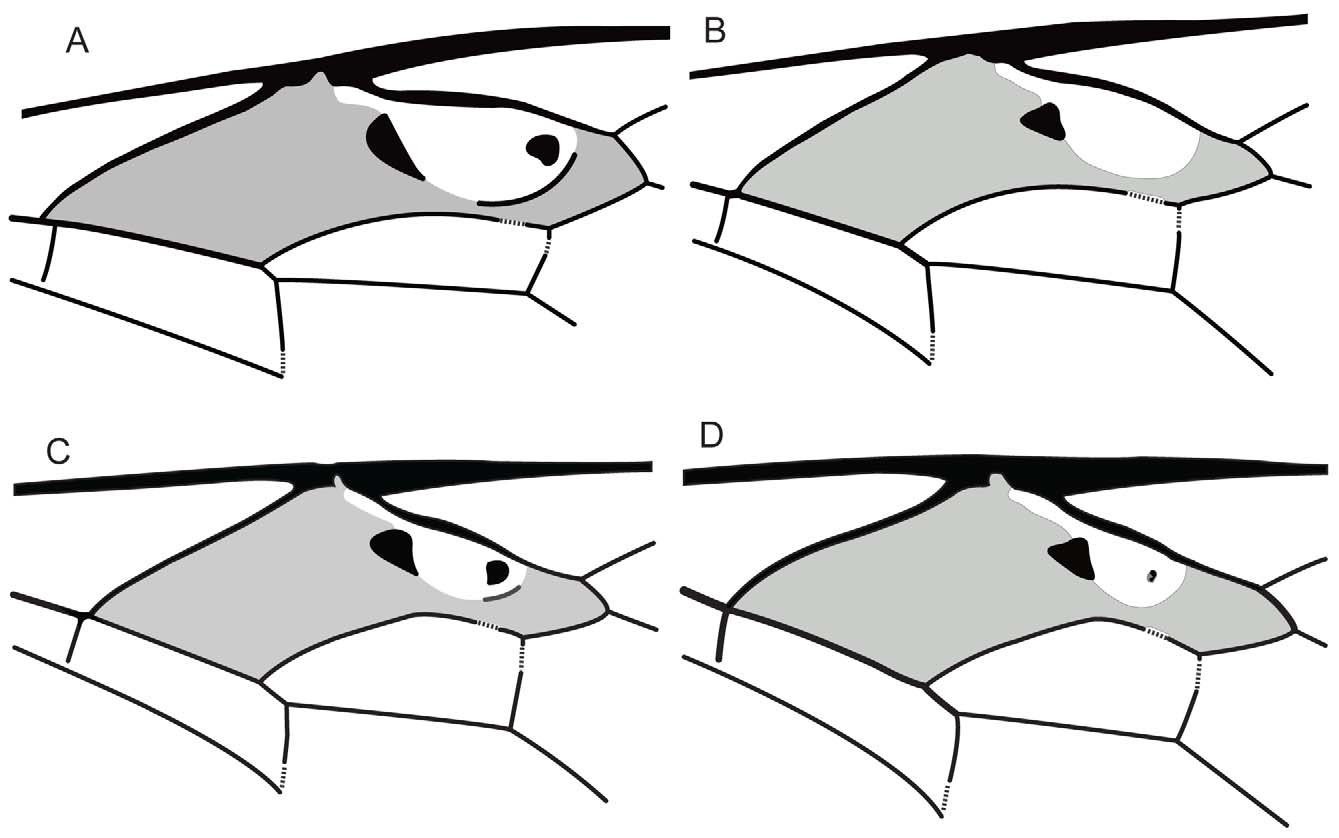
1 Fore wing vein cu-a reaching M+Cu distad of the base of Rs&M ( Fig. 2 View FIGURE 2 A).............. E. cheoi Fernández-Triana, 2005 View in CoL
1’ Fore wing vein cu-a reaching M+Cu coincident or proximal to the base of Rs&M ................................... 2
2(1’) Central sclerite of fore wing discosubmarginal cell absent ( Fig. 2 View FIGURE 2 B)....................... E. neotropicus Hooker, 1912 View in CoL
2’ Central sclerite of fore wing discosubmarginal cell present, distinctly pigmented or with only an almost indistinct, unpig- mented thickening present in the fenestra.................................................................. 3
3(2’) Central sclerite of fore wing discosubmarginal cell strongly sclerotized, thicker than Rs+2r, subcircular to D-shaped ( Fig. 2 View FIGURE 2 C); notauli entirely absent................................................................ E. purgatus ( Say, 1835) View in CoL
3’ Central sclerite on fore wing discosubmarginal cell weakly sclerotized, thinner than Rs+2r, irregularly shaped ( Figs 2 View FIGURE 2 D, 4A); notauli weak but discernible............................................................................. 4
4(3’) Upper mandibular tooth 3 or more times as long as lower tooth; fore wing distal sclerite absent ( Fig. 2 View FIGURE 2 D); 5‒6 hamuli on hind wing vein R1 ........................................................................ E. doylei Gauld, 1988 View in CoL
4’ Upper mandibular tooth 1.3 times as long as lower tooth; fore wing distal sclerite present ( Fig. 4 View FIGURE 4 A); 8‒9 hamuli on hind wing vein R1 .................................................................................... E. diae sp n.

No known copyright restrictions apply. See Agosti, D., Egloff, W., 2009. Taxonomic information exchange and copyright: the Plazi approach. BMC Research Notes 2009, 2:53 for further explanation.