
Odontomolgus cognatus, Cheng, Yu-Rong, Ho, Ming-Jay & Dai, Chang-Feng, 2016
|
publication ID |
https://doi.org/ 10.11646/zootaxa.4174.1.19 |
|
publication LSID |
lsid:zoobank.org:pub:05CD698B-A523-42C6-96F0-7D97156BA447 |
|
DOI |
https://doi.org/10.5281/zenodo.5675442 |
|
persistent identifier |
https://treatment.plazi.org/id/11452501-CD23-D836-E9D4-FE7347CCF870 |
|
treatment provided by |
Plazi |
|
scientific name |
Odontomolgus cognatus |
| status |
sp. nov. |
Odontomolgus cognatus sp. nov.
( Figs 4–6 View FIGURE 4 View FIGURE 5 View FIGURE 6 )
Type host. Pavona explanulata ( Lamarck, 1816) (family Agariciidae ).
Location in host. Surface of colony.
Type locality. Yenliao Bay , Taiwan.
Etymology. The specific name is derived from the Latin “ cognatus ”, meaning relative, and refers to the close relationship of this new species to its congener, Odontomolgus actinophorus ( Humes & Frost, 1964) .
Type material. Three females and two males obtained from washings of a coral colony collected at five m depth on 12 August 2010. The female holotype (NTUIO-COPE 3), male allotype (NTUIO-COPE 4), and paratypes (NTUIO-COPE s5; one female and one male) are deposited in the Institute of Oceanography , National Taiwan University, Taipei, Taiwan .
Description of female. Body ( Fig. 4 View FIGURE 4 A–B) relatively broad. Body length of dissected specimen 1.70 mm (1.69– 1.72 mm) and greatest width 0.62 mm (0.61–0.62 mm), based on three specimens. Cephalosome partially delimited from first pedigerous somite by dorsal suture line ( Fig. 4 View FIGURE 4 A–B). Urosome ( Fig. 4 View FIGURE 4 A–C) 5-segmented. Genital double-somite ( Fig. 4 View FIGURE 4 C) with transverse suture dorsally but fused laterally and ventrally; anterior half with lateral expansions in dorsal view. Genital apertures ( Fig. 4 View FIGURE 4 A–C) located dorsally in first third of double-somite. Three postgenital somites unornamented and equal in length. Caudal ramus ( Fig. 4 View FIGURE 4 C–D) 185 × 55µm, with six naked setae. Surface of body with small setules ( Fig. 4 View FIGURE 4 A). Egg sacs not observed.
Antennule ( Fig. 4 View FIGURE 4 E) 7-segmented; armature: 4, 13, 6, 3, 4 + 1 aesthetasc, 2 + 1 aesthetasc, 7 + 1 aesthetasc; all setae naked. Antenna ( Fig. 4 View FIGURE 4 F) 4-segmented; armature: 1, 1, 3, I; measurements (length × width) of segments 78 × 63 µm, 156 × 63 µm, 63 × 34 µm, 47 × 25 µm, respectively; terminal claw 63 µm long and slightly curved. Mandible ( Fig. 4 View FIGURE 4 G) with prominent proximal notch; inner margin distinctly bilobate; convex side with a digitate process; terminal lash slender, with spinules on both sides. Maxillule ( Fig. 4 View FIGURE 4 H) armed with three plumose apical setae. Maxilla ( Fig. 4 View FIGURE 4 I) 2-segmented, first segment unarmed; second segment terminating in distal lash bearing setules; with one nodular tubercle near base of lash, one inner seta transformed to globular tubercle covered with minute setules, one spiniform anterior seta, and one small proximal seta. Maxilliped ( Fig. 4 View FIGURE 4 J) 3-segmented, syncoxa unarmed; basis with two unequal setae (larger one about 21µm) along medial margin; small endopodal segment with one relatively large spiniform process, one small subapical inner seta, and one bipinnate spine.
Legs 1–4 ( Fig. 5 View FIGURE 5 A–D) with 3-segmented exopods and endopods (except for leg 4 endopod being 2- segmented). Armature formula of spines (in Roman numerals) and setae (in Arabic numerals) as follows:
Coxa Basis Exopod Endopod
Leg 1 0-1 1-0 I-0; I-1; IV+4 0-1; 0-1; I+5 Leg 2 0-1 1-0 I-0; I-1; IV+5 0-1; 0-2; III+3 Leg 3 0-1 1-0 I-0; I-1; IV+5 0-1; 0-2; III+2 Leg 4 0-1 1-0 I-0; I-1; IV+5 0-1; II Leg 5 ( Fig. 5 View FIGURE 5 E) 190 × 75 µm, consisting of an unornamented free segment bearing two unequal apical setae and a small adjacent dorsal (= outer basal) seta.
Leg 6 ( Fig. 4 View FIGURE 4 C) represented by two small setae arising from operculum closing off genital aperture.
Description of male. Body ( Fig 6 View FIGURE 6 A–B) resembling that of female. Body length 1.61 mm (1.54–1.68 mm) and greatest width 0.51 mm (0.48–0.54 mm), based on two specimens. Urosome ( Fig. 6 View FIGURE 6 A) 6-segmented. Caudal ramus as in female.
Antennule, antenna, mandible, maxillule, and maxilla as in female, except for antennule with three additional long aesthetascs (positions indicated by dots in Fig. 4 View FIGURE 4 E) and antenna with additional spinules on medial surface of basis ( Fig. 6 View FIGURE 6 B). Maxilliped ( Fig. 6 View FIGURE 6 C) consisting of three segments and terminal claw; syncoxa broadest, unarmed; basis with two unequal inner setae and longitudinal rows of small spinules as figured; endopodal segment very short and unarmed; terminal claw evenly curved, with two unequal setae at its base.
Legs 1–4 as in female except for leg 1 ( Fig. 6 View FIGURE 6 D); third endopodal segment of leg 1 armed with two spines and four setae instead of one spine and five setae in female.
Leg 5 ( Fig. 6 View FIGURE 6 A) a small free segment with two setae and one adjacent dorsal (= outer basal) seta as in female. Leg 6 ( Fig. 6 View FIGURE 6 A) represented by two small setae on posteroventral operculum on genital somite.
Remarks. Odontomolgus is one of the most speciose genera in the Anchimolgidae , currently accommodating 17 valid species ( Walter & Boxshall 2014). Kim (2006) pointed out that the transformation of the inner seta on the second segment of the maxilla into a hairy globule or a mucus-like structure is not a diagnostic feature of the genus Odontomolgus since it is only displayed by three other species so far: O. actinophorus , O. mucosus Kim, 2006 and O. unioviger Kim, 2006 . Odontomolgus cognatus sp. nov. can be readily distinguished from these three congeners by the larger body size (1.70 mm) and the armature of the third exopodal segment of leg 4 being IV+5 instead of III+5. The remaining dissimilarities among these species are summarized in Table 2 View TABLE 2 .
O. O. cognatus O. mucosus O. mucosus O. unioviger
actinophorus (Taiwan) (Moluccas)
Body size ♀ (mm) 1.42 1.70 1 0.91 0.82 Armature of maxillule 3 plumose setae 3 plumose 1 naked and 3 4 naked setae 1 naked and 3
setae plumose setae plumose setae
Armature of third exopodal segment III+ 5 IV + 5 III + 5 III + 5 III +5 of leg 4
Armature of caudal ramus plumose setae naked setae plumose setae plumose setae plumose setae
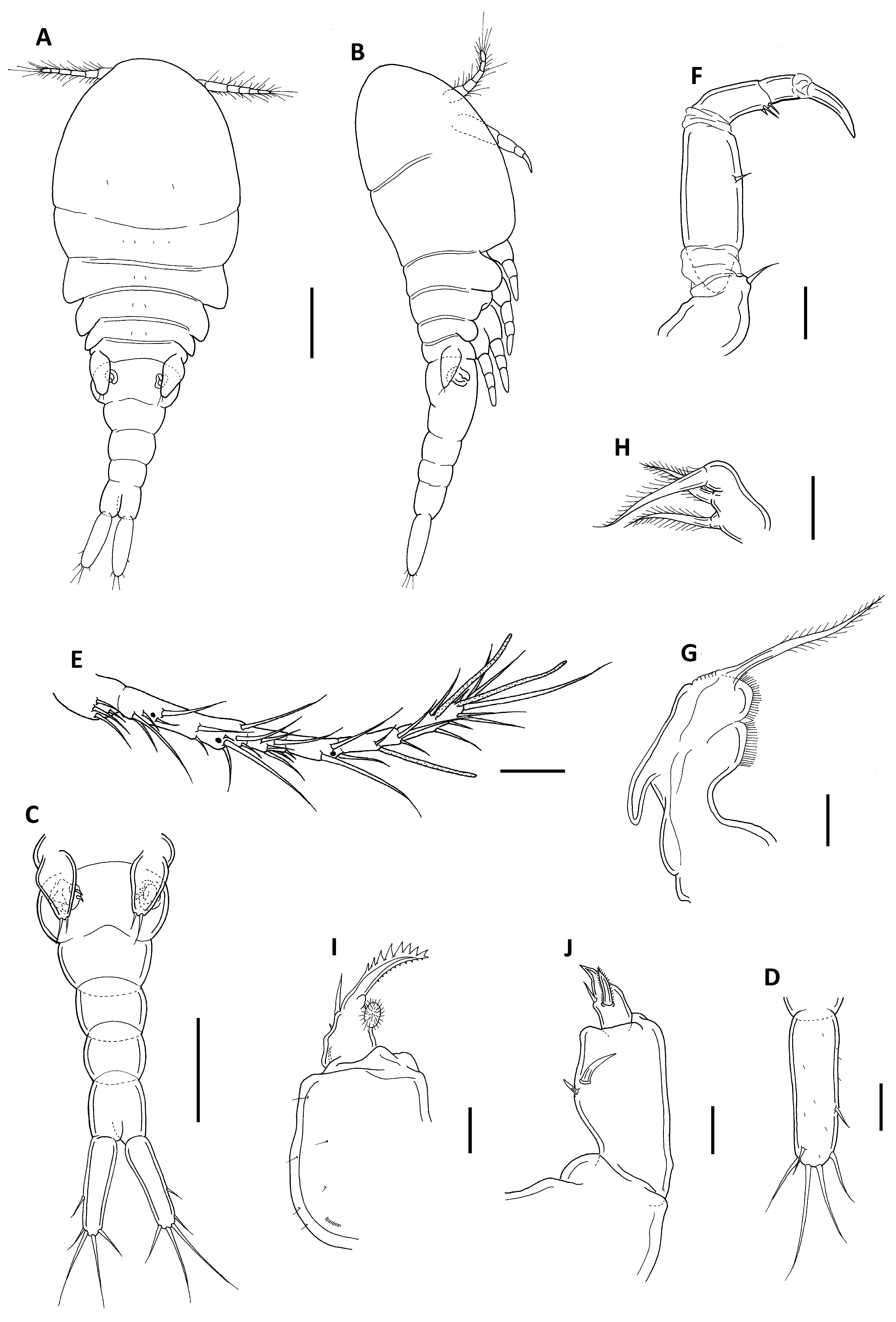
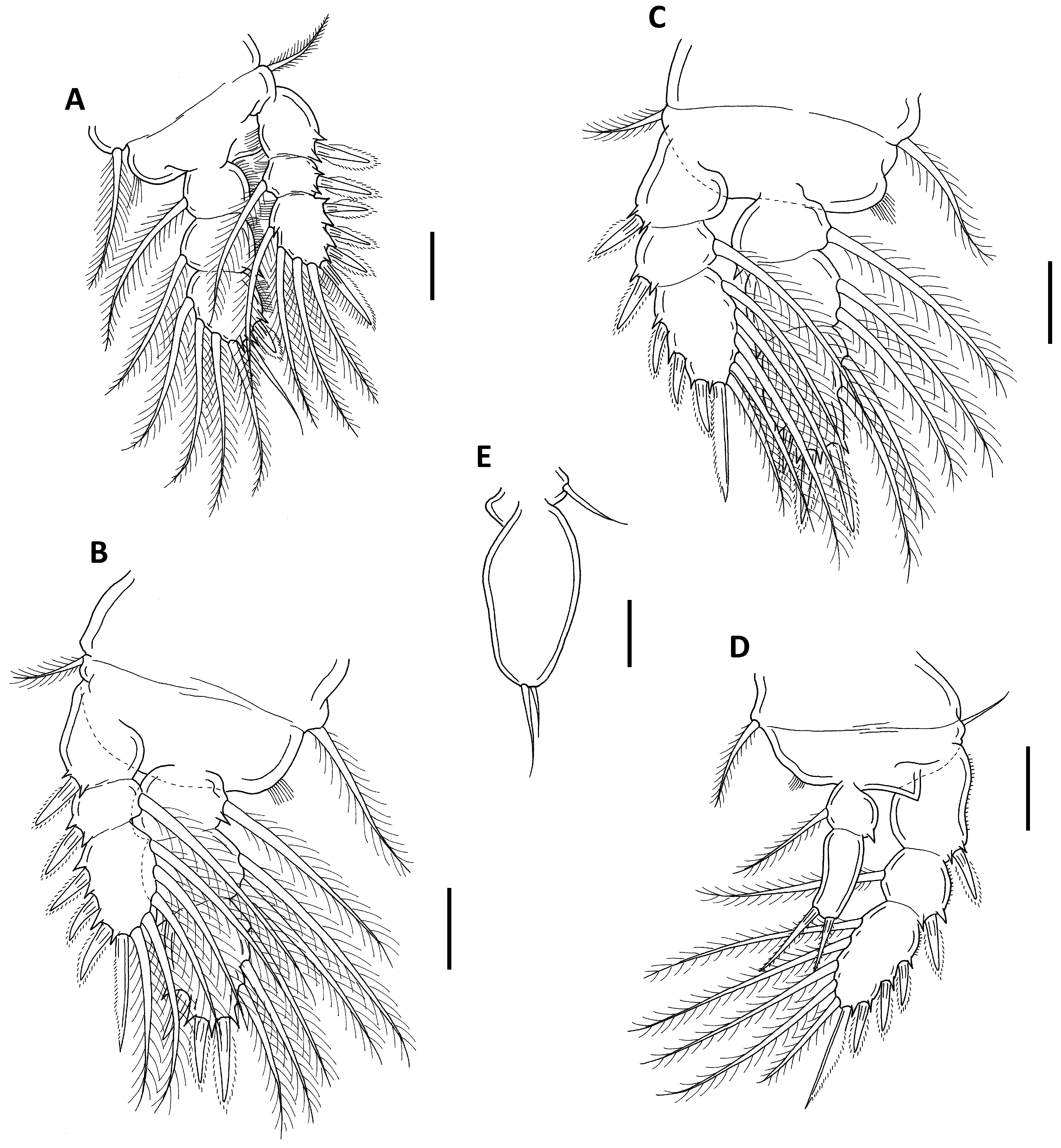
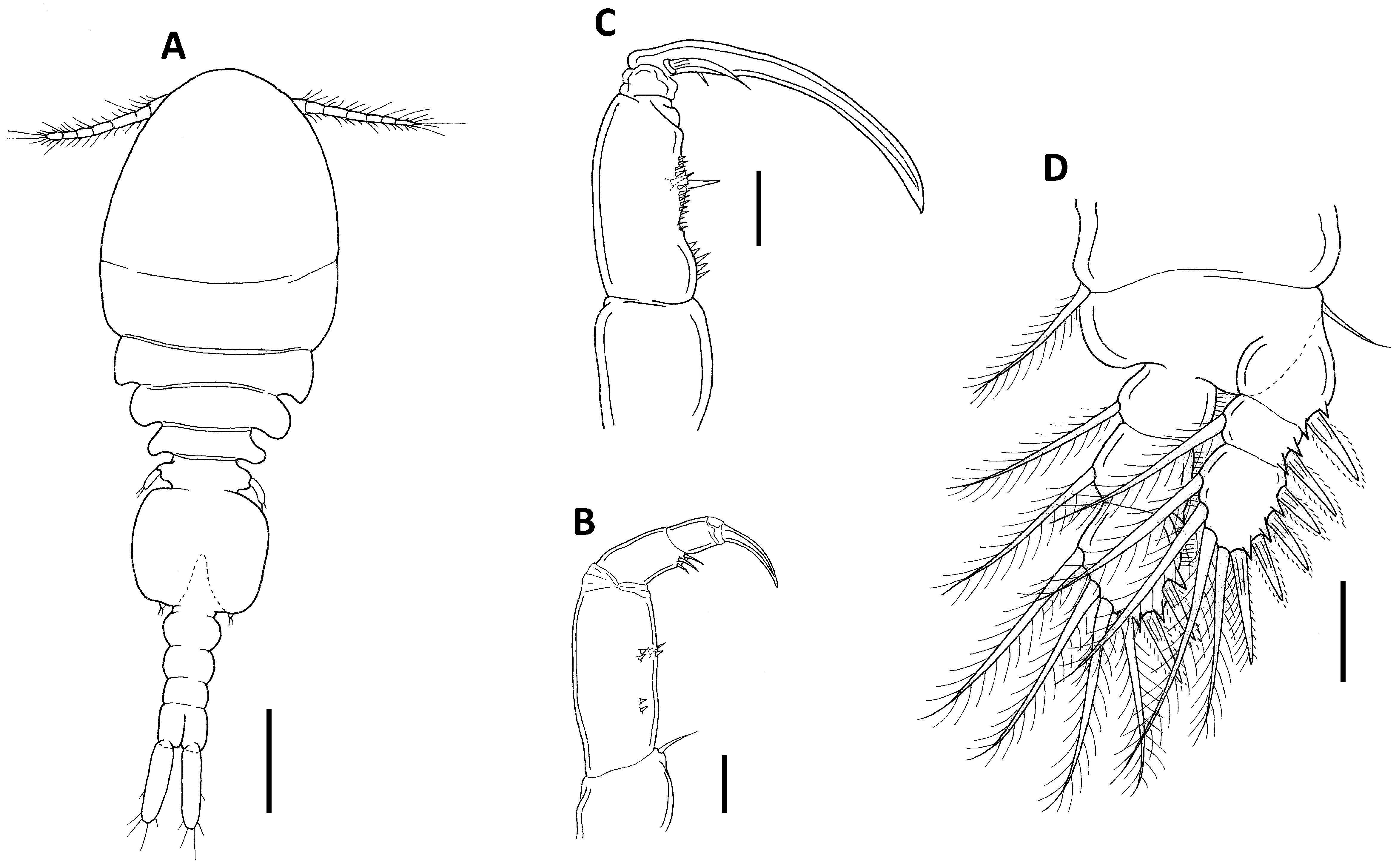
TABLE 2. Differences between Odontomolgus actinophorus, O. cognatus, O. mucosus (Taiwan), O. mucosus (Moluccas) and O. unioviger.
| Length × width of caudal ramus (µm) 139 × 40 | 185 × 55 | 87 × 25 | 96 × 22 | 67 × 32 |
|---|---|---|---|---|
| Shape of the genital double-somite rounded | rounded | rounded | rounded | quadrangular |
| Spinules on leg 5 exopod present | absent | present | absent | present |
| Length × width of leg 5 exopod (µm) 141 × 67 | 190 × 75 | 65 × 13 | 76 × 13 | 58 × 17 |
No known copyright restrictions apply. See Agosti, D., Egloff, W., 2009. Taxonomic information exchange and copyright: the Plazi approach. BMC Research Notes 2009, 2:53 for further explanation.
|
Kingdom |
|
|
Phylum |
|
|
Class |
|
|
Order |
|
|
Family |
|
|
Genus |