
Sociellus subgeminus, Cheng, Yu-Rong, Ho, Ming-Jay & Dai, Chang-Feng, 2016
|
publication ID |
https://doi.org/ 10.11646/zootaxa.4174.1.19 |
|
publication LSID |
lsid:zoobank.org:pub:05CD698B-A523-42C6-96F0-7D97156BA447 |
|
DOI |
https://doi.org/10.5281/zenodo.5675448 |
|
persistent identifier |
https://treatment.plazi.org/id/11452501-CD2E-D830-E9D4-FC6743C3FAEC |
|
treatment provided by |
Plazi |
|
scientific name |
Sociellus subgeminus |
| status |
sp. nov. |
Sociellus subgeminus sp. nov.
( Figs 8–10 View FIGURE 8 View FIGURE 9 View FIGURE 10 )
Type host. Pavona explanulata ( Lamarck, 1816) (family Agariciidae ).
Location in host. Surface of colony.
Type locality. Yenliao Bay , northern Taiwan.
Etymology. The specific name “ subgeminus ” refers to the similarity between body shape of this new species and its congener, Sociellus geminus Kim, 2006 .
Type material. Eighteen females and eight males obtained from washings of a coral colony collected at five m depth on 12 August 2010. The female holotype (NTUIO-COPE 7), male allotype (NTUIO-COPE 8), and paratypes (NTUIO-COPE s9; 15 females and five males) are deposited in the Institute of Oceanography , National Taiwan University, Taipei, Taiwan .
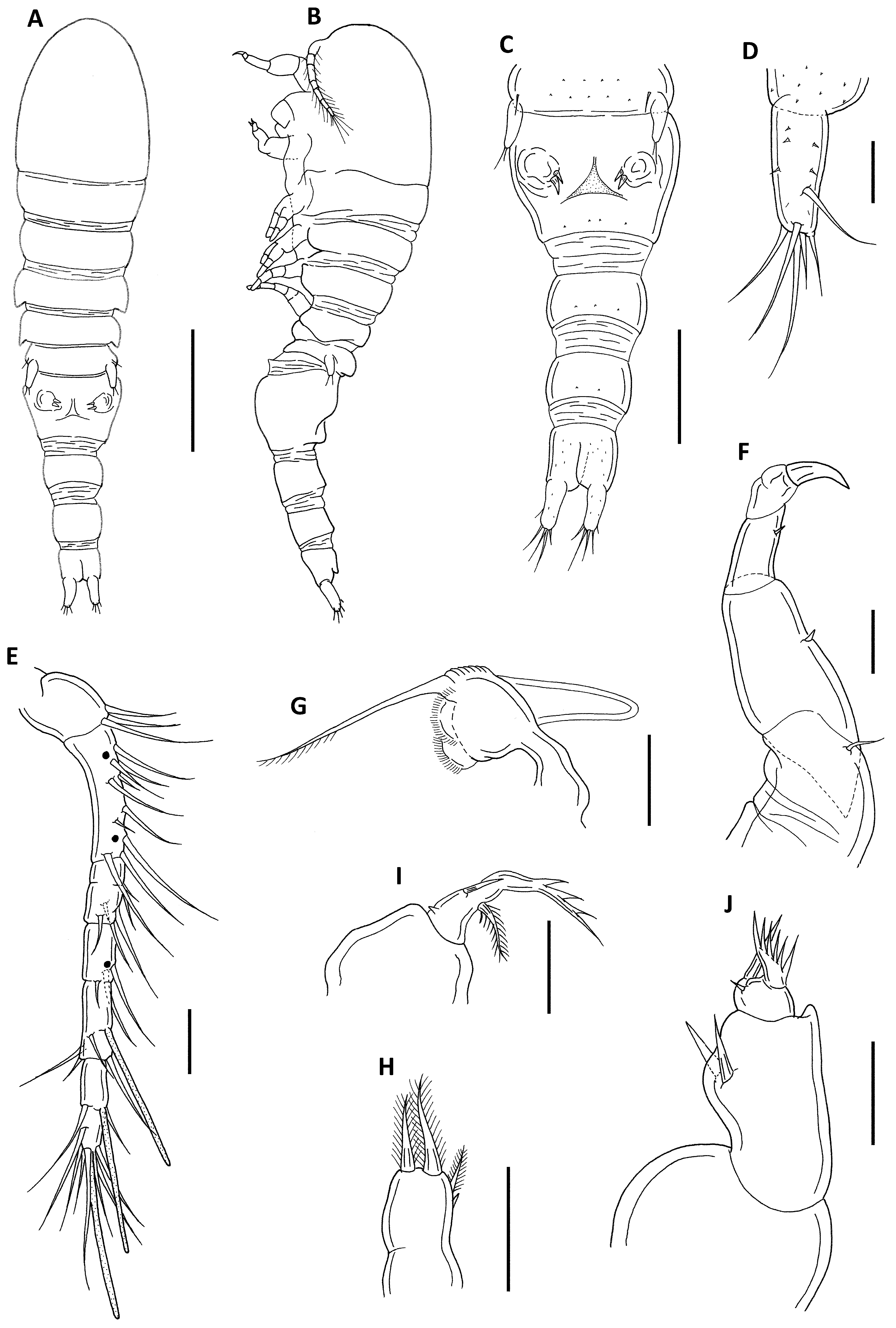
Description of female. Body ( Fig. 8 View FIGURE 8 A–B) elongate and slender. Length 0.94 mm (0.92–0.96 mm) and greatest width 0.23 mm (0.19–0.24 mm), based on five specimens. Cephalosome delimited from first pedigerous somite by dorsal furrow. Cephalosome 273 × 233 µm, long than wide. Measurements (length × width) of first to fifth pedigerous somites 75 × 206 µm, 64 × 187 µm, 56 × 193 µm, 54 × 163 µm, and 50 × 150 µm, respectively. Urosome ( Fig. 8 View FIGURE 8 C) 5-segmented. Genital double-somite in dorsal view 130 × 160 µm. Genital apertures located dorsally near middle of double-somite. Three postgenital somites 56 × 100 µm, 62 × 78 µm, and 50 × 68 µm, respectively. Anal operculum projecting posteriorly ( Fig. 8 View FIGURE 8 B). Caudal ramus ( Fig. 8 View FIGURE 8 D) 50 × 18µm, with six naked setae and several minute spinules. Surface of body with small setules ( Fig. 8 View FIGURE 8 C). Egg sacs not observed.
Antennule ( Fig. 8 View FIGURE 8 E) 7-segmented; armature: 3, 10, 6, 3, 4 + 1 aesthetasc, 2 + 1 aesthetasc, 7 + 1 aesthetasc; all setae naked. Antenna ( Fig. 8 View FIGURE 8 F) 4-segmented; armature formula 1, 1, 1, I.
Mandible ( Fig. 8 View FIGURE 8 G) with prominent proximal notch; inner margin distinctly bilobate; convex side with strongly tapering process; terminal lash slender with spinules. Maxillule ( Fig. 8 View FIGURE 8 H) with two plumose apical setae, one plumose subapical seta, and one minute spiniform element laterally. Maxilla ( Fig. 8 View FIGURE 8 I) 2-segmented, first segment unarmed; second segment with lash showing three small serrations along convex margin, one small spiniform element, one relatively large anterior seta, and one plumose inner sea. Maxilliped ( Fig. 8 View FIGURE 8 J) 3-segmented, syncoxa unarmed; basis with two setae along medial margin; small endopodal segment with one large spiniform process bearing spinules along outer side, one spine and one minute subapical seta.
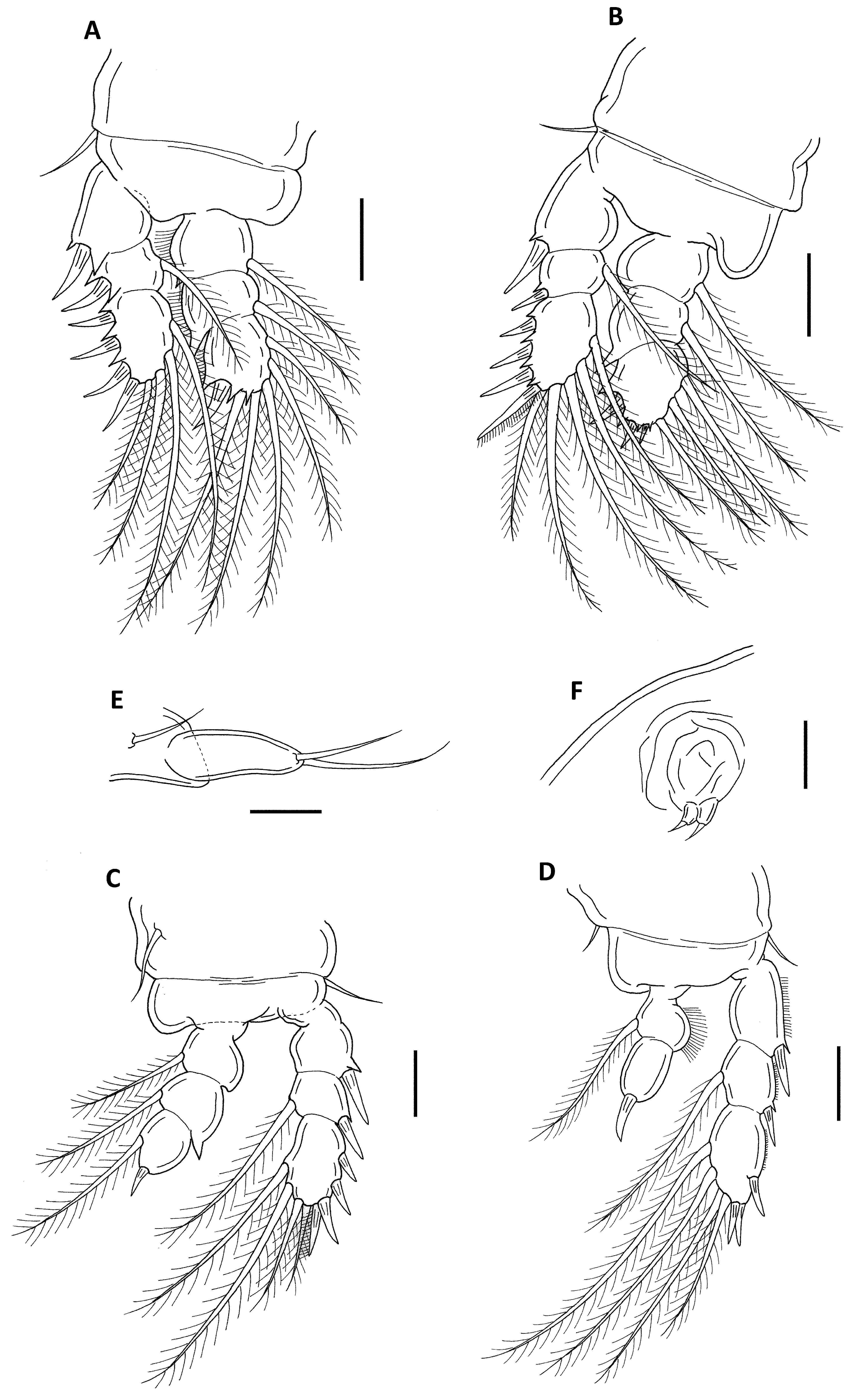
Legs 1–4 ( Fig. 9 View FIGURE 9 A–D) with 3-segmented exopods and endopods (except for leg 4 endopod being 2- segmented). Legs 1–2 lacking inner coxal seta. Formula of spines (in Roman numerals) and setae (in Arabic numerals) as follows:
Leg 5 ( Fig. 9 View FIGURE 9 E) with unornamented free segment, bearing two apical setae and small adjacent dorsal (= outer basal) seta.
Leg 6 ( Fig. 9 View FIGURE 9 F) represented by two small setae arising from operculum closing off genital aperture.
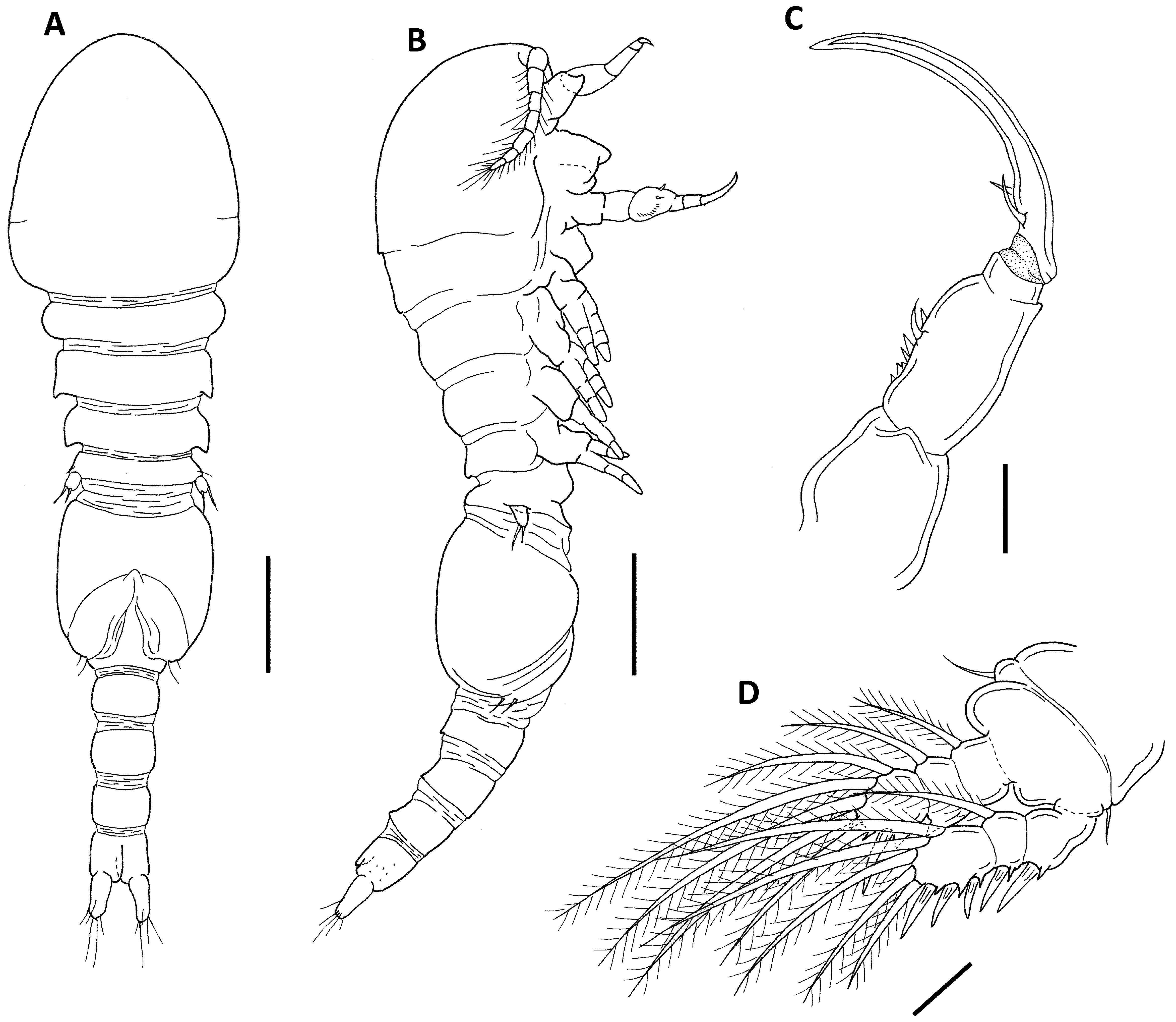
Description of male. Body ( Fig. 10 View FIGURE 10 A–B) elongate, slender, similar in shape to that of female. Length 0.74 mm (0.71–0.79 mm) and greatest width 0.19 mm (0.18-0.19 mm), based on five specimens. Caudal ramus as in female.
Antennule, antenna, mandible, maxillule and maxilla as in female except for antennule with three additional long aesthetascs (positions indicated by dots in Fig. 8 View FIGURE 8 E). Maxilliped ( Fig. 10 View FIGURE 10 C) 4-segmented, syncoxa broadest, unarmed; basis with two unequal setae and spinules near medial margin; first endopodal segment very short and unarmed; second endopodal segment represented by large claw, bearing two setae at its base.
Legs 1–4 as in female except for leg 1 ( Fig. 10 View FIGURE 10 D); third exopodal segment of leg 1 armed with three spines and five setae instead of four spines and four setae in female.
Leg 5 ( Fig. 10 View FIGURE 10 A–B) small free segment (exopod) with two setae and one adjacent dorsal (= outer basal) seta as in female.
Leg 6 ( Fig. 10 View FIGURE 10 A–B) represented by two small setae on posteroventral operculum on genital somite. Remarks. The genus Sociellus currently contains only two species: Sociellus torus Humes, 1992 from the Great Barier Reef, northeastern Australia and Sociellus geminus from the Moluccas. Both species utilize the scleractinian host Gardineroseris planulata ( Humes 1992; Kim 2006). Sociellus subgeminus sp. nov. differs from its two congeners by the differences summarized in Table 3.
S. geminus S. torus S. subgeminus sp. nov.
Body size (mm) 0.97 0.75–0.82 0.92–0.96 Armature of second segment of antenna 3 setae 3 setae 1 seta
Armature of maxillule 4 setae 2 setae 4 setae Armature of first endopodal segment of leg 1 0-0 0-0 0-1
Armature of third endopodal segment of leg 1 I +2 or I+ 3 I + 2 I +5
Armature of third exopodal segment of leg 2 IV + 4 IV + 4 IV +5
Armature of first endopodal segment of leg 2 0-0 0-0 0-1
Armature of second endopodal segment of leg 2 0-2 0-2 0-1
Armature of third exopodal segment of leg 3 IV + 2 IV + 1 IV +4
Leg 3 endopod 3-segmented 2-segmented 3-segmented Armature of third exopodal segment of leg 4 III +3 or IV+ 3 III + 2 III +4
Leg 4 endopod 2-segmented 1-segmented 2-segmented Humes & Boxshall (1996) placed Sociellus in the Rhynchomolgidae , presumably because the type species S. torus displayed a mandible with a linear inner margin (cf. Humes 1992: Fig. 10 View FIGURE 10 ). Kim (2006) proposed that the genus Sociellus should be placed in the family Anchimolgidae since the second species of the genus, S. geminus , exhibits the typical anchimolgid type of mandible with a distinctly bilobate inner margin, suggesting that Humes’ (1992) description is inaccurate. The position of Sociellus in the Anchimolgidae is further corroborated by the mandibular morphology of S. subgeminus sp. nov. The presence of a large process on the convex margin of the mandible in all three members of Sociellus suggests that the genus is closely related to the genera of the Odontomolgus -group.
No known copyright restrictions apply. See Agosti, D., Egloff, W., 2009. Taxonomic information exchange and copyright: the Plazi approach. BMC Research Notes 2009, 2:53 for further explanation.