
Calagasma margarita Bergroth, 1914
|
publication ID |
https://doi.org/10.11646/zootaxa.4170.2.6 |
|
publication LSID |
lsid:zoobank.org:pub:43CD8644-9C39-40C9-96B7-4423B200E70C |
|
DOI |
https://doi.org/10.5281/zenodo.6080891 |
|
persistent identifier |
https://treatment.plazi.org/id/115487BC-FFD8-FF97-FF6B-DBAAAB2CFD84 |
|
treatment provided by |
Plazi |
|
scientific name |
Calagasma margarita Bergroth, 1914 |
| status |
|
Calagasma margarita Bergroth, 1914
Calagasma margarita Bergroth, 1914: 432 –433 (description); Rolston, 1987: 69 –70 (synonymy).
Materiel examined (n: 6). French Guiana, 1 ♂ syntype in Bergroth’s collection at the MZHF. There is no locality label associated with this specimen, but only a handwritten label " Calagasma margarita B." ( Fig. 1 View FIGURE 1. A D, also available at http://id.luomus.fi/GV.39157) . French Guiana, Roura ( Montagne des Chevaux RN 2 pK 22): 24.I.2010, 1 ♀, glass flight interception trap ; 20.I.2013, 1 ♀ ( Fig. 3 View FIGURE 3 D), Lumivie® trap with blue LED (Light- Emitting Diode); 24.II.2013, 1 ♀, Lumivie® trap with blue LED; 26.VII.2014, 1 ♂ ( Figs. 1 View FIGURE 1. A E & F, 3A, B, C), GemLight® trap with one UV LED and one green LED. Régina (Les Nouragues, Inselberg) : 7.I.2013, 1 ♂, glass flight interception trap, SEAG leg. (Roland Lupoli collection). The interception and automatic light traps collect the insects in a liquid medium which were harvested weekly all year long ( Dalens & Touroult, 2014). The insects were sorted by family by the Société Entomologique Antilles-Guyane ( SEAG) as part of their French Guiana insect identification program.
Measurements. Total length: 8.4 mm (8.1–8.6); pronotum width (at humeral angles): 5.7 mm (5.4–6.0); abdomen width: 6.7 mm (6.4–6.9); head length: 1.2 mm; head width across the eyes: 1.9 mm; pronotum length: 1.7 mm. Antennomers I: 0.5 mm, II: 2.4 mm, III: 2.6 mm, IV: 2.4 mm.
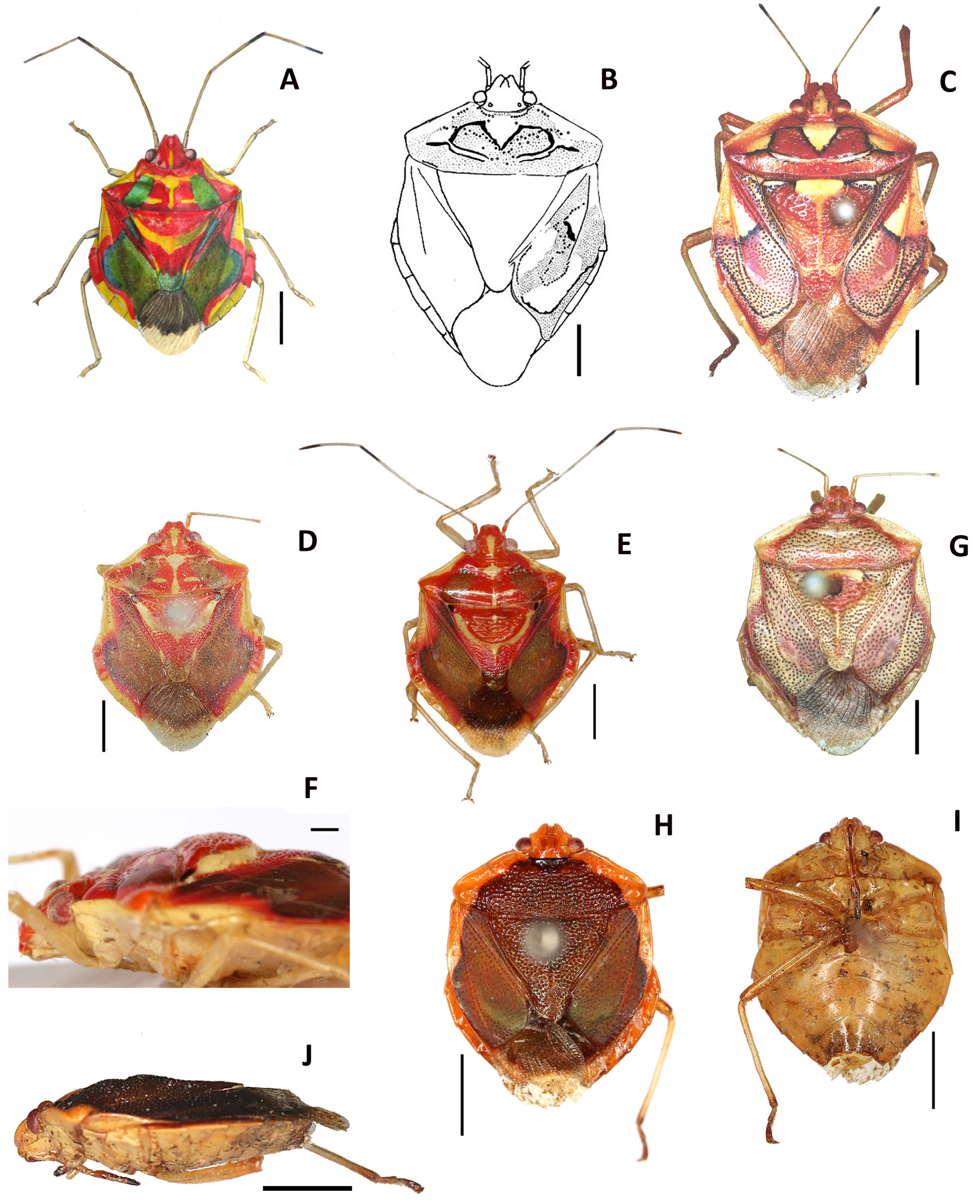
Diagnosis. Background dorsal color red, with two dorsolateral greenish-brown symmetrical spots on the pronotum. More than half of the basal portion of each corium matt greenish-brown, which gives it the appearance of leather texture, the same color as the two pronotal spots ( Fig. 1A View FIGURE 1. A , D, E). U-shaped impunctate calloused yellow spot present on the central part of the scutellum ( Fig. 1A View FIGURE 1. A ), marking a slope in the scutellum between the raised apical part and the lower basal part which are both red with concolorous punctation. Character not visible in dorsal view ( Fig. 1A View FIGURE 1. A ) but in lateral view ( Fig. 1 View FIGURE 1. A F). Antennomers I rufous and relatively shorter, not exceeding the apex of the head, antennomers II yellow, antennomers III and IV yellow with black spots at the tip. Antennomers II shorter than antennomers III. Mandibular plates parallel along lateral margins. Anterolateral margins of the pronotum slightly concave. Callus raised upward. Venter brownish yellow as the legs. Tibiae cylindrical.
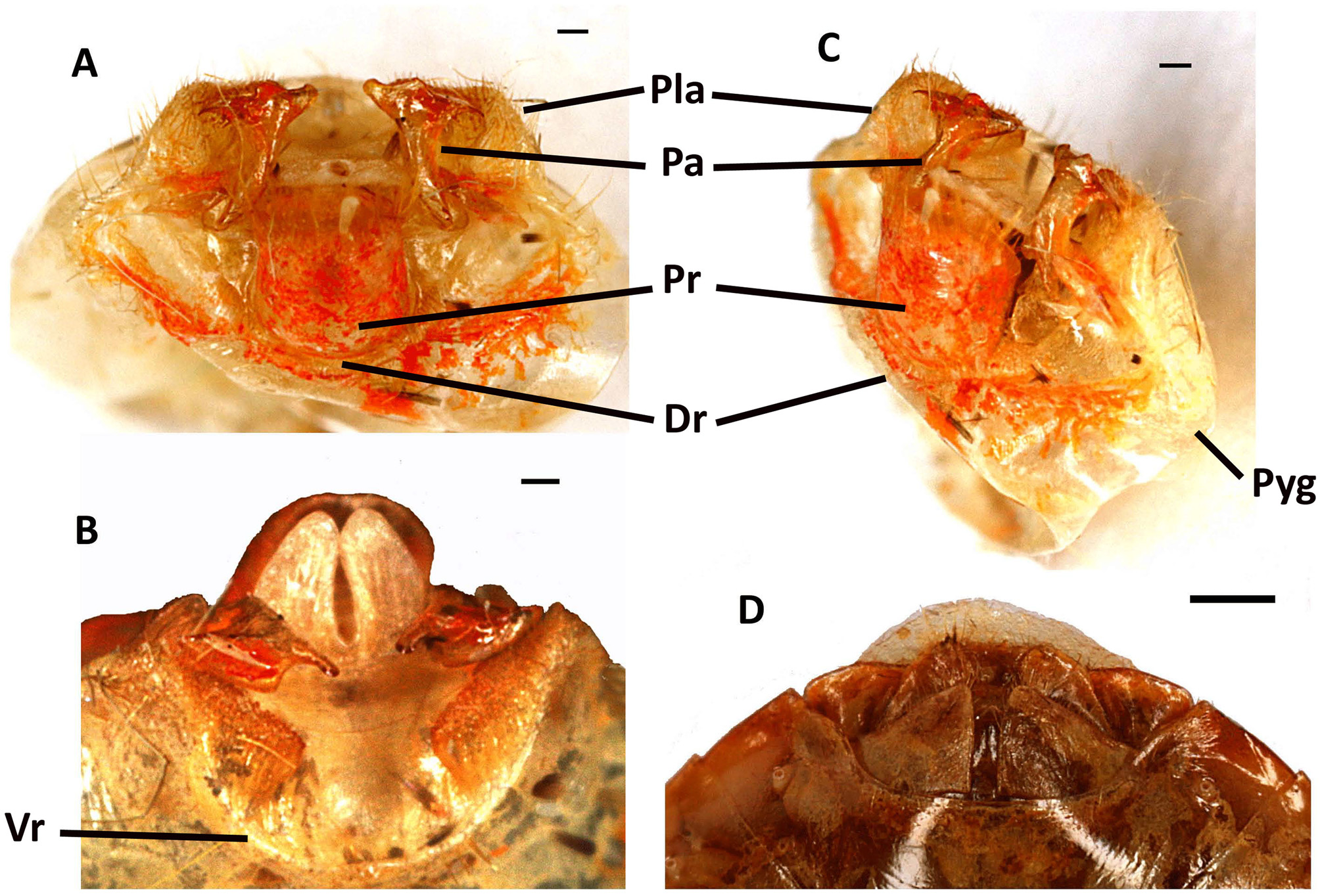
Female genital plates with posterior margins of laterotergites 8 almost flat and slightly rounded on their outer edge ( Fig. 3 View FIGURE 3 D). Similarly, posterior margins of laterotergites 9 with a rounded end, and gonocoxites 8 triangular.
Pygophore soft, thin and slightly sclerotized, mainly yellow with red spots ( Fig. 3 View FIGURE 3 A, B, C). Parameres and proctiger rufous. Ventral rim and dorsal rim poorly developed descending very low, just forming a cuticular fold, lined with red for dorsal rim, leaving proctiger and parameres completely visible in the dorsal view. Posterolateral angles of pygophore ear-shaped and covered with setae that continue along dorsal rim. Parameres posteroventrally directed, shaped like carpenter's hammers and positioned flat in pygophore, with the widest parts face to face in the dorsal view, protruding slightly on the ventral side. Proctiger cylindrical. Yellow bilobed process located just behind proctiger ( Fig. 3 View FIGURE 3 B).
Comments. The characteristics of the six examined C. margarita specimens resemble the description of Bergroth (1914) and not that of Spinola (1837) and Rolston (1987). The comparison between the specimen collected in French Guiana 26.VII.2014 ( Fig. 1 View FIGURE 1. A E), the syntype from MZHF ( Fig. 1 View FIGURE 1. A D) and the Bergroth drawing ( Fig. 1A View FIGURE 1. A ) emphasizes the care and precision of Bergroth in his description.
Although the background color of C. margarita is red, but a brighter red than E. histrio , the overlying pattern is completely different. Bergroth did not mention the three yellowish-white triangular spots bordered with black on the pronotum of E. histrio ( Fig. 1 View FIGURE 1. A C). Similarly, the dorsolateral greenish-brown symmetrical spots on the pronotum and corium of C. margarita ( Fig. 1A View FIGURE 1. A ) are not present on the pronotum and corium of E. histrio ( Fig. 1 View FIGURE 1. A C), whereas in E. histrio the basal part of the corium is mainly pink and sporadically punctated by black dots, with a black line bordering the outline of the corium ( Fig. 1 View FIGURE 1. A C). The U-shaped yellow spot on the central part of the scutellum of C. margarita ( Fig. 1A View FIGURE 1. A ) is absent in E. histrio which has only a yellow spot on the basal edge of the scutellum ( Fig. 1 View FIGURE 1. A C). The enhanced apical part of the scutellum is also absent in E. histrio . C. margarita is about 25–30 % smaller than E. histrio . The body of C. margarita is more circular than the body of E. histrio which is more oval. Also, the anterolateral borders of the pronotum are convex in E. histrio whereas they are concave in C. margarita . C. margarita antennomers II have no black spot at their tips ( Fig. 1A View FIGURE 1. A ) although E. histrio antennomers II have black spots. Spinola and Rolston also stated that antennomers II are longer than antennomers III in E. histrio , whereas the reverse is true in C. margarita ( Fig. 1A View FIGURE 1. A ). Rolston mentioned that antennomers I surpasse the apex of the head in E. histrio . This is apparent in the specimen of UFPR ( Fig. 1 View FIGURE 1. A C). In contrast, in C. margarita , antennomers I are relatively shorter and do not exceed the apex of the head ( Fig. 1A View FIGURE 1. A ). Mandibular plates are parallel in C. margarita ( Fig. 1A View FIGURE 1. A ) whereas they are strongly reflexed along the lateral margins in E. histrio . The tibiae of C. margarita are actually cylindrical ( Fig. 1 View FIGURE 1. A E), whereas they are triangular with a wider apex in E. histrio . Female genital plates ( Fig. 3 View FIGURE 3 D) are clearly different from those of E. histrio ( Figs. 2A View FIGURE 2. A & B). The posterior margins of laterotergites 8 are almost flat and slightly rounded on their outer edge in C. margarita whereas they form an acute right angle in E. histrio . Similarly, the posterior margins of laterotergites 9 in C. margarita have a rounded end whereas they form a 45° acute angle in E. histrio . The gonocoxites 8 are subrectangular in E. histrio whereas they are triangular in C. margarita . Then, all these data confirm that C. margarita is not a junior synonym of E. histrio but a valid species.
Distribution. FRENCH GUIANA, Régina and Roura.

No known copyright restrictions apply. See Agosti, D., Egloff, W., 2009. Taxonomic information exchange and copyright: the Plazi approach. BMC Research Notes 2009, 2:53 for further explanation.
|
Kingdom |
|
|
Phylum |
|
|
Class |
|
|
Order |
|
|
Family |
|
|
Genus |
Calagasma margarita Bergroth, 1914
| Lupoli, Roland 2016 |
Calagasma margarita
| Rolston 1987: 69 |
| Bergroth 1914: 432 |