
Epipedus histrio Spinola, 1837
|
publication ID |
https://doi.org/10.11646/zootaxa.4170.2.6 |
|
publication LSID |
lsid:zoobank.org:pub:43CD8644-9C39-40C9-96B7-4423B200E70C |
|
DOI |
https://doi.org/10.5281/zenodo.6080887 |
|
persistent identifier |
https://treatment.plazi.org/id/115487BC-FFDC-FF93-FF6B-D9ECAA83FE8E |
|
treatment provided by |
Plazi |
|
scientific name |
Epipedus histrio Spinola, 1837 |
| status |
|
Epipedus histrio Spinola, 1837
Epipedus histrio Spinola, 1837: 315 –316; Rolston, 1987: 69 –70; Grazia & Campos, 2010
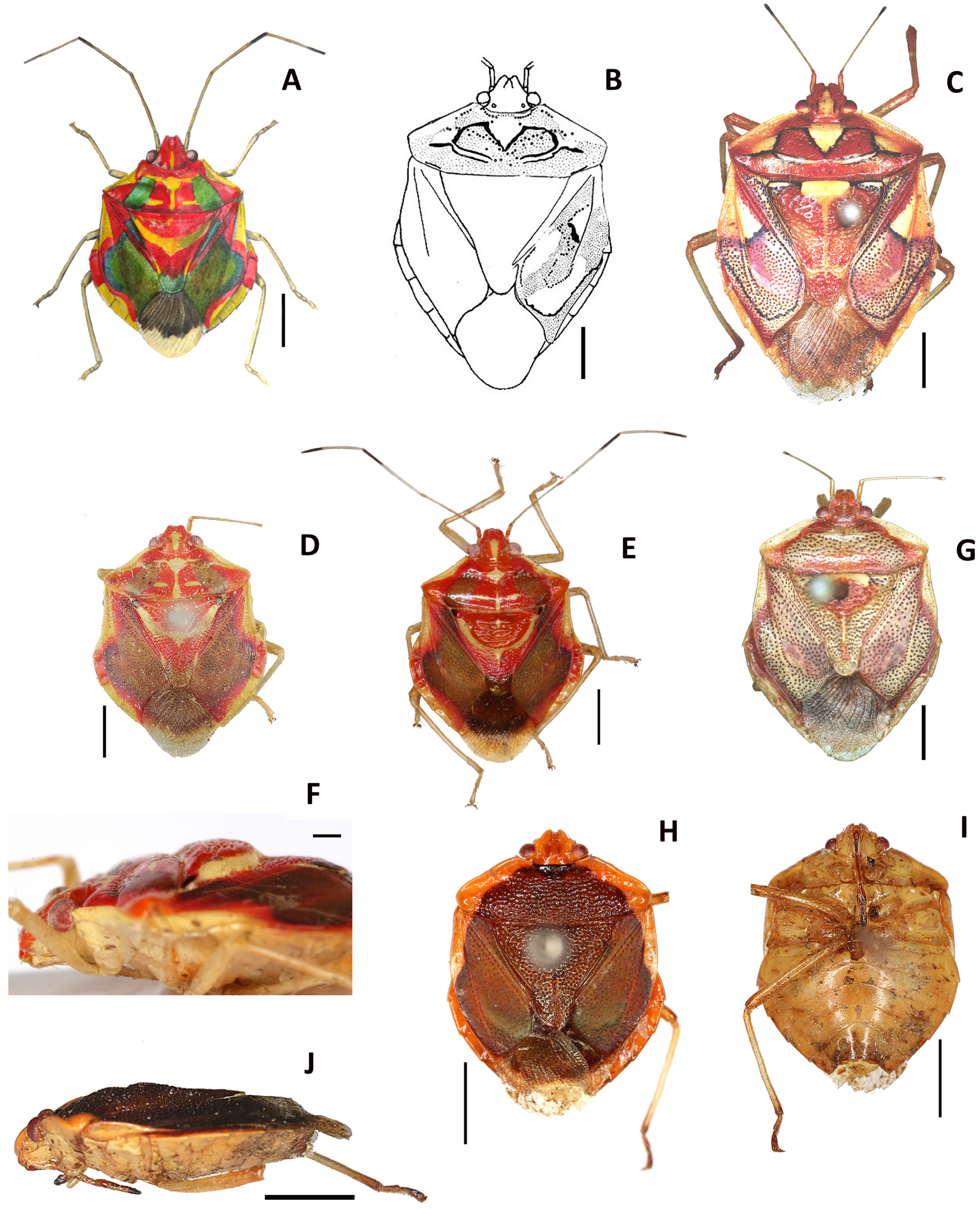
Material examined (n: 1). Brazil, Rio de Janeiro ( Corcovado Mountain ): 1961, 1 ♀ Alvarenga & Seabra leg. ( UFPR) ( Fig. 1 View FIGURE 1. A C).
Measurements. Total length: 11.6 mm; pronotum width: 7.1 mm; abdomen width: 8.2 mm; head length: 1.3 mm; head width across the eyes: 2.2 mm; pronotum length: 2.2 mm; antennomers I: 0.7 mm; II: 3.1 mm; other antennomers are missing. Measurements were performed on photos digitalized with a precision scale, by expanding, measuring and comparing them on a computer screen. Spinola mentioned a body length of 5 lines, i.e. 11.3 mm and a width of 3.5 lines, i.e. 7.9 mm, and Rolston a body length of 12.2 mm, a width of 8.5 mm. and the following lengths of antennomers: I: 0.8 mm, II: 3.6 mm, III: 2.6 mm, IV: 2.5 mm.
Diagnosis. Global dorsal color mostly rufous with eight contrasting brownish yellow spots bordered by black lines ( Fig. 1 View FIGURE 1. A C). Clypeus and vertex brownish yellow, bordered on each side by a black line. Mandibular plates rufous and reflexed along lateral margins, converging over clypeus apically. Clypeus slightly longer than mandibular plates. Antennomers I and II rufous. Spinola (1837) stated that antennomers II are longer than antennomers III and this was confirmed by Rolston (1987). Distal tenth of antennomers II fuscous. Antennomers I surpassing apex of head. Anterolateral margins of the pronotum convex. Pronotum with extensive rufous markings and concolorous punctures partially enclosed in V-shaped black lines, forming three V-shaped brownish yellow spots. Scutellum flat, mostly rufous with concolorous punctation. Impunctate brownish yellow macule covering most of the mesial half of the scutellar base. At the base of the scutellum on each side, macule and border along most of frenal margins brownish yellow, much of this border outlined in black. Median brownish yellow impunctate carina, starting from the basal tumescence and then disappearing before the apex. Corium greyish rufous with black deep punctations uniformly distributed, except in a large brownish yellow impunctate spot bordered by a black line in the apical part of exocorium and mesocorium, and in a rufous impunctate area in central mesocorium. Distal part of the corium surrounded by a narrow black submarginal line. Costal margin of each corium strongly reflexed basally, posterior margin sigmoid, costal angle acute. Connexivum yellow, impunctate, slightly exposed, less than half as broad as exocorium. Venter greyish yellow as well as legs. All tibiae broadly, shallowly sulcate apically otherwise cylindrical (right protibia and left metatibia visible in Fig. 1 View FIGURE 1. A C) as mentioned by Spinola (1837). Female genital plates with distal ends of the laterotergites 8 & 9 forming acute angles ( Fig. 2 View FIGURE 2. A B).
Comments: The description by Spinola (1837) of colorful patterns and markings on the dorsal side of E. histrio are consistent with the descriptions and drawings ( Fig. 1 View FIGURE 1. A B) by Rolston (1987) and the specimen of UFPR ( Fig. 1 View FIGURE 1. A C). Spinola wrote that head, pronotum, scutellum and cories are scarlet red with some yellowish-white spots. He mentioned three yellowish-white triangular spots bordered with black on the pronotum. These spots are not easy to see on the black and white figure of Rolston ( Fig. 1 View FIGURE 1. A B) although they are clearly visible and contrasted in the specimen of UFPR ( Fig. 1 View FIGURE 1. A C). This specimen has dimensions and shape of the body similar to those observed by Spinola and Rolston. Spinola mentioned that the first antennomer is reddish in color and that the others are yellowish with a black spot at the tip. This is the case for the specimen of UFPR ( Fig. 1 View FIGURE 1. A C) for antennomers I and II. On the other hand, the female genital plates of this specimen ( Fig. 2 View FIGURE 2. A B) are very similar to those of the specimen in the Rolston drawing ( Fig. 2A View FIGURE 2. A ) particularly the acute shapes of laterotergites 8 & 9.
Distribution. BRAZIL, Rio de Janeiro.

No known copyright restrictions apply. See Agosti, D., Egloff, W., 2009. Taxonomic information exchange and copyright: the Plazi approach. BMC Research Notes 2009, 2:53 for further explanation.
|
Kingdom |
|
|
Phylum |
|
|
Class |
|
|
Order |
|
|
Family |
|
|
Genus |
Epipedus histrio Spinola, 1837
| Lupoli, Roland 2016 |
Epipedus histrio
| Rolston 1987: 69 |
| Spinola 1837: 315 |