
Zancleopsis dichotoma ( Mayer, 1900 )
|
publication ID |
https://doi.org/ 10.1163/18759866-BJA10023 |
|
publication LSID |
lsid:zoobank.org:pub:86E35163-0808-44CD-A1F8-D7FB607EFC1B |
|
DOI |
https://doi.org/10.5281/zenodo.8357094 |
|
persistent identifier |
https://treatment.plazi.org/id/12088786-8E24-FF81-FD57-61FAE7E4FA65 |
|
treatment provided by |
Felipe |
|
scientific name |
Zancleopsis dichotoma ( Mayer, 1900 ) |
| status |
|
Zancleopsis dichotoma ( Mayer, 1900) View in CoL View at ENA
Gemmaria dichotoma Mayer, 1900: 35 View in CoL , pl. 17 fig. 40.
Downloaded from Brill.com 10/07/2022 07:01:58 PM
via free access
Zancleopsis dichotoma View in CoL View at ENA . Hartlaub, 1907: 115, fig. 105; Schuchert & Collins, 2021 (small form): 272, fig. 22.
Examined material: Sample SN 035, Singapore, 28/11/2017, polyps in ethanol and formalin. – Sample BFLA4170 , off Florida, 09/08/2019, medusa in ethanol and in situ photos. – Sample BFLA4171 , off Florida, 09/08/2019, medusa in ethanol and in situ photos. –S ample BFLA4248 , off Florida, 23/11/2019, medusa in ethanol and in situ photos .
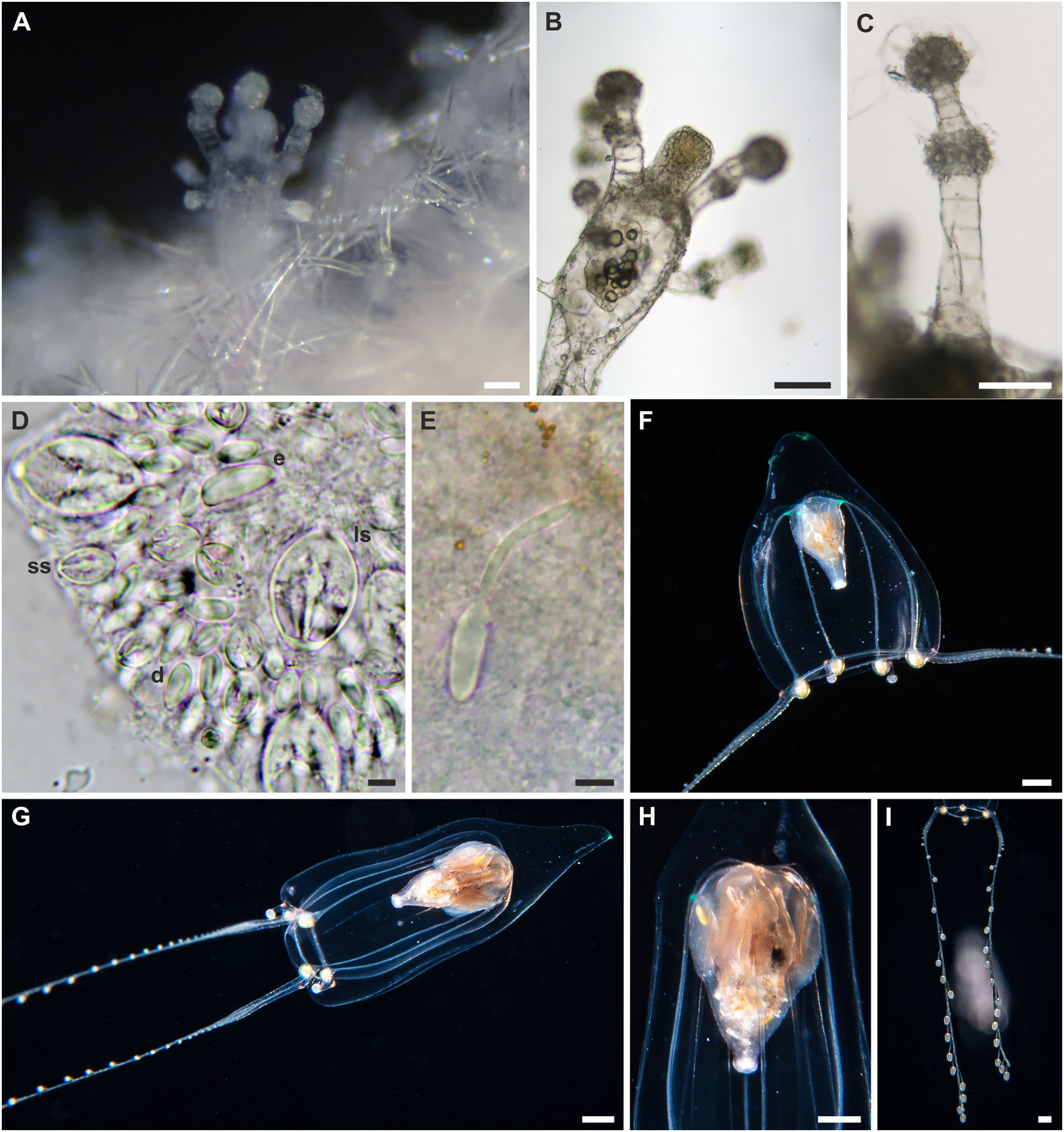
Description: Polyp. Colonies monomorphic, living in association with sponges ( fig. 7A View FIGURE 7 ). Hydrorhiza tubular and covered by a thin perisarc, embedded by the sponge host. Pedicels short and completely embedded in the sponge host, unbranched, covered by a thin perisarc. Hydranth slightly pyriform, up to 0.7 mm long, with variable diameter (110–170 Μm) ( fig. 7A, B View FIGURE 7 ). Hypostome proboscis-like ( fig. 7B View FIGURE 7 ). Tentacles organised in two alternating whorls of 4–5 tentacles ( fig. 7A View FIGURE 7 ). Each tentacle with terminal and sub-terminal capitula (diameter: 60–100 Μm in the distal whorl; 40–50 Μm in the proximal whorl) ( fig. 7C View FIGURE 7 ). Tentacles about 260–400 Μm long in the distal whorl, shorter in the proximal whorl (about 150–220 Μm). Nematocyst clusters 50–100 Μm distant from one other. Living hydranths transparent. Desmonemes, microbasic euryteles, small and large stenoteles ( figs. 7D, E View FIGURE 7 ) occurring simultaneously and concentrated in the capitula, as well as scattered in the hydrorhiza; microbasic euryteles also in the hydranth.
Polyp cnidome. i) Desmonemes (undischarged: 7–9 × 4 Μm; discharged capsule: 6 × 3 Μm). ii) Microbasic euryteles (undischarged: 14–15 × 5–6 Μm; discharged capsule: 13–14 × 5–6 Μm; shaft: 17–20 Μm). iii) Large stenoteles (undischarged: 22–23 × 14–16 Μm; discharged capsule: 13 × 20 Μm). iv) Small stenoteles (undischarged: 9–10 × 6–7 Μm; discharged capsule: 7 × 9 Μm).
Adult medusa (from Schuchert & Collins, 2021, small form only). Total bell height up to 3 mm, 1/4 to 1/3 of the height taken by pointed apical process ( fig. 7F, G View FIGURE 7 ); umbrella bell-shaped to conical, relatively thick walls, with shallow interradial subumbrellar pockets, tip of apical process green. Manubrium height about half the subumbrellar height when gonads start to develop, pear-shaped, short tubular oral part, mouth rim with four perradial white regions, upper part of manubrium (stomach) ochre coloured, with about 10 longitudinal, shallow gonad folds, folds mostly adradial, irregular ( fig. 7H View FIGURE 7 ). Radial canals not forming mesenteries, smooth. Tentacle bulbs all equally developed, almost spherical, placed adaxial of origin of tentacles, white or faintly yellow. Two long, opposite tentacles, much extendable/contractible, with up to 25 short, abaxial, side branches ending in capitula, size of capitula gradually increasing towards distal ( fig. 7I View FIGURE 7 ). The other tentacle pair very short, ending in spherical nematocyst knob. In young animals these short tentacles either missing or just beginning to develop. All tentacle bases with a red ocellus on abaxial side.
Adult medusa cnidome (preserved tissue). i) Desmonemes (8.5 × 5 µm). ii) Small stenoteles (18–21 × 14–17).iii) Larger stenoteles (24–26 × 22–23 µm). iv) Macrobasic euryteles (15–16 × 6–7 µm.
Distribution: Atlantic Ocean (Florida) and Indo-West Pacific ( Singapore).
Remarks: Schuchert & Collins (2021) found that Z. dichotoma medusae were genetically similar to A. cabela polyps. However, they found two genetically divergent morphs, one smaller (bell height of 3 mm) and the other larger (bell height up to 15 mm). The small morph better agrees with the original description of Z. dichotoma by Mayer (1900), and it was here linked through genetic data to Astrocoryne -like polyps from Singapore, supporting the synonymisation of the genus Astrocoryne with Zancleopsis .
| PM |
Pratt Museum |
No known copyright restrictions apply. See Agosti, D., Egloff, W., 2009. Taxonomic information exchange and copyright: the Plazi approach. BMC Research Notes 2009, 2:53 for further explanation.
|
Kingdom |
|
|
Phylum |
|
|
Class |
|
|
Order |
|
|
Family |
|
|
Genus |
Zancleopsis dichotoma ( Mayer, 1900 )
| Maggioni, Davide, Schuchert, Peter, Arrigoni, Roberto, Hoeksema, Bert W., Huang, Danwei, Strona, Giovanni, Seveso, Davide, Berumen, Michael L., Montalbetti, Enrico, Collins, Richard, Galli, Paolo & Montano, Simone 2021 |
Zancleopsis dichotoma
| Hartlaub, C. 1907: 115 |
Gemmaria dichotoma
| Mayer, A. G. 1900: 35 |