
Tuberculobasis, Machado, Angelo B. M., 2009
|
publication ID |
https://doi.org/ 10.5281/zenodo.187806 |
|
DOI |
https://doi.org/10.5281/zenodo.4391359 |
|
persistent identifier |
https://treatment.plazi.org/id/1312774B-FFD4-FFE5-FF04-FB4BFE04FE83 |
|
treatment provided by |
Plazi |
|
scientific name |
Tuberculobasis |
| status |
gen. nov. |
Tuberculobasis View in CoL gen. nov.
Type-species: Leptobasis mammilaris Calvert, 1909 , by present designation.
Etymology: From the Latin tuberculum – a tubercle and – basis, a Greek feminine noun for pedestal or foundation; a common suffix used in Coenagrionidae . The name Tuberculobasis refers to the presence of paired tubercles on mesepisterna of males and most females.
Distribution. South America ( Brazil, Colombia, Peru, Suriname, Venezuela) ( Fig. 101).
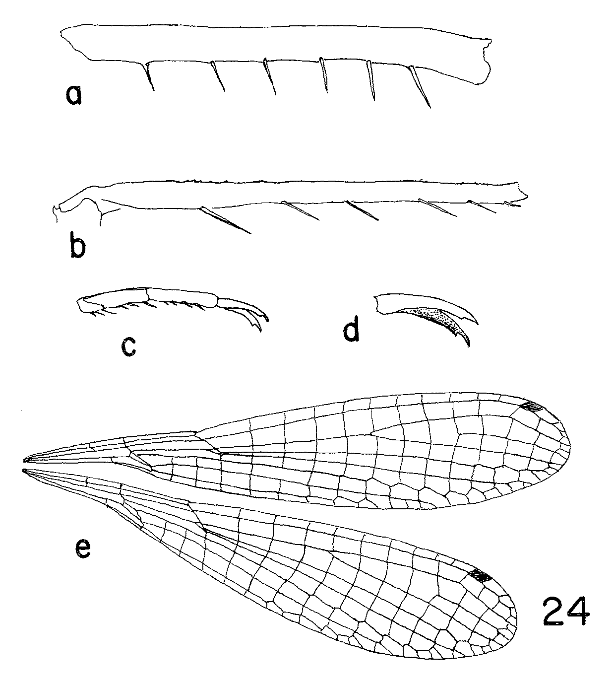
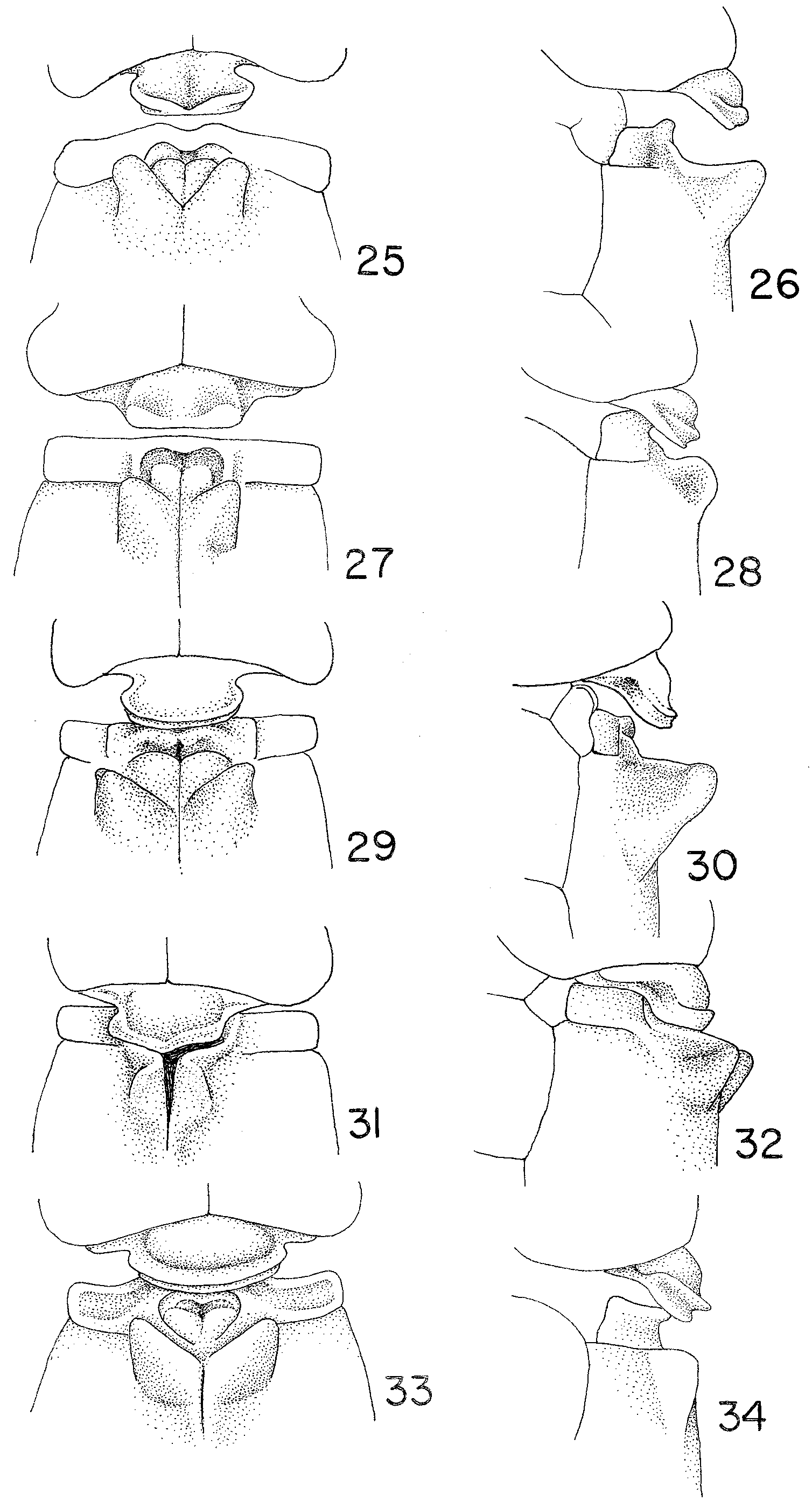
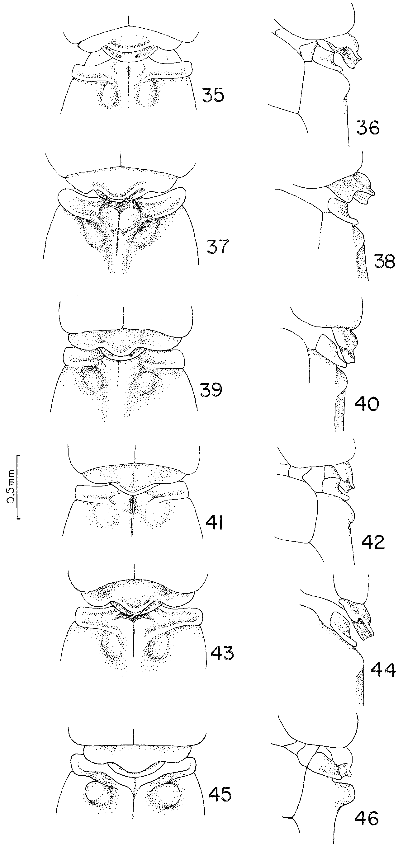
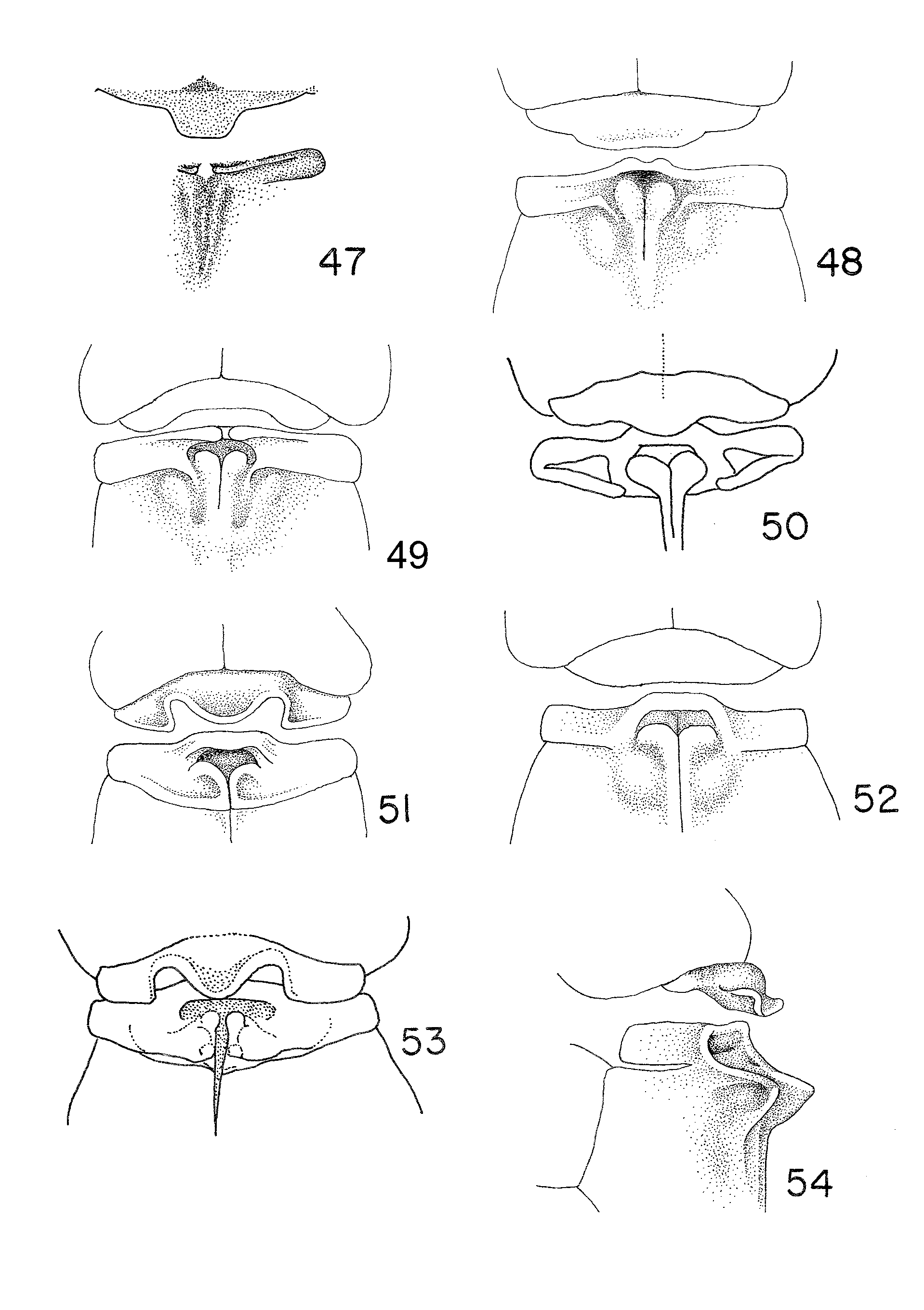
Generic characterization. Small to medium size damselflies (HW 15.5–22.0 mm). Top of head black or brown (reddish yellow in T. mammilaris ), with blue postocular spots (sometimes absent in T. mammilaris ). Thorax usually with three longitudinal pale blue or silver stripes: antehumeral, metepisternal, and sometimes metepimeral. S1–6 variously brownish or yellowish, S7–10 red, yellow or orange brown. Frons rounded. Hind prothoracic lobe in males with median lobe well-developed projected caudally and two-lipped ( Figs 25–45 View FIGURES 25 – 34 View FIGURES 35 – 46 ), in females single-lipped ( Figs 47–56 View FIGURES 47 – 54 View FIGURES 55 – 56 ). Mesepisterna with an antero-medial pair of tubercles present in all males and 64% of known females. Cercus in lateral view much shorter than S 10 in both sexes. Metafemoral spines on distal 1/2 as long as width of femur ( Fig. 24 View FIGURE 24 a); metatibial spurs shorter than intervening spaces ( Fig. 24 View FIGURE 24 b). Supplementary tooth of tarsal claw well-developed ( Figs 24 View FIGURE 24 c, d). Wings ( Fig. 24 View FIGURE 24 e) petiolated distally to Ac for a distance as long as Ac length (88.25%) or 1/2 as long ( Fig. 24 View FIGURE 24 e). R 3 in FW originating closer to Px 5–6 (more frequently at 6) in HW closer to Px 4–6 (more frequently at 5). Penis distal segment with distal 1/3 laterally expanded into a large lateral terminal lobe and with two lateral lobes on each side ( Figs 89–95 View FIGURES 87 – 94 View FIGURES 95 – 100 ). No terminal fold. Internal fold with a filiform process directed disto-laterally ( Figs. 89–93 View FIGURES 87 – 94 ). Male cercus domeshaped (Fig. 68), the distal part sharply decumbent forming a ventral process (ventro-medial in T. arara and T. costalimai ), which is perpendicular to the long axis of cercus. Female sternum S8 with a vulvar spine (except in T. cardinalis , see under that species). Ovipositor extending to about level of tip of cercus ( Figs 95–99 View FIGURES 95 – 100 ). Ventral border of lateral valve of ovipositor with one row of subtriangular denticles fine tipped directed ventro-posteriorly associated with a long seta tapering into a fine tip ( Fig. 2 View FIGURE 2 b).
Larvae unknown.


No known copyright restrictions apply. See Agosti, D., Egloff, W., 2009. Taxonomic information exchange and copyright: the Plazi approach. BMC Research Notes 2009, 2:53 for further explanation.