
Doriprismatica marinae Matsuda and Gosliner, 2018
|
publication ID |
https://doi.org/ 10.11646/zootaxa.4444.5.1 |
|
publication LSID |
lsid:zoobank.org:pub:6A536780-96AE-42B0-913E-C05767BC63EC |
|
DOI |
https://doi.org/10.5281/zenodo.5981644 |
|
persistent identifier |
https://treatment.plazi.org/id/FB55BFD1-FC47-48FE-9BC3-62C3EDA1E0C6 |
|
taxon LSID |
lsid:zoobank.org:act:FB55BFD1-FC47-48FE-9BC3-62C3EDA1E0C6 |
|
treatment provided by |
Plazi |
|
scientific name |
Doriprismatica marinae Matsuda and Gosliner |
| status |
sp. nov. |
Doriprismatica marinae Matsuda and Gosliner View in CoL sp. nov.
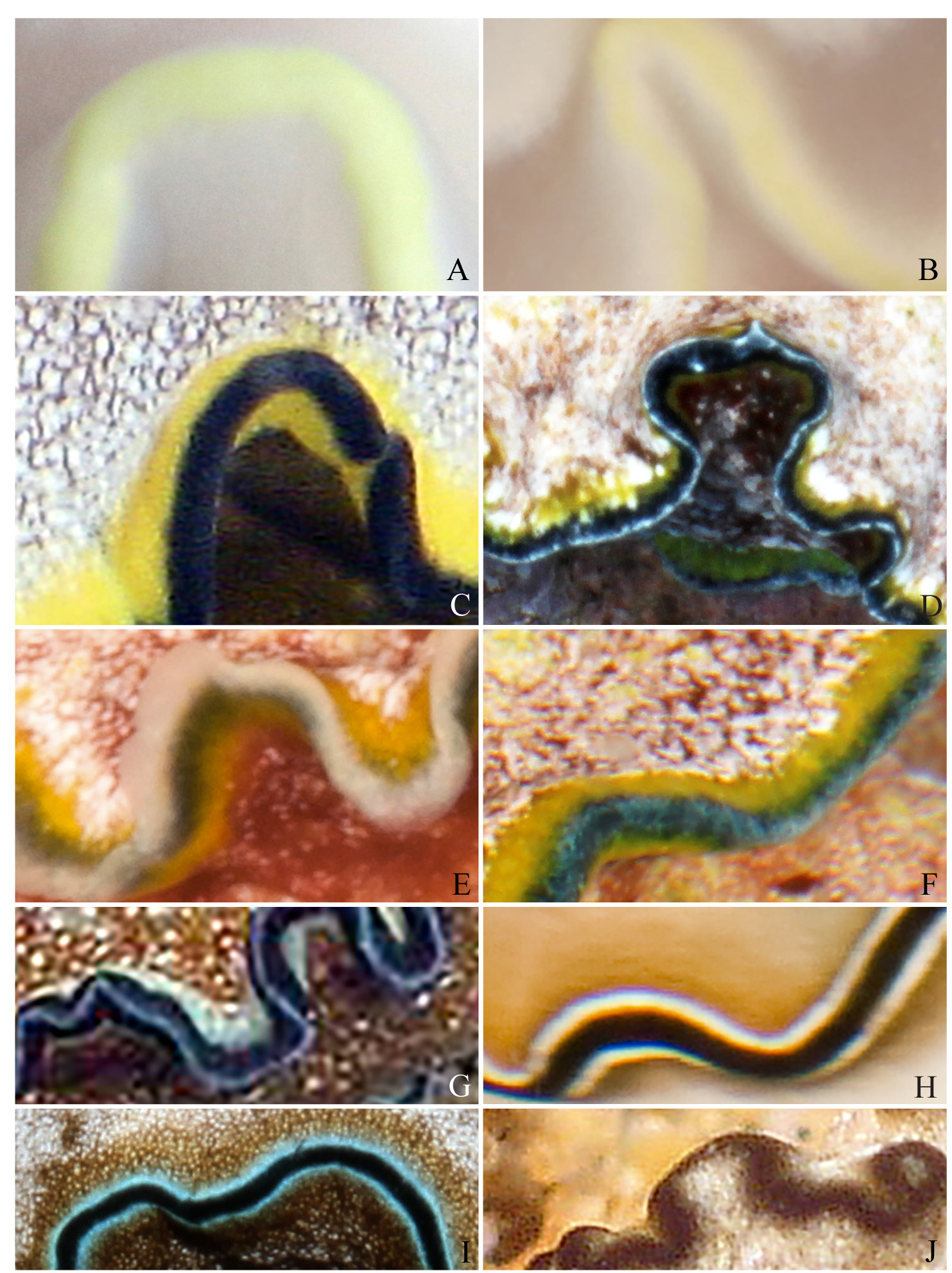
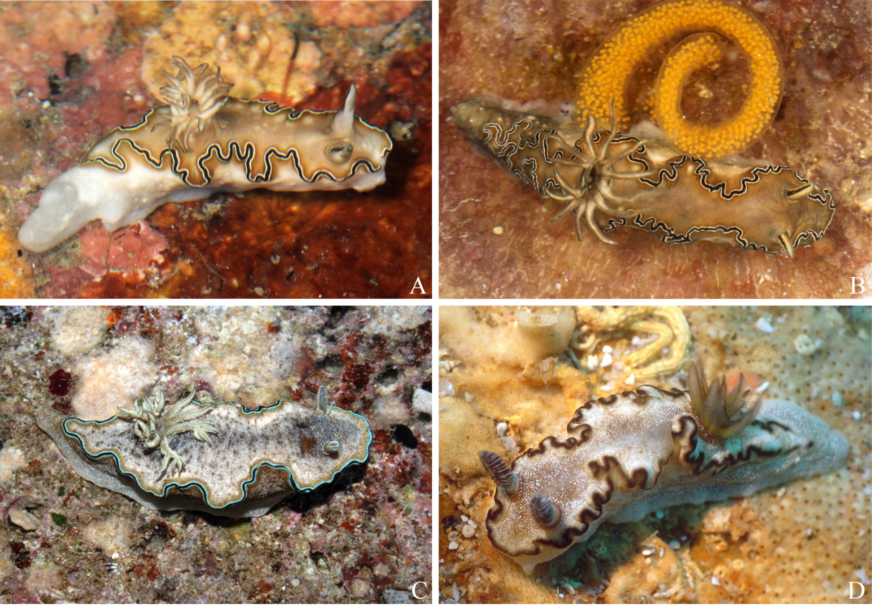
Figures (2J, 11D, 13F, G, 14F–L)
Doriprismatica sp. 6 Gosliner et al. 2015: p. 240, upper right photo.
Doriprismatica sp. C Matsuda & Gosliner 2017.
Type material. Holotype: CASIZ-194050, one specimen, dissected, 7.5 mm preserved, Madagascar, South Madagascar, “Pointe Evatra, crique fond rocheux et gazon d’algues”, 30 April and 6 May 2010, Expedition Atimo Vatae-South Madagascar Expedition 2010, 3-8 meters, orig. fixative 95% EtOH. This specimen was tissue sampled (foot) for DNA analysis in Matsuda & Gosliner (2017), GenBank: KT600691 View Materials ( COI).
Etymology. Doriprismatica marinae is named after Marina Poddubetskaia , who found and collected the type specimen.
Distribution. Madagascar.
External morphology. Doriprismatica marinae has a long mantle that tapers posteriorly ( Fig. 11D View FIGURE 11 ) and terminates gradually above the posterior end of the foot. The mantle is a warm brown color that is covered in white dots that are denser towards the edge of the mantle. The mantle is thrown into permanent and semi-permanent undulations that are outlined by one brown marginal mantle band ( Fig. 2J View FIGURE 2 ). There are two pairs of permanent folds, that are equally spaced between the rhinophores and the gills. The gill forms an arch around the anus opening posteriorly. The 10 lamellae are unipinnate and unifid apically and a cream color with two thin brown bands running up the outer edge and joining at the tip. The base of the rhinophores are a cream color and the rhinophores themselves are the same color brown as the mantle band with 12 white lamellae. The short, rounded oral tentacle is visible in Fig. 11D View FIGURE 11 . The genital opening is on the right side of the body just behind the rhinophores.
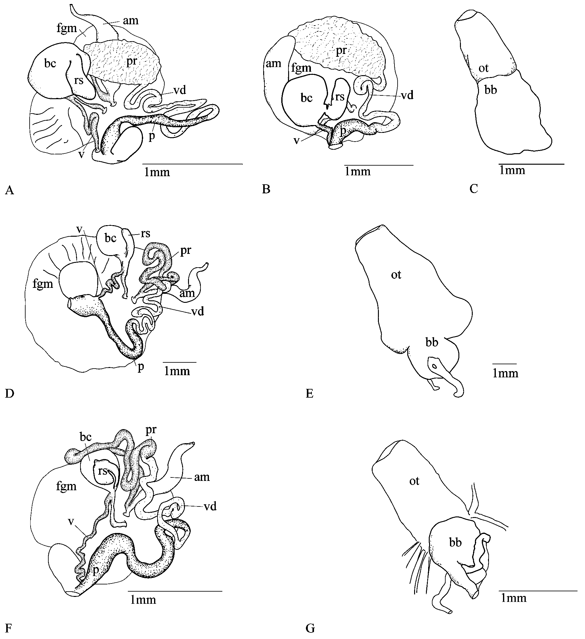
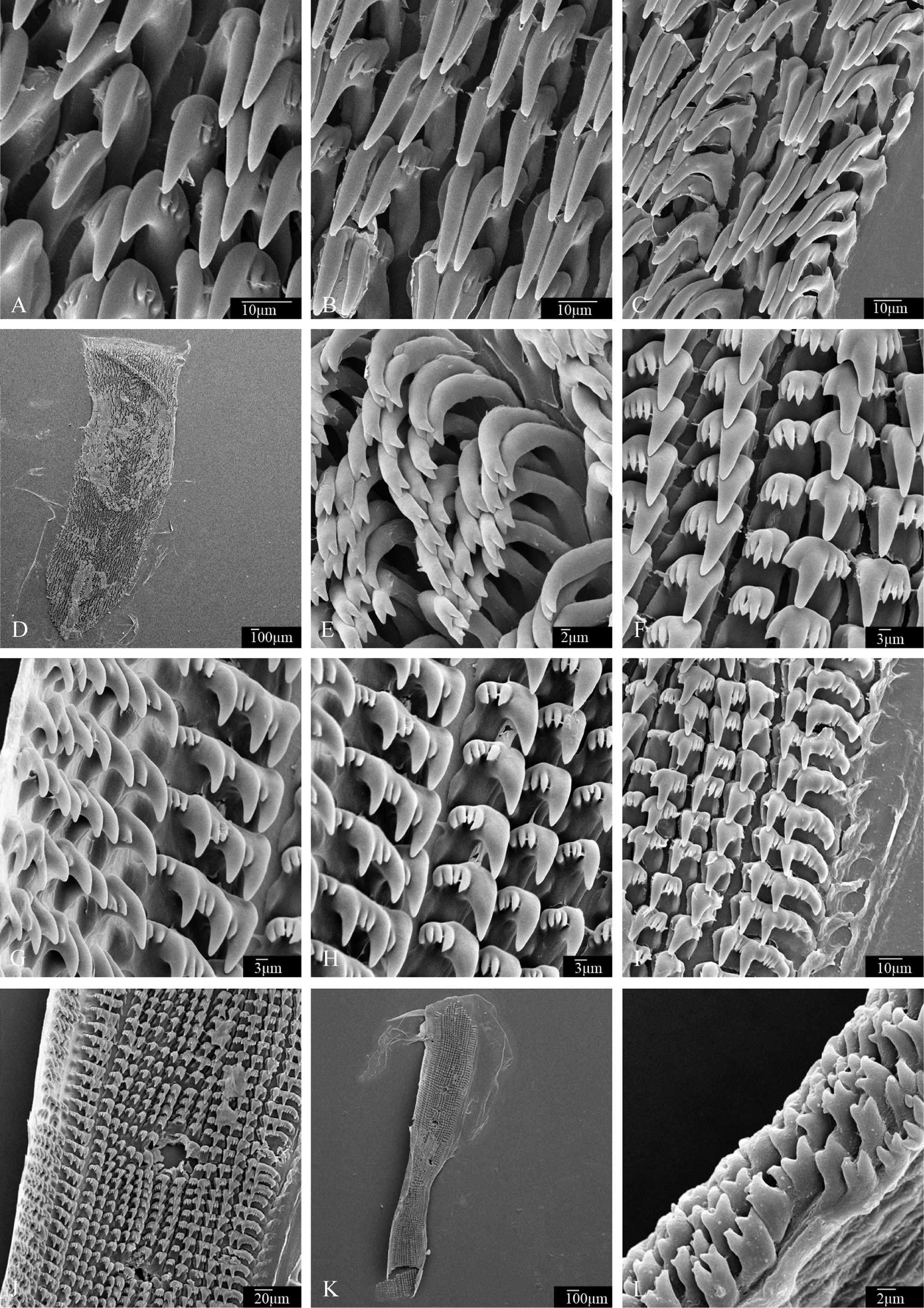
Internal anatomy. Radular structure ( Fig. 14F–L View FIGURE 14 ). The buccal mass is about twice as long as oral tube ( Fig. 13G View FIGURE 13 ). The radular ribbon is long and narrow ( Fig. 14K View FIGURE 14 ) and there are more teeth on the left side of the rachidian tooth ( Fig. 14J View FIGURE 14 ) (135x 17.1.5). The rachidian tooth ( Fig. 14F View FIGURE 14 ) is short and stubby with a short central cusp that is bifid in some cases. It has one denticle on the right side, and 2–4 denticles on the left that are all similar in length to the rachis. The mid-laterals on the left ( Fig. 14H View FIGURE 14 ) have a long and pointed curved central cusp that has 3–4 short well-defined denticles on the outer edge and none on the inner. The denticles are similar in size to the central cusp of the rachis, and are packed next to each other adding girth to each tooth. The outer denticles on the left side ( Fig. 14G View FIGURE 14 ) begin to fuse together to form a single tooth that has three long curved central cusps with denticles packed between them. The first lateral tooth on the right side of the rachis has a long curved central cusp with one denticle on the inner edge and 3–4 on the outer edge. These denticles are similar in shape and size to the denticles on the left side. The next two inner lateral teeth only have denticles on the outer edge ( Fig. 14I View FIGURE 14 ), the third lateral has only one denticle on the outer edge, and the fifth lateral (also the outermost tooth) is fused. This outer lateral has two central cusps with 2–3 denticles in between them, and an undeveloped third central cusp on the outermost edge with 2–3 denticles between it and the middle central cusp. The jaw rodlets are short and stubby and are bifid with some instances where two bifid tips fuse before connecting to the jaw plate ( Fig. 14L View FIGURE 14 ).
Reproductive system ( Fig. 13F View FIGURE 13 ). The bursa copulatrix is approximately double the size of the receptaculum seminis sac. The vagina is medium in length and connects with the receptaculum seminis duct before reaching the bursa copulatrix and sperm sac. The penial bulb is long and convoluted leading to a shorter muscular vas deferens. A long, twisted prostate gland connects to the base of the ampulla adjacent to the albumen gland.
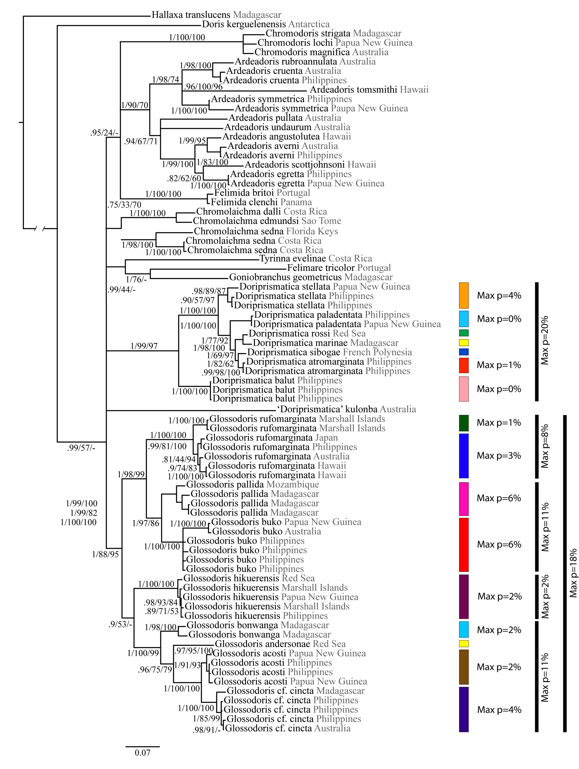
Remarks. Doriprismatica marinae is distinct morphologically and molecularly from the other Doriprismatica . A detailed comparison of D. marinae with its sister taxon D. rossi is warranted. The general body color is similar in that both species have opaque white spots, but D. marinae has only a single mantle band whereas the others, D. rossi and D. paledentata , have three distinct bands. Doriprismatica rossi also lacks the black pigment on the gill rachis that is evident in D. marinae . Also, the rhinophore color distinguishes these two species. The gill branches of D. marinae are held vertically, whereas those of D. rossi are horizontal to the mantle surface and include secondary branches at the tips of some of the larger gill branches. The long body shape that starts with a rounded anterior that tapers posteriorly is similar to D. atromarginata , and differs from D. rossi , which has a wellrounded, distinct posterior end of the mantle rather than one that gradually merges with the posterior end of the foot. The buccal bulb to oral tube ratio is less in D. marinae . The jaws of D. marinae bear short bifid rodlets in contrast to the uniformly elongate rodlets of D. rossi . The radula of D. marinae is markedly asymmetrical with far more teeth on the left side of the radula and a rachidian row of teeth is present. In contrast, D. rossi lacks rachidian teeth and has a symmetrical radula. In D. marinae , the radular teeth all have a very short primary cusp whereas D. rossi has far more elongate cusps on all of its teeth. Although the reproductive systems of these two species are quite similar, D. marinae has a longer vagina than does D. rossi . More specimens, beyond the single specimens of D. marinae and D. rossi studied here, are needed to more fully understand the range of variation in jaw, radular structure, and reproductive anatomy, but the differences observed above are consistent with species-specific differences found in other clearly distinct species of Doriprismatica and are not likely to reflect intraspecific variation. The fact that these differences span, external coloration, body shape, gill morphology, buccal armature and reproductive anatomy strongly suggests that these two species are distinct. The ABGD analysis as outlined in Matsuda & Gosliner (2017) also supports the distinctness of D. marinae and D. rossi , despite the relatively low genetic divergence of 2% for the COI gene ( Fig. 5 View FIGURE 5 ).
No known copyright restrictions apply. See Agosti, D., Egloff, W., 2009. Taxonomic information exchange and copyright: the Plazi approach. BMC Research Notes 2009, 2:53 for further explanation.