
Alafrasca, Lu, Si-Han & Qin, Dao-Zheng, 2014
|
publication ID |
https://doi.org/ 10.11646/zootaxa.3779.1.4 |
|
publication LSID |
lsid:zoobank.org:pub:90BE22AF-B587-45F5-BEF0-63C5B57592C5 |
|
DOI |
https://doi.org/10.5281/zenodo.6131448 |
|
persistent identifier |
https://treatment.plazi.org/id/147F87A1-D07C-FFAA-EEF3-8689FAA3FA28 |
|
treatment provided by |
Plazi |
|
scientific name |
Alafrasca |
| status |
gen. nov. |
Alafrasca View in CoL gen. nov.
Type species: Alafrasca sticta sp. nov., here designated.
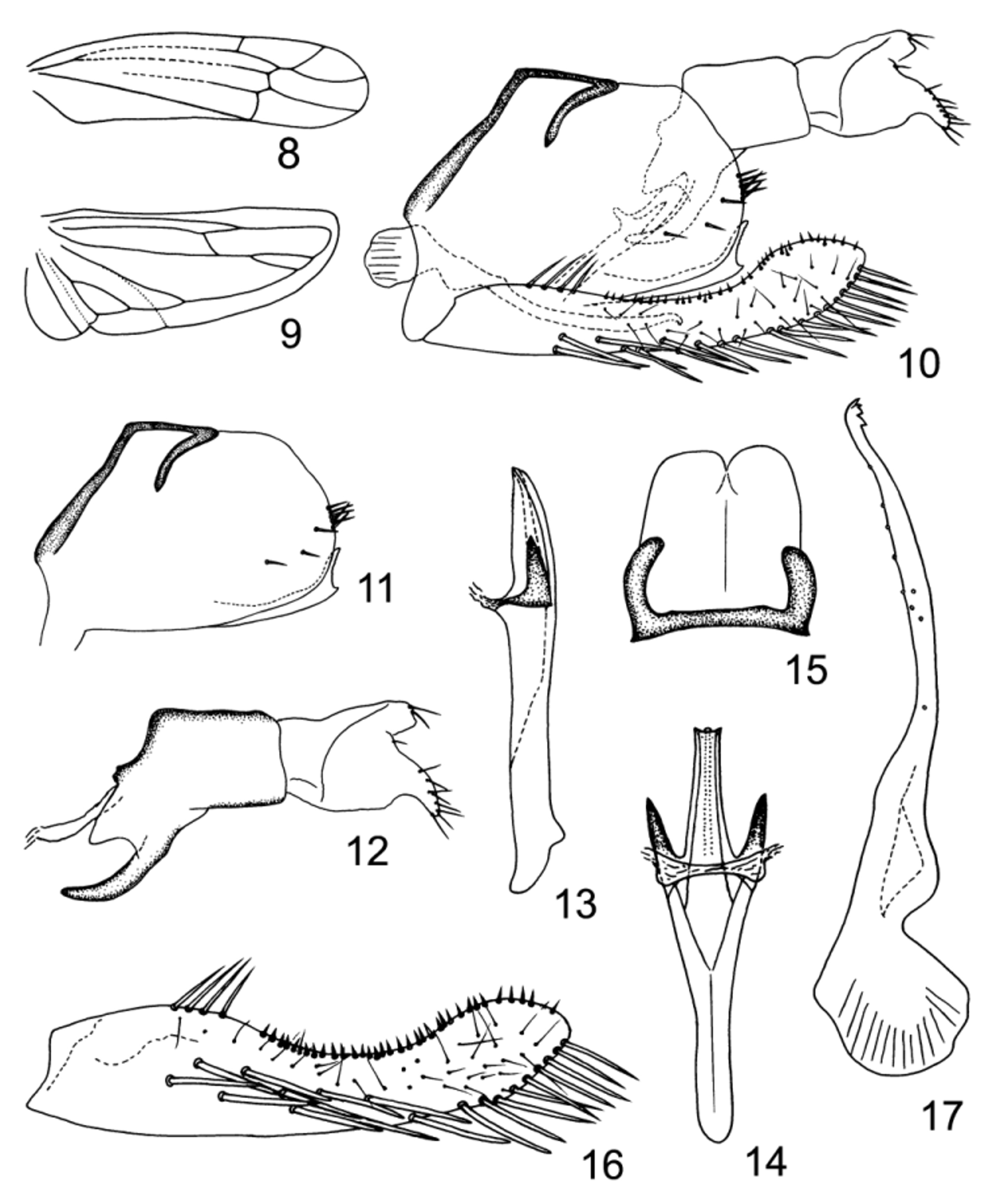
Description. Body robust. Head as wide as pronotum ( Figs. 2, 3 View FIGURES 1 – 7 ). Vertex short, rounded anteriorly, anterior and posterior margins subparallel ( Figs. 2, 3 View FIGURES 1 – 7 ), profile of transition to face rounded ( Fig. 1 View FIGURES 1 – 7 ), coronal suture distinct, extending to anterior margin of vertex ( Figs. 2, 3 View FIGURES 1 – 7 ). Face broad, lateral frontal suture present, ante- and frontoclypeal areas strongly swollen ( Figs. 1, 4 View FIGURES 1 – 7 ). Ocelli present. Pronotum large ( Figs. 2, 3 View FIGURES 1 – 7 ). Mesonotum basally with black spot in middle, scutoscutellar sulcus distinct ( Figs. 2, 3 View FIGURES 1 – 7 ). Forewing narrow, rounded apically, apical cells occupying more than one-third of total length, 3rd cell shortly stalked, c and r cells nearly equal in width, narrower than m and cua cells; veins RP, MP’ arise from r cell and MP”+CuA’ from m cell ( Fig. 8 View FIGURES 8 – 17 ). Hindwing with CuA branched, point of branching distad of coalescence of CuA with MP” ( Fig. 9 View FIGURES 8 – 17 ).
Abdominal apodemes well developed, apically parallel sided ( Fig. 7 View FIGURES 1 – 7 ). Male pygofer elongated, terminally with few rigid microsetae on each side of lobe, ventral appendage present ( Figs. 5 View FIGURES 1 – 7 , 10, 11 View FIGURES 8 – 17 ), dorsal bridge short ( Fig. 6 View FIGURES 1 – 7 ). Anal tube process well developed, branched apically ( Figs. 5 View FIGURES 1 – 7 , 10, 12 View FIGURES 8 – 17 ). Subgenital plate far exceeding pygofer side, all categories of setae present; setae of basal group located at dorsal margin in basal 1/3, lateral macrosetae arranged in two rows and merged into a single row distally, reaching apex of plate, short marginal microsetae occupying more than half length of anterior margin ( Figs. 5 View FIGURES 1 – 7 , 10, 16 View FIGURES 8 – 17 ). Paramere apophysis with prominent dentifer and few sensory pits in apical half ( Fig. 17 View FIGURES 8 – 17 ). Connective lamellate ( Fig. 15 View FIGURES 8 – 17 ). Aedeagal shaft tubular, with pair of basal processes, preatrium long and trough-like; without dorsal apodeme ( Figs. 13, 14 View FIGURES 8 – 17 ).
Remarks. Alafrasca is similar to Alebroides Matsumura , Apheliona Kirkaldy , Alebrasca Hayashi & Okada , Matsumurama Thapa , Nikkotettix Matsumura , Pradama Dworakowska , Schizandrasca Anufriev , Shumka Dworakowska and Znana Dworakowska in having veins MP’ and RP in the forewing stalked (in Nikkotettix and Znana , 3rd apical cell stalked or sessile), both arising from r cell and CuA in the hindwing branched apically. However, the new genus differs from Alebroides Matsumura in having the branching of CuA in the hindwing distad of the coalescence of CuA with MP” ( Fig. 9 View FIGURES 8 – 17 ), from Apheliona , Shumka and Znana in having the head as wide as the pronotum and the coronal suture extending to the anterior margin of the vertex ( Figs. 2, 3 View FIGURES 1 – 7 ), from Alebrasca , Matsumurama and Pradama in having the lateral macrosetae of the subgenital plate arranged in two rows and merged into a single row distally ( Figs. 5 View FIGURES 1 – 7 , 10, 16 View FIGURES 8 – 17 ), from Nikkotettix in the absence of a ventral process at the base of the aedeagal shaft ( Figs. 13, 14 View FIGURES 8 – 17 ) and the subgenital plate having stout setae in the basal group ( Figs. 5 View FIGURES 1 – 7 , 10, 16 View FIGURES 8 – 17 ), from Schizandrasca in having a well-developed aedeagal preatrium. The new genus also differs from Shumka in lacking large blackish patches on the anterior margin of the vertex ( Figs. 2, 3 View FIGURES 1 – 7 ) and the manubrium of the connective not bilobate ( Fig. 15 View FIGURES 8 – 17 ).
Etymology. The generic name is an arbitrary combination of letters, and is regarded as feminine.

No known copyright restrictions apply. See Agosti, D., Egloff, W., 2009. Taxonomic information exchange and copyright: the Plazi approach. BMC Research Notes 2009, 2:53 for further explanation.