
Setophoma sp.
|
publication ID |
https://doi.org/ 10.1016/j.phytochem.2014.12.020 |
|
DOI |
https://doi.org/10.5281/zenodo.10530031 |
|
persistent identifier |
https://treatment.plazi.org/id/150F87E0-B22A-FFC0-CD47-4E88FD90FC5F |
|
treatment provided by |
Felipe |
|
scientific name |
Setophoma sp. |
| status |
|
2.1. Dereplication of Setophoma sp. micro-extracts
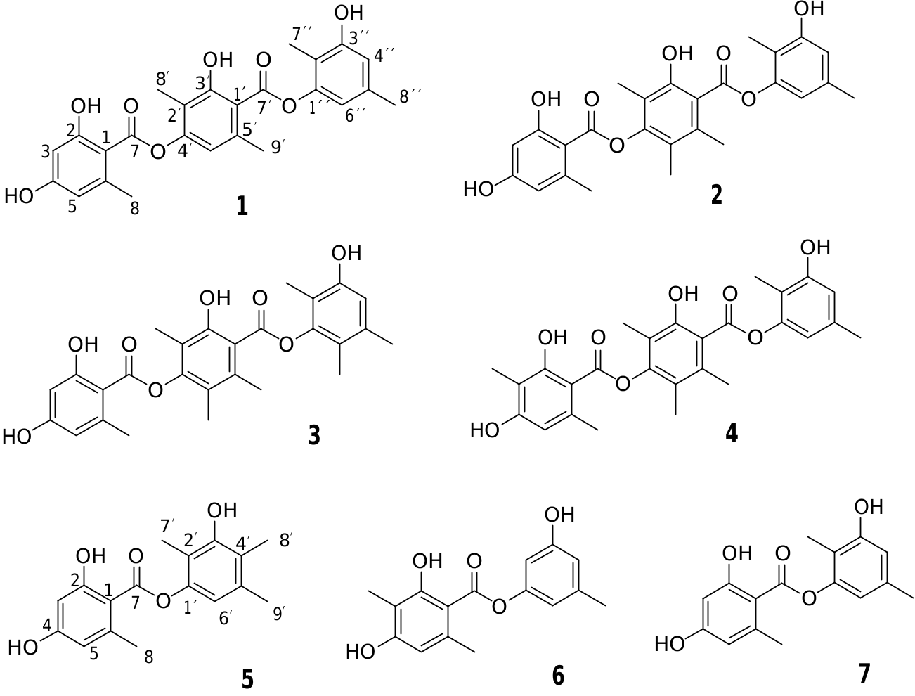
The micro-extracts obtained from cultivation of Setophoma sp. in nine solid media (rice, PDA, MEA, MEAox, CYA, YES, OAT, DRYES, and WATM) were dereplicated through UHPLC–DAD–HRMS data analysis. The base peak chromatograms (BPC) of rice, PDA, MEA, MEAox and OAT media showed, in general, the production of the same metabolites according to the UV profiles and accurate masses of detected peaks. The main detected peaks displayed pseudomolecular ions [M — H] — at m/z 451.1393 (1), 465.1557 (2), 479.1702 (3), 479.1699 (4), 301.1087 (5), 287.0930 (6), and 287.0932 (7), respectively, clearly suggesting the presence of likely isomers for the four last ions (Fig. S2, Suppl. Data). The mono-isotopic masses were then submitted to the AntiBase 2012 query (±5 ppm) ( Laatsch, 2012) which afforded some known candidates. The obtained data from the investigated ions, in particular the UV and the HRMS/MS spectra, were thoroughly compared with the chemical profiles of the AntiBase candidates. Notable similarity was found for the detected compounds to the candidates lecanorins B and C, thielavins Q and R produced by the same strain ( de Medeiros et al., 2013). Moreover, the observed maximum UV absorptions for the investigated unknown peaks were in the range of 272–280 nm, while the fragmentation profile afforded common fragment ions such as m/z 149, 163 and 177 (Figs. S38–S44). Indeed, except for the suggested depsides, the obtained spectroscopic features did not fit to the structures of the remaining known candidates. Hence, they were discarded and the detection of new metabolites produced by the microorganism was assumed.
Although the chemical profile of Setophoma sp. in the rice, PDA, MEA, MEAox and OAT media had showed spectroscopic resemblances, the first medium seemed to be optimal for the microorganism cultivation due to the relative intensity of some target compounds (Fig. S3). The other tested media did not yield the same depside production as displayed in Fig. S1 View Fig . Therefore, the rice medium was chosen to scale up fungal growth.
| S |
Department of Botany, Swedish Museum of Natural History |
No known copyright restrictions apply. See Agosti, D., Egloff, W., 2009. Taxonomic information exchange and copyright: the Plazi approach. BMC Research Notes 2009, 2:53 for further explanation.
|
Kingdom |
|
|
Phylum |
|
|
Class |
|
|
Order |
|
|
Family |
|
|
Genus |