
Polymastia norfanzii, Ekins & Erpenbeck & Wörheide & Hooper, 2023
|
publication ID |
https://doi.org/ 10.11646/zootaxa.5369.1.3 |
|
publication LSID |
lsid:zoobank.org:pub:F906AFDC-DA4E-4ADB-9835-BC4B7692F1FD |
|
DOI |
https://doi.org/10.5281/zenodo.10247567 |
|
persistent identifier |
https://treatment.plazi.org/id/6B12CAAA-B341-4860-9E45-F33DBB995B5D |
|
taxon LSID |
lsid:zoobank.org:act:6B12CAAA-B341-4860-9E45-F33DBB995B5D |
|
treatment provided by |
Plazi |
|
scientific name |
Polymastia norfanzii |
| status |
sp. nov. |
Polymastia norfanzii View in CoL sp. nov. Ekins, Erpenbeck & Hooper urn:lsid:zoobank.org:act:6B12CAAA-B341-4860-9E45-F33DBB995B5D
Figures 1 View FIGURE 1 , 2 View FIGURE 2 & 10 View FIGURE 10 , Table 5 View TABLE 5
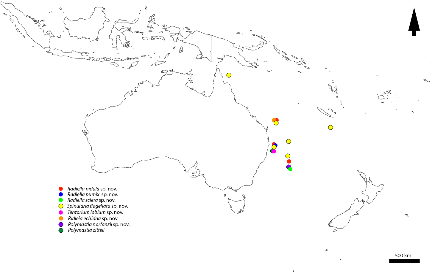
Material examined: Holotype QM G339367 , Lord Howe Plateau , Pacific Ocean Seamounts, Australia -34.2315, 162.6765, 515–700 m, Sherman Sled, Coll. NORFANZ expedition on RV Tangaroa , 85-014, MF336, 26/V/2003. GoogleMaps
Etymology: named in honour of the NORFANZ expeditions which enabled the collection of these new species.
Diagnosis: Polymastia with a single invaginated non-contractile papilla, choanosomal tracts of large tylostyles, forming bouquets below the ectosome. Smaller tylostyles also in the ectosome and scattered in the choanosome, lacking stellate bundles of tylostyles in the choanosome.
Morphology: The sponge is dome shaped 11 mm in diameter and 5 mm in height. It was originally attached to a rock. It is cream in colour and has a hispid surface ( Fig. 10 A View FIGURE 10 ). The upper surface bears a single non-contractile papilla 2 mm in length and approximately 0.5 mm in width, which is invaginated into the upper surface ( Fig. 10 B, C View FIGURE 10 ). There is a single oscule on the summit of the papilla of 0.1 mm in diameter ( Fig. 10 A–C View FIGURE 10 ).
TABLE 5. (Continued)
Skeleton: Aquiferous canals are obvious throughout the choanosome and continue up through the papillae ( Fig 10 B, C View FIGURE 10 ). The choanosomal skeleton is composed of longitudinal tracts of principal styles, with smaller tylostyles scattered between the tracts ( Fig. 10 C, D View FIGURE 10 ). These tracts originate in the base of the sponge and form bouquets beneath the ectosome and then protrude through the ectosome where they form the outer hispid layer. The walls and exterior face of the single papilla are protected and supported by the tylostyles ( Fig. 10 C View FIGURE 10 ). The ectosomal skeleton is composed of the tightly packed bouquets of ascending styles forming a dense palisade along with the smaller tylostyles ( Fig. 10 E View FIGURE 10 ).
Spicules. Principal styles 499–(1226)–2560 × 6.1–(17.4)–30.2 μm, (n=62) ( Fig. 10 F,G View FIGURE 10 ), small tylostyles 92.7– (140)–272 × 2.9–(4.5)–8.7 μm, (n=52) ( Fig. 10 H View FIGURE 10 ).
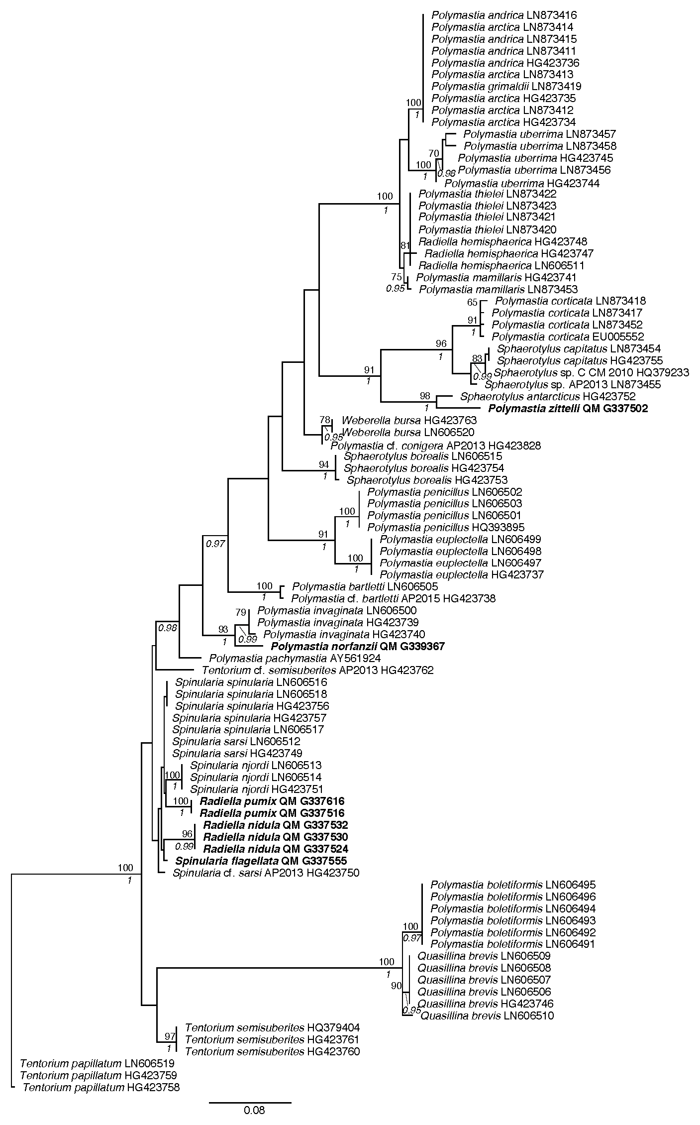
Molecular data: 28S-C region barcode of holotype QM G339367 (ENA Accession number OY741341), This sample is genetically different from all other samples analysed in this study. It forms the sister to a supported P. invaginata clade .
Remarks: This new species is different from P. invaginata firstly by the absence of the stellate bundles of the small tylostyles, that have been consistently referred to as occurring throughout the choanosome. This new species has scattered loose tylostyles in the choanosome and only at the termination of the aquiferous channels do there appear to be tylostyles formed into stellate clusters acting as filters ( Fig. 10 I View FIGURE 10 ). Polymastia norfanzii sp. nov. is also differentiated from P. invaginata by the presence of strictly tylostyles in the choanosomal bundles, rather than the dominating larger styles of the central papillae, surrounded by the invagination caused by partial retraction of the papilla, lacks the contractile ability of the papillae in P. invaginata . This is corroborated by the large number of spicules both internally providing support for the papillae, and externally providing protection ( Fig. 10 C View FIGURE 10 ). The contractile and retractile papilla on P. invaginata have a smooth exterior surface devoid of spicules.
The differences in molecular results obtained in this study compared to previous studies along with the morphological differences mentioned above, separate this new species from P. invaginata . It also raises the possibility that several species may be currently under P. invaginata , as more specimens are recovered in the future, this will most likely elucidate more species.

| QM |
Queensland Museum |
| RV |
Collection of Leptospira Strains |
No known copyright restrictions apply. See Agosti, D., Egloff, W., 2009. Taxonomic information exchange and copyright: the Plazi approach. BMC Research Notes 2009, 2:53 for further explanation.
|
Kingdom |
|
|
Phylum |
|
|
Class |
|
|
Order |
|
|
Family |
|
|
Genus |