
Macrostomus tarsalis, Rafael, José Albertino & Cumming, Jeffrey M., 2006
|
publication ID |
https://doi.org/10.5281/zenodo.171981 |
|
DOI |
https://doi.org/10.5281/zenodo.6493802 |
|
persistent identifier |
https://treatment.plazi.org/id/163B5B4D-346C-FFEF-FEF7-CA3DFB5747A3 |
|
treatment provided by |
Plazi |
|
scientific name |
Macrostomus tarsalis |
| status |
sp. nov. |
Macrostomus tarsalis View in CoL , n. sp.
( Figs. 29–39 View FIGURES 29 33 View FIGURES 34 39 , 40 View FIGURE 40 )
Diagnosis. Prosternum yellow; anterolateral part of scutum, including postpronotal lobe without pruinosity; female hind leg with ventral pinnate scales at distal quarter of femur and some dorsal near middle of femur, dorsal along dorsal face and ventral at basal half of hind tibia; male hind tarsomere 3 widened in dorsal view; cell dm rather acute; M1, M2 and A1 rather distinct.
Description. Male. Head lost. Thorax black with prosternum yellow; grayblue pruinose with anterolateral part of scutum, including postpronotal lobe without pruinosity. 3 dorsocentral setae, the mid one weaker; 3 notopleural setae; 4–5 antepronotal setae; 2 proepisternal setae being one strong; 7–8 katatergital setae. Legs, predominantly yellow with apex of mid femur and apex of mid tibia, distal half of hind femur, entire hind tibia and all tarsi, black; tarsomere 2 inconspicuously yellow medially. Slightly stronger setae: mid tibia with 1 anterodorsal near base, 2–3 ventral very short along distal threequarter; hind femur with 2 anterior at basal threequarter and at distal threequarter, no anteroventral setae. Slender setae: hind tibia with 4 dorsal; hind tarsomere 1 with 1 dorsal. Hind tarsomere 3 modified, widened in dorsal view. Wing ( Fig. 29 View FIGURES 29 33 ) browninfuscated at costal cell, pterostigma and apex of cell r2+3; cell dm rather acute; M1, M2 and A1 rather distinct. Halter light yellow to white. Terminalia ( Figs. 30–33 View FIGURES 29 33 ). Tergite 8 ( Fig. 30 View FIGURES 29 33 ) with large broad projection and wide basal median cleft; sternite 8 ( Fig. 31 View FIGURES 29 33 ) with posterolateral projection. Epandrium ( Fig. 32 View FIGURES 29 33 ) with anterodorsal and posterodorsal lobes of subequal height. Anterior cercus elongate; posterior cercus complex, slightly bifid distally with long slender setae at margin; with a wide ventral protuberance with elongate digitiform yellow setae distally, internally with elongate protuberance with short distal yellow setae and a smaller and thinner simple protuberance near base of the latter. Hypandrium ( Fig. 33 View FIGURES 29 33 ) with large sinus at apex and a unique protuberance inside sinus, many long setae on distal posterior half.
Female. Frons wider than anterior ocellus width; face approximately anterior ocellus width, without pruinosity; 2 pairs of ocellar setae; postgena setae rather long. Prosternum yellow and propleuron partially yellow. 3–4 dorsocentral setae; 3–4 antepronotal setae (dorsocentral and antepronotal setae variable in the same specimen). Slightly stronger setae: mid tibia with 1 anterodorsal near base, 1 posterodorsal near middle, 2 posteroventral at base and apex of median third and 1 anteroventral at apex of median third. Pinnate scales on hindleg ( Fig. 34 View FIGURES 34 39 ): ventral at distal quarter and some dorsal thinner near middle of femur; dorsal along dorsal face and ventral at basal half of tibia. Wing ( Fig. 35 View FIGURES 34 39 ) wider than in male, brown infuscated at costal and r1 cells and apex of r2+3 cell.
Terminalia ( Figs. 36–39 View FIGURES 34 39 ). Tergite 8 and sternite 8 as depicted in Figs. 36 View FIGURES 34 39 . Tergite 8 ( Fig. 38 View FIGURES 34 39 ) with large distal cleft. sternite 8 ( Fig. 37 View FIGURES 34 39 ) rather membranous ventrally. Genital fork ( Fig. 39 View FIGURES 34 39 ) elongate with short lateral arms not projected laterally. Genital chamber membranous.
Size. Body length not measured; wing 4.8 mm.
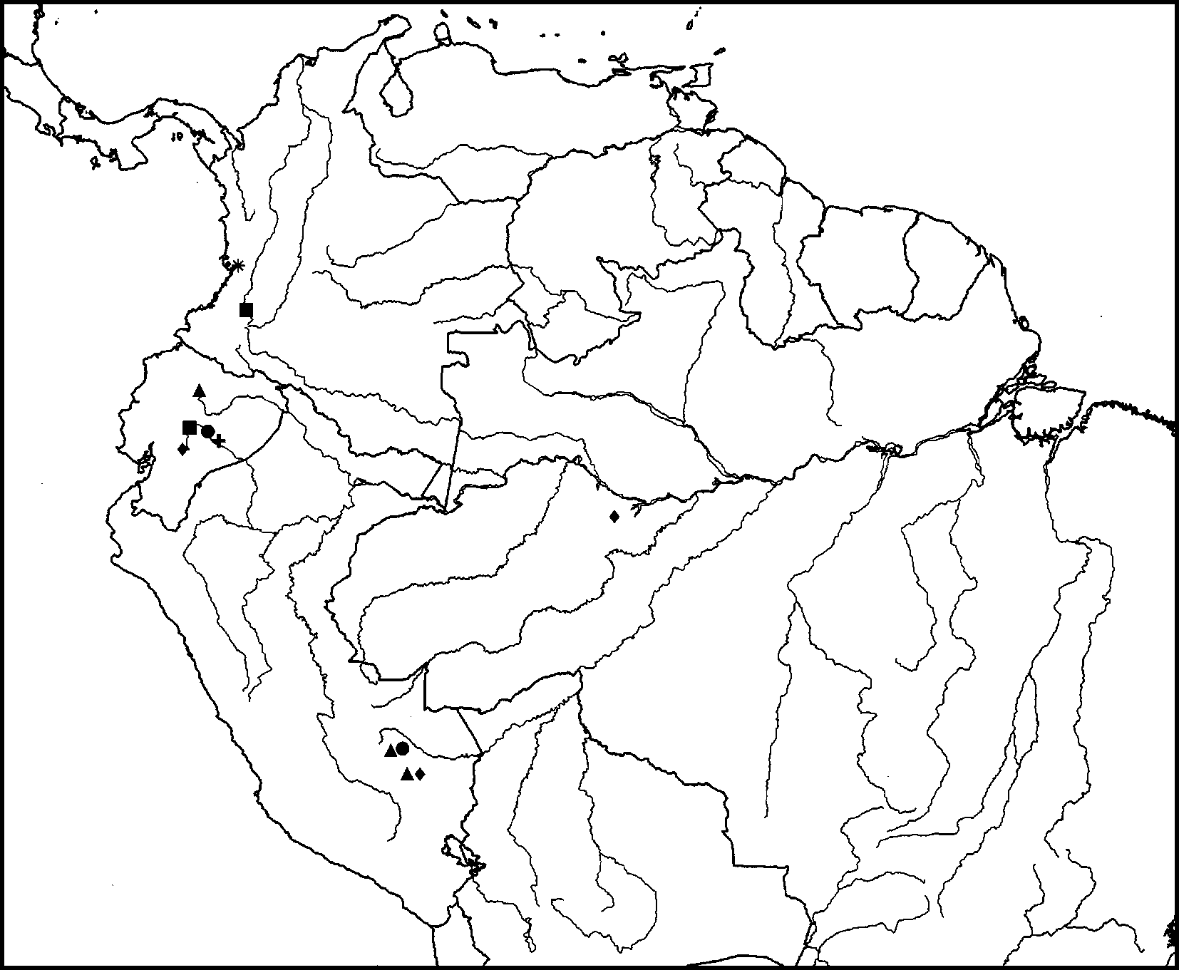
Geographical distribution. Ecuador ( Fig. 40 View FIGURE 40 ).
Material. Holotype ɗ: ECUADOR, NapoPastaza, 2–8 mi [les] N[orth] of Puyo, 953 m, II.9.1955 (sic), E.I. Schlinger & E.S. Ross collectors (CAS). Paratype. Same data and same pin as the holotype (1Ψ CAS).
Holotype condition. Head lost. Right wing mounted in microslide; terminalia in glycerine together with female terminalia of paratype.
Etymology. From Greek tarsos = tarsus, in reference to the shape of the hind tarsomere 3.
Discussion. As mentioned previously, M. tarsalis apparently belongs to the same group as M. alpinus , M. ciliaticosta and M. lineatus , on the basis of these species possessing a rather membranous area medially on the hypandrium. M. tarsalis is distinct from the remaining species in having tergite 8 with a large broad distal projection and a wide basal median cleft, sternite 8 with a posterolateral projection, and the hypandrium with a large sinus at the apex and many long setae on the distal posterior half.


No known copyright restrictions apply. See Agosti, D., Egloff, W., 2009. Taxonomic information exchange and copyright: the Plazi approach. BMC Research Notes 2009, 2:53 for further explanation.
|
Kingdom |
|
|
Phylum |
|
|
Class |
|
|
Order |
|
|
Family |
|
|
Genus |