
Robertsella meridionalis, Gouvêa, Ariane, 2013
|
publication ID |
https://doi.org/10.11646/zootaxa.3734.1.8 |
|
publication LSID |
lsid:zoobank.org:pub:8465E63F-E373-4AF9-BECF-250821B2A313 |
|
DOI |
https://doi.org/10.5281/zenodo.6156123 |
|
persistent identifier |
https://treatment.plazi.org/id/165C879F-EF62-5879-FF04-C0F93EBADAAD |
|
treatment provided by |
Plazi |
|
scientific name |
Robertsella meridionalis |
| status |
sp. nov. |
Robertsella meridionalis n. sp.
( Figs. 1A – D View FIGURE 1. A – D ; 2A–D; 3A, C; 4A – B)
Type material. Brazil: Santa Catarina, RV Soloncy Moura, stn 4, from 27o03.120"S – 46o31.339"W to 27o03.101"S – 46o31.367"W, otter trawl, 8.xi. 2009, 400 m: holotype male, cl 22 mm, cw 30 mm (MZUSP 21772).
Comparative material. Robertsella mystica Guinot, 1969 (see below).
Description. Carapace wider than long, maximal width at fourth anterolateral tooth, minutely granular near anterolateral margins, coarsely granular posterolaterally; mesogastric, cardiac, intestinal regions smooth, poorly defined by faint grooves; hepatic region little more distinct. Ventrally, granulation neatly more distinct on suborbital, subhepatic and branchiostegal regions. Pterygostomian region coarsely granular near moulting suture, minutely granular near mxp3, with dense, coarse granules on low crest (scrapper) above inhalant channel. Branchiostegal margin lined with granules, above and, along margin row of strong granules. Fronto-orbital width little more than half of maximal width of carapace; frontal margin V-shaped, with medial cleft; frontal margin smooth; just behind frontal margin inconspicuous scattered granules and pits. Supraorbital margin interrupted by 2 small notches, margin serrated with low, acute granules, serration coarsest in lateral half, continued to outer orbital tooth (first anterolateral). Inner supraorbital tooth separated from front by wide, shallow gap, low, blunt; outer orbital tooth very low, lobe-like. Suborbital margin lined with small granules, mesially with broadly triangular tooth directed toward front, laterally with broad lobe, V-shape wide cleft between suborbital lateral lobe and outer orbital tooth. Anterolateral margin projecting in 5 distinctly granular teeth (outer orbital included), increasing successively in size posteriorly from first to fourth tooth: second tooth triangular, blunt; third very strong, broad triangular; fourth spiniform, sharp; fifth (last anterolateral tooth) very small but distinct, acute. Gap between anterolateral teeth 3 – 4 much wider than between teeth 2 – 3. Posterolateral margin well defined and rather straight behind last anterolateral tooth, poorly defined posteriorly, about as long as anterolateral margin. Limit between epistome, endostome well defined, forming pronounced, sinuous lip, interrupted by 2 V-shaped, deep notches, one at each side of deep mesial notch. Ocular peduncle cylindrical, very scattered granular dorsally, freely movable, thick, constricted subproximally, fully retractable into orbital cavity; cornea occupying much less than half-length of whole eye; ommatidia dark, poorly recognizable. Antennules prominent; proximal article thickest laterally, with semicircular, transverse row of granules; second article smooth, elongated, subcylindrical, articulated with proximal article at mesial end of antennular fossa; third article nearly equal in length to second, swollen distally, tapered to proximal articulation with second article, terminally with long marginal setae at either side of dorsal flagellum. Antennal article 2+3 immovable, filling orbital gap; articles 4, 5 freely movable, subcylindrical. Thoracic sternum minutely granular anteriorly, granulation stronger posteriorly; sterno-abdominal cavity deep, slightly granular anteriorly, lateral margins with scattered granules, more discernible posteriorly. Abdominal locking system functional in male, thoracic sternal button small, directed forward, placed about mid-length between thoracic sternal sutures 4/5 and 5/6. Third maxillipeds inserted wide apart from each other; ischium smooth, pitted, with faint longitudinal furrow, mesial margin granular; merus distinctly shorter than ischium, evenly granular; palp smooth. Chelipeds heterochelous, right P1 strongest. Merus of major P1 trigonal, dorsal margin with strong, blunt, subdistal tooth; surfaces evenly covered with fine granules, granulation strongest dorsally. Carpus with acute spine on inner margin; surfaces finely granular, granules stronger anteriorly and ventrolaterally. Propodus stout, surfaces minutely granular, granules forming reticular patterns. Fingers black, closing terminally only; cutting edges bluntly dentate, teeth becoming more coalescent posteriorly; teeth stronger on fixed finger. Minor cheliped similar to major cheliped except for its slender fingers. Ambulatory legs (P2–P5 detached from body) long, slender, similar to each other. Meri P2–P5 meri spinous dorsally, granular ventrally. P2 – P5 carpi with dorso-lateral, longitudinal, smooth depression, on each side a row of distinct granules; ventral surface smooth. P2–P5 propodi with scattered granular proximally. P2–P5 dactyli corneous-tipped, 6 longitudinal rows of dense, long setae, 2 dorsal, 2 ventral, 2 lateral. Abdomen of 4 somites and telson; abdominal somites 1–2 markedly short, somite 1 much broader than somite 2; somites 3–5 fused together; abdominal somite 3 expanded laterally, almost completely covering penis; abdominal suture 3/4 faint, still recognizable; suture 4/5 indistinct, its position indicated laterally only by faint cleft. Telson semicircular. G1 distinctly curved outward, just reaching to thoracic sternal suture 4/5. G2 very short, just reaching to sternal suture 7/8.
Etymology. The epithet is derived from the Latin noun meridies (“south”) suffixed with - alis, meridionalis , adjective for “southern”, in allusion to the southern type locality of this species.
FIGURE. 3 A, C, Robertsella meridionalis sp. n., male holotype cl 22 mm, cw 30 mm (MZUSP 21772). B, D, Robertsella mystica Guinot, 1969 , male cl 18 mm, cw 23 mm (USNM 170418). A–B, lateral view of the first gonopod. C–D, mesial view of the first gonopod. Scale bars 0.4 mm.
FIGURE. 4 A–B, Robertsella meridionalis sp. n., male holotype cl 22 mm, cw 30 mm (MZUSP 21772). C–D, Robertsella mystica Guinot, 1969 , male cl 18 mm, cw 23 mm (USNM 170418). A, C, lateral view of carapace. B, D, second left pereiopod. Note pereiopod coarsely tuberculate ventrally in B and smooth in D. Scale bars 5 mm.
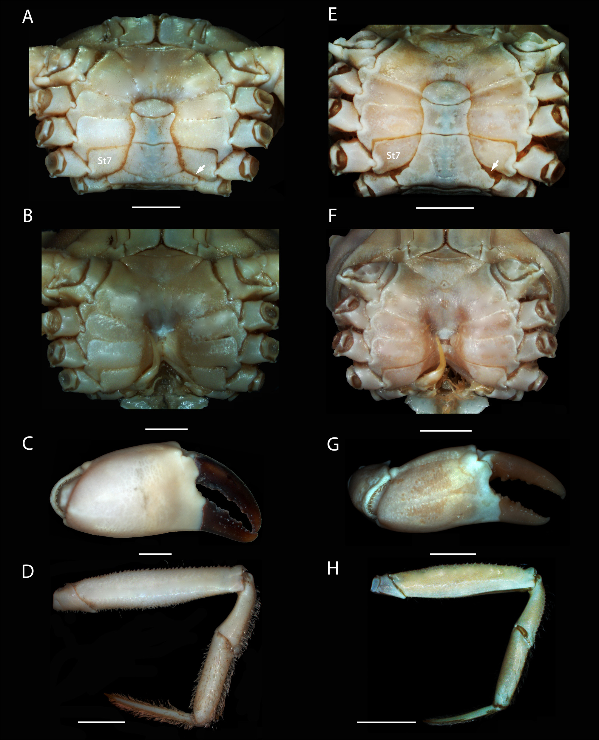
Remarks. Robertsella was until now known from one species, R. mystica , from the northwestern Atlantic (South Carolina, Gulf of Mexico, Florida, and Dry Tortugas). Although Robertsella meridionalis n. sp. is represented by only one adult male, it can be distinguished from its northern counterpart by a suite of carapace and appendage characters. In Robertsella meridionalis n. sp. (i) the well-developed lateral expansion of the third abdominal somite is in contact with thoracic sternite 7 and thus covers the gonopore and almost the entire penis ( Figs. 1D View FIGURE 1. A – D , 2A View FIGURE 2. A – D ) (much shorter abdominal lateral expansion that is not in contact with sternite 7 thus leaving the gonopore and most of the proximal part of penis uncovered in R. mystica ; Figs. 1 View FIGURE 1. A – D H, 2E); (ii) abdominal suture 3/4 is evident along its entire length, less so near the axis only ( Figs. 1D View FIGURE 1. A – D , 2A View FIGURE 2. A – D ) (abdominal suture 3/4 evident at lateral notch only in R. mystica ; Figs. 1 View FIGURE 1. A – D H, 2E); (iii) when the abdomen is closed, the abdominal suture 5/6 is located well above the thoracic sternal suture 6/7 ( Figs. 1D View FIGURE 1. A – D , 2A View FIGURE 2. A – D ) (abdominal suture 5/6 and thoracic sternal suture 6/7 aligned to each other in R. mystica ; Figs. 1 View FIGURE 1. A – D H, 2E); (iv) the lateral margins of the thoracic sternite and episternite 7 are straight, aligned to each other and subparallel in relation to the body axis ( Fig. 1D View FIGURE 1. A – D ) (sternal and episternal margins distinctly convex in R. mystica ; Fig. 1 View FIGURE 1. A – D H); (v) the portion of the thoracic sternite 8 not covered by the lateral expansion of the third abdominal somite is proportionally larger than in R. mystica ( Figs. 2A View FIGURE 2. A – D , E); (vi) the meri of pereiopods 2–5 are densely and coarsely tuberculate ventrally( Figs. 2D View FIGURE 2. A – D , 4B) (ventral surface of P2–P5 meri almost smooth in R. mystica ; Figs. 2 View FIGURE 2. A – D H, 4D); (vii) the cheliped ischial ridge (pars stridens of the stridulatory mechanism, see below) is densely and coarsely tuberculate ( Fig. 1C View FIGURE 1. A – D ) (ischial ridge less tuberculate in R. mystica ); (viii) pterygostomial and branchiostegal regions are densely and coarsely tuberculate (Fig. 4A) (pterygostomial and branchiostegal regions much less tuberculate in R. mystica ; Fig. 4C); (ix) the tubercles in both the branchiostegal margin and the transversal row of tubercles above the branchiostegal margin are much denser and stronger than in R. mystica (Figs. 4A, C); and (x) frontal region of carapace strongly deflexed ( Fig. 1A View FIGURE 1. A – D ) (frontal region distinctly less deflexed in R. mystica ; Fig. 1 View FIGURE 1. A – D E).
Robertsella meridionalis n. sp. and R. mystica probably produce sound by stridulation. Both species have an ischial ridge on the mesial side of the right and left chelipeds (par stridens, see Guinot-Dumortier & Dumortier 1960) that rubs against a scrapper (plectrum) on the carapace, a curved tuberculate edge located above the inhalant water opening. The chelipeds basis-ischium is fused together into a single article providing additional leverage for the rasping movement.

No known copyright restrictions apply. See Agosti, D., Egloff, W., 2009. Taxonomic information exchange and copyright: the Plazi approach. BMC Research Notes 2009, 2:53 for further explanation.
|
Kingdom |
|
|
Phylum |
|
|
Class |
|
|
Order |
|
|
InfraOrder |
Brachyura |
|
Family |
|
|
Genus |