
Thiotricha lumnitzeriella Kyaw, Ueda, & Hirowatari, 2021
|
publication ID |
https://doi.org/ 10.11646/zootaxa.4980.2.5 |
|
publication LSID |
lsid:zoobank.org:pub:8FE34466-8401-4AD9-8DE7-C15C3DBBB5C9 |
|
DOI |
https://doi.org/10.5281/zenodo.4985749 |
|
persistent identifier |
https://treatment.plazi.org/id/1660F241-FFD1-3749-FF58-15D026B8F807 |
|
treatment provided by |
Plazi |
|
scientific name |
Thiotricha lumnitzeriella Kyaw, Ueda, & Hirowatari |
| status |
sp. nov. |
Thiotricha lumnitzeriella Kyaw, Ueda, & Hirowatari View in CoL , new species
[Japanese name: Hirugimodoki-kibaga]
Figs 1 View FIGURE 1 , 6A View FIGURE 6 , 7 View FIGURE 7 , 9 View FIGURE 9 , 11 View FIGURE 11 , 13A–C View FIGURE 13 , 14 View FIGURE 14 .
Thiotricha sp.1: Murphy, 1990: 162 .
Polyhymno sp.: Ueda, 2011: 645 .
Polyhymno sp. 4: Ueda, 2013: 298 .
Type material. Holotype: JAPAN: Okinawa Pref.: 1♂, [Iriomotejima], Komi, Taketomi, 8 vii 2017 larva, T. Hirowatari, S. Yagi, K.M.M.Kyaw, Host: Lumnitzera racemosa , 30 vii 2017 em., KM –92, in ELKU.
Paratypes: JAPAN: Kagoshima Pref.: 1♂, Amami-oshima, Mt. Yui-dake, Setouti-tyo Town , 30 ix 2014, S. Sameshima leg. ( KGU) ; Okinawa Pref.: 4♂, 3♀, same locality, collecting date, collector, and host plant as holotype, 16 vii–2 viii 2017 em, gen slide no. KM – 93 (♂), 94 (♀), 120 (♀), 121 (♀), 129 (♂) ( ELKU) ; 1♂, 4♀, same locality and host plant as holotype, 25 vi 2019 (larva), 16–19 vii 2019 em., K.M.M. Kyaw ( ELKU) ; 3♂, 2♀, Hosidate mangrove, Taketomi-cho, Iriomote-jima , 14 viii 2001 em., F. Komai, gen slide no. TU –746 (♂),748 (♂),758 (♀), Host Plant: Lumnitzera racemosa (OPU) ; 1♂, 3♀, same locality, collector, and host plant, 16 viii 2001 em., gen slide no. TUW–13 (♂),111 (♀), 119 (♀) ( OPU) ; 2♂, same locality and host plant, 3 x 2001, 17–31 x 2001 em., T . Saito , gen slide no. KM – 11 (♂), 85 (♀), 86 (♂), 110 (♀) ( OPU) ; 4♂, 3♀, same locality, collector, and host plant, 3 x 2001, 1–16 xi 2001 em., gen slide no. KM – 95 (♂), 98 (♂), 109 (♀), 130 (♂) ( OPU) ; 1♀, same locality and collector, 21 x 2000 ( OPU) ; 1♀, Uehara, Taketomi-cho, Iriomote-jima , 20 x 2000, T . Saito ( OPU) .
Diagnosis. Thiotricha lumnitzeriella is similar to T. clinopeda Meyrick, 1918 , T. symphoracma Meyrick, 1927 , and T. tethela Bradley, 1961 in wing pattern by sharing an oblique dark fuscous streak that extends from the costa and the dark fuscous line to tornus. However, it differs from the latter two species by having the small yellowish brown and triangular-shaped area between apical spot, costal streak, a short blackish line at outer margin of tornus and a fuscous scale on termen. Additionally, the male genitalia of T. lumnitzeriella are similar to those of T. clinopeda by sharing almost all characters except in the shape of phallus, but the phallus of the latter is moderately pointed basally and rounded in T. lumnitzeriella .
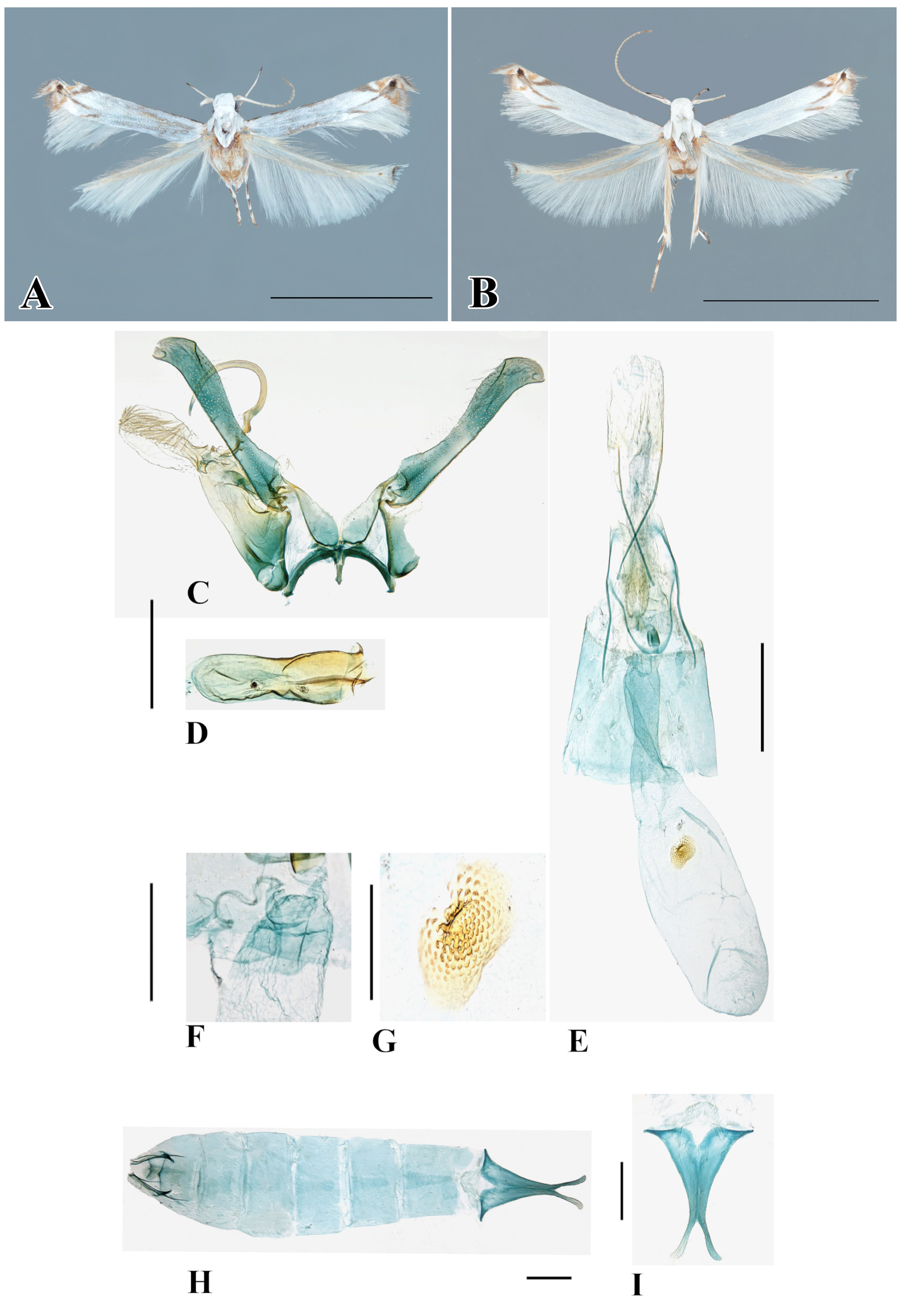
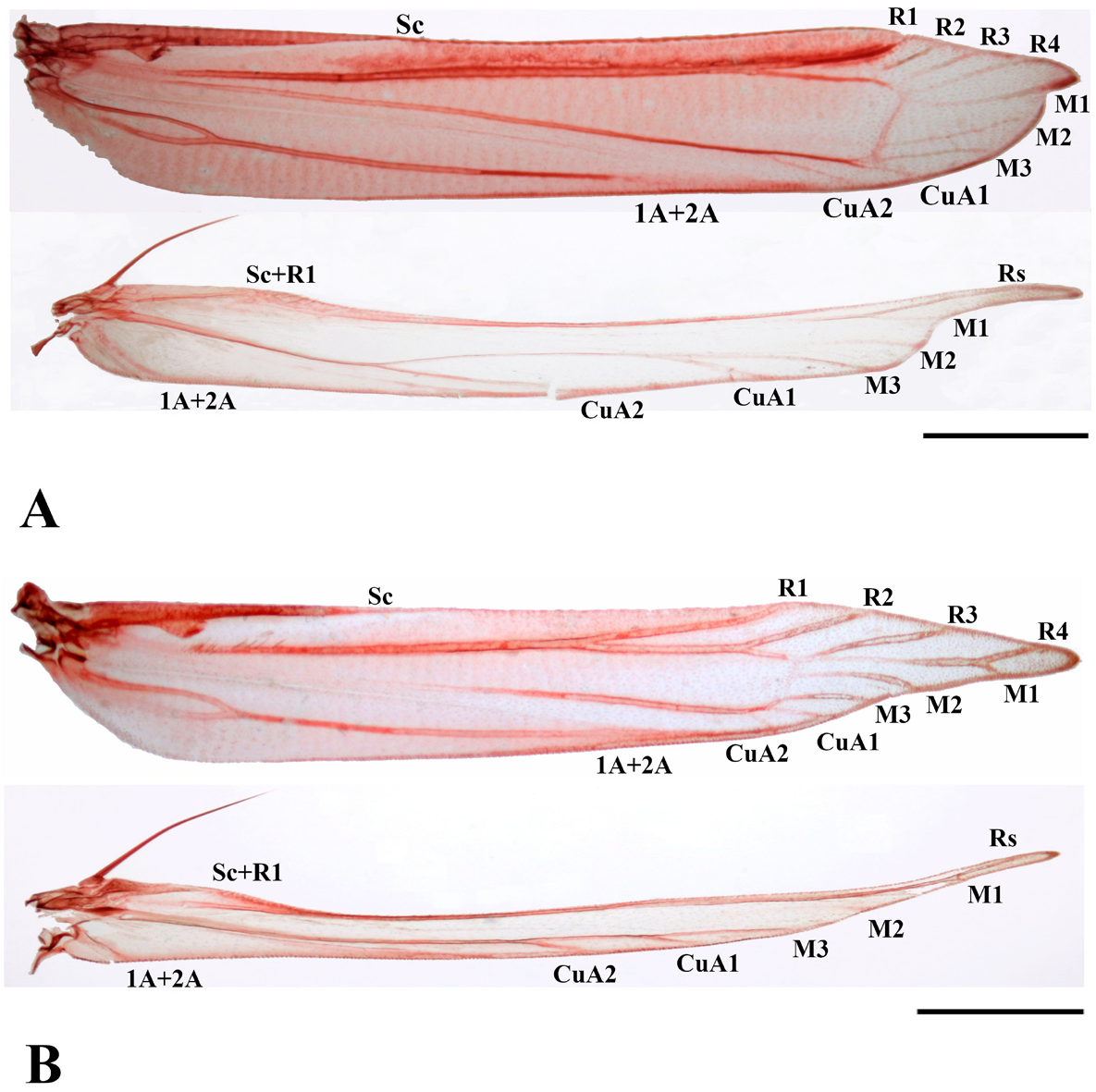
Description. Adult. Male (holotype: Fig. 1A View FIGURE 1 ). Forewing length 2.7 mm in holotype, 2.6–3.4 mm (n=12) in paratypes. Wing expanse 6.2 mm in holotype, 5.5–7.2 mm (n=12) in paratypes. Head glossy white. Antenna filiform, scape elongate, white, sparsely speckled with brown scales; flagellum white before middle, and then grayish brown beyond middle, with rather long and fine cilia ventrally. First palpomere of labial palpus white, shortest; second thickened, slightly suffused with brown scales on entire surface; third as long as second, but more slender, sharply acute, with black scales medially on dorsal surface. Mesothorax shiny white; tegula shiny white, with blackish scales along anterior margin. Legs white; fore femur with grayish black on outer surface, tibia and all tarsomeres entirely grayish black; mid-tibia slightly suffused with an oblique grayish black stripe at midpoint on outer surface, all tarsomeres white on inner surface, first three tarsomeres grayish black ringed with white apically and last two segments grayish black entirely; hind femur and tibia concealed with white, stiff and stout bristles above and below, with an oblique black stripe laterally on outer surface near posterior margin, first and second tarsomeres white on inner surface and grayish black on outer surface, ringed with white apically; remaining three grayish black entirely. Forewing with R4 stalked with M1; R5 absent (or coincident with R4); CuA1 and CuA2 separated; retinaculum represented by a hook arising from Sc; a group of needle-shaped scales attached around 1A+2A on the underside; ground color white to whitish gray, apex obtuse, termen obliquely rounded, apex confluent with following lines, a very oblique wedge-shaped dark fuscous streak from costa at 3/4, a small yellowish brown and inverted triangularshaped area between apical spot and costa streak, each area intercepted by a whitish line, apex conveying a dark fuscous line to tornus and a short blackish line at outer margin of tornus towards the posterior; a black spot at apex, a fuscous scale on termen beneath apex, a small yellowish brown scale beneath it, brown cilia at inner rim of apex and black hairs at outer margin, running into a hook-shaped apically. Termen, cilia brown at anterior rim, grayish-black at posterior edge, well-fringed long and white beyond tornus. Hindwing narrower than forewing, whitish brown, with a tiny black dot apically; apex sharply produced, well-fringed white cilia, around apex with a short black post median line.
Female ( Fig. 1B View FIGURE 1 ). Forewing length 2.8–3.3 mm (n=11). Wing expanse 6.0– 7.5 mm (n=11). Similar to male, but retinaculum represented by a hook arising from Sc, with a series of curved liner scales along Radius and two rows of apical curved liner scales along Sc and Radius.
Abdomen ( Fig. 1H, I View FIGURE 1 ). Terga and sterna 1–7 unmodified as in Fig. 1H View FIGURE 1 . Tergum-8 concave, emarginate anteriorly and a few tubercles arising on surface; sternum 8 expanded basally, narrowly elongate, and tapered towards apex becoming funnel-shaped, then bifurcate at posterior end as in Figs 1H, I View FIGURE 1 .
Male genitalia ( Fig. 1C, D View FIGURE 1 ). Uncus significantly elongates and nearly more than half length of tegumen, domeshaped, dense with numerous long and delicate setae from apex beyond middle on the ventral surface. Tegumen also elongates, as long as 1.5 times of uncus. Gnathos long, extremely curved from base, C-shaped, slightly acute at tip. Anellus lobe with a pair of small membranous thumb-like lobes with delicate and rather long setae, distinct and some short setae also appearing on surface apically. Costal process of valva quite broadened basally, uniformly extends from base, then dilates again with a slightly rounded apical margin, sparsely suffused with long hairs along lateral and upper surface, but compactly at approximately 1/3 of apex, short and robust spines also on lateral inner surface of apex. Vinculum narrow; median process of vinculum producing somewhat long and pear-shaped lobe; thin with short and delicate setae on median process. Saccus a short and stout processes. Phallus partially sclerotized and rounded on basal portion, widened equally from base towards distal portion, a tulip-shaped at tip, with a heavily sclerotized margin at distal half.
Female genitalia ( Fig. 1 E–G View FIGURE 1 ). Papillae anales bilobed and subrectangular-shaped, with long and short setae on entire surface. Apophyses anteriores almost equal in length to apophyses posteriores. Lamella antevaginalis sclerotized and rounded basally. Ostium near posterior margin of 8 th sternite. Ductus bursae short, with a widened surface area, approximately half length of corpus bursae. Ductus seminalis arising from posterior end of ductus bursae ( Fig. 1F View FIGURE 1 ). Corpus bursae, particularly large and ovate; signum situated at middle posteriorly, depressed, and pentagonal shaped with many minute denticles.
Distribution. Japan, Ryukyus (Amami-oshima Island, Iriomote-jima Island); Singapore.
Host plant. Lumnitzera racemosa Willd. (Combretaceae)
Etymology. The species name is derived from the host plant “ Lumnitzera ” and that the suffix of the species epithet is derived from the Latin, “ ella ” meaning small, referring to the small size.
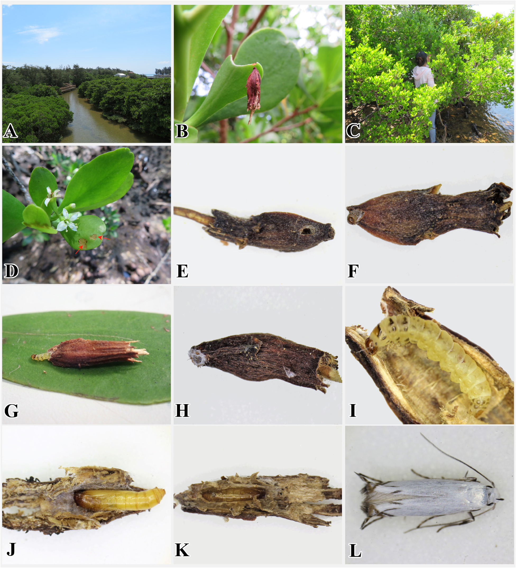
Biology ( Fig. 7 View FIGURE 7 ). The larvae of this species also make a portable case using the flower bud of its host plant. It penetrates the flower bud ( Fig. 7E View FIGURE 7 ) and then lives and feeds inside it. After consuming the flower bud, the larvae move while carrying its case and then attaches to the underside of a leaf ( Fig. 7B View FIGURE 7 ). While the larva is attached to the leaf surface, it consumes the leaf tissues for growth and development until pupation. Consequently, the leaf surface becomes transparent, and small white patches appear on the host plant leaf ( Fig. 7D View FIGURE 7 ). When the larva is ready to pupate, it encloses the tip of the case with its silk ( Fig. 7H View FIGURE 7 ) and then finally develops into the adult stage and leaves the pupal exuviae inside the case ( Fig. 7K View FIGURE 7 ).
Larva ( Fig. 7G, I View FIGURE 7 ). Length 3.5–4.0 mm (n=8). Head subglobular. Body pale yellow in early instars and yellowish-brown in later instars. Prothoracic shield yellowish-brown, with dark brown on caudal margin. Thoracic leg short, pale yellowish. Pinaculum more or less rounded, dark brown on T1–T3 and A1, A2, A8, and A9; paler on remaining abdominal segments. Anal shield heavily sclerotized, yellowish-brown ( Fig. 9C View FIGURE 9 ). Anal fork deeply emarginated mesially, forming two lateral lobes ( Fig. 9D View FIGURE 9 ). Anal prolegs with many minute spines on dorsal surface. Crochets in a circle, uniordinal, 12–15 in number on planta, 10–13 on anal planta.
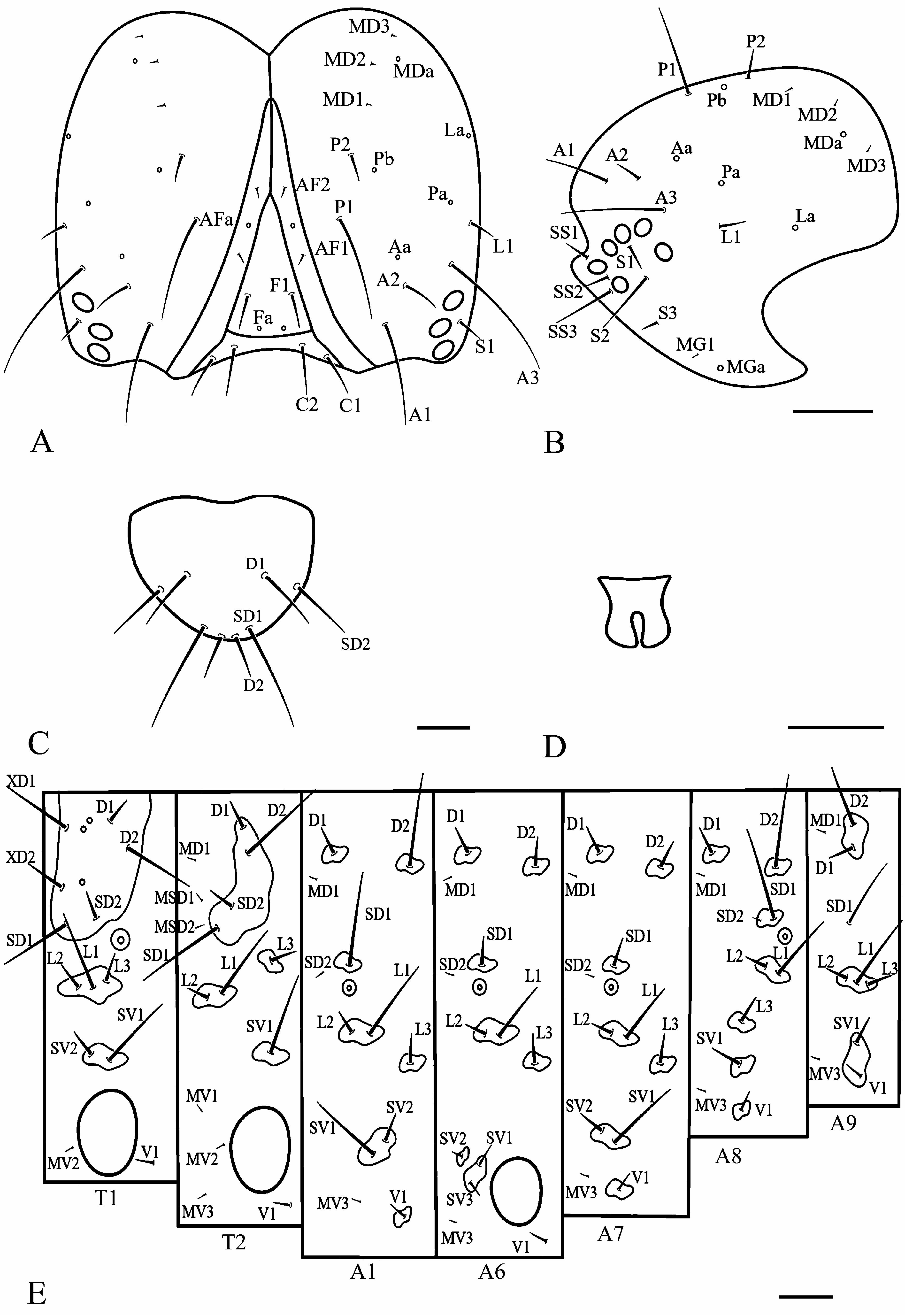
Chaetotaxy ( Fig. 9 View FIGURE 9 ) Head ( Fig. 9A, B View FIGURE 9 ): epicranial suture shorter than frontoclypeus, AF1 about equal in length with AF2; C2 slightly longer than C1; P1 dorsolateral to AF1 about 5X longer than P2; P2 dorsolateral to AF2 and above P1; MD1-MD3 setae forming nearly in a line at the posterior margin of head capsule; mouthparts semihypognathous; genal area with six stemmata, forming a subsemicircular pattern; A1 dorsoanterior to stemma-3, slightly shorter than A3; A2 dorsolateral to A1, and shorter than A1 and A3; L1 dorsoposterior to stemma-1; distance between L1 from A3 slightly longer than distance between A3 from A2; S1 below stemma-3, short as A2; S2 longer than S1 and S3, near the opening of the stemmatal semicircle; S3 about 1/2 shorter than S1, and ventroposterior to stemma-6; SS1 near mandibular condyle, same length as SS2; SS2 between SS1 and SS3; SS3 about 3X longer than SS1 and SS2; MGa present, close to MG1.
Thorax: Prothorax ( Fig. 9E View FIGURE 9 ). Shield with SD1 ventrolateral to XD1 and XD2, along anterior margin; XD2 less than twice the distance from XD1 than from SD1; XD1 about 2X longer than XD2; SD2 and D1 about equal in lengths, both setae less than about 2½–3X length of SD1 and D2; SD2 about 1½ the distance from XD2 than from SD1; L-group trisetose, on same pinaculum, anteroventral to spiracle; L1 longest; L2 and L3 short, about equal in lengths; SV-group bisetose, on same pinaculum; SV1 about 2–2½X longer than SV2; MV1 absent; MV2 approximate to anterolateral coxal margin; V1 approximate to mesoposterior coxal margin.
Meso and metathorax ( Fig. 9E View FIGURE 9 ). with D1, D2, SD1, and SD2 on same pinaculum; D2 about 3½–4X length of D1; SD1 about 3½–4X length of SD2; MD1 anteroventral to D2; MSD 1 in line with MSD2, anterior to and slightly above SD2; MSD2 slightly anterior to SD1; L1 about 2½–3X length of L2, both on a same pinaculum, slightly anterior to D-SD group pinaculum; L3 slightly longer than L2, in vertical line with SV1; MV1, MV2 and MV3 anterior to coxa; with MV2 approximate to anterolateral coxal margin, and MV3 slightly above V1.
Abdomen ( Fig. 9E View FIGURE 9 ). A1 with D2 about 3½–4X longer than D1 on A1; D1 and D2 about equal in lengths on A2 (not shown); D1 dorsoanterior to D2; MD1 slightly ventral to D1 and D2; SD1 about equal in length with D2 on A1 and with D1 on A2; SD2 minute, anteroventral to SD1 and on different pinaculum; SD1 above spiracle; SD2 anteroventral to SD1; L-group trisetose; L1 and L2 on same pinaculum, in vertical line with SD group and below spiracle; L1 about 3½–4X longer than L2 and L3 on A1 and about 2½–3X longer than L2 and L3 on A2 (not shown); L3 slightly longer than L2; SV-group bisetose on A1, SV1 about 2½–3X longer than SV2 and on same pinaculum on A1; SV-group trisetose and on same pinaculum on A2 (not shown); MV3 dorsoanterior to V1; A3–A6 as above A2, except each segment bearing a pair of protuberant prolegs; planta bearing uniordinal, uniserial crochets, in a circle (not shown); SV-group setae near base of proleg; SV1 and SV3 on same pinaculum, SV2 on separate pinaculum; A7 as above except with SV-group bisetose on same pinaculum, and MV3 dorsoanterior to V-group pinaculum, and V1 ventral to SV pinaculum; A8 as above except with SD1 about equal in length with D2; D 2 in vertical line with SD1, minute SD2 on same pinaculum as SD1, SD-group pinaculum slightly anterior to spiracle, spiracle slightly dorsal to all spiracles on A1–A7; L1 about 2½–3X length of L2; L1 and L 2 in vertical line with D2 and SD1; L3 anteroventral to L1 and L2; SV-group unisetose; A9 as above except with D1 ventral to D2 and on same pinaculum; MD1 anteroventral to D2; SD1 hair-like, slightly longer than D2; L-group trisetose on same pinaculum, L1 longest; L2 and L3 about equal in lengths; SV1 and V1 on same pinaculum.
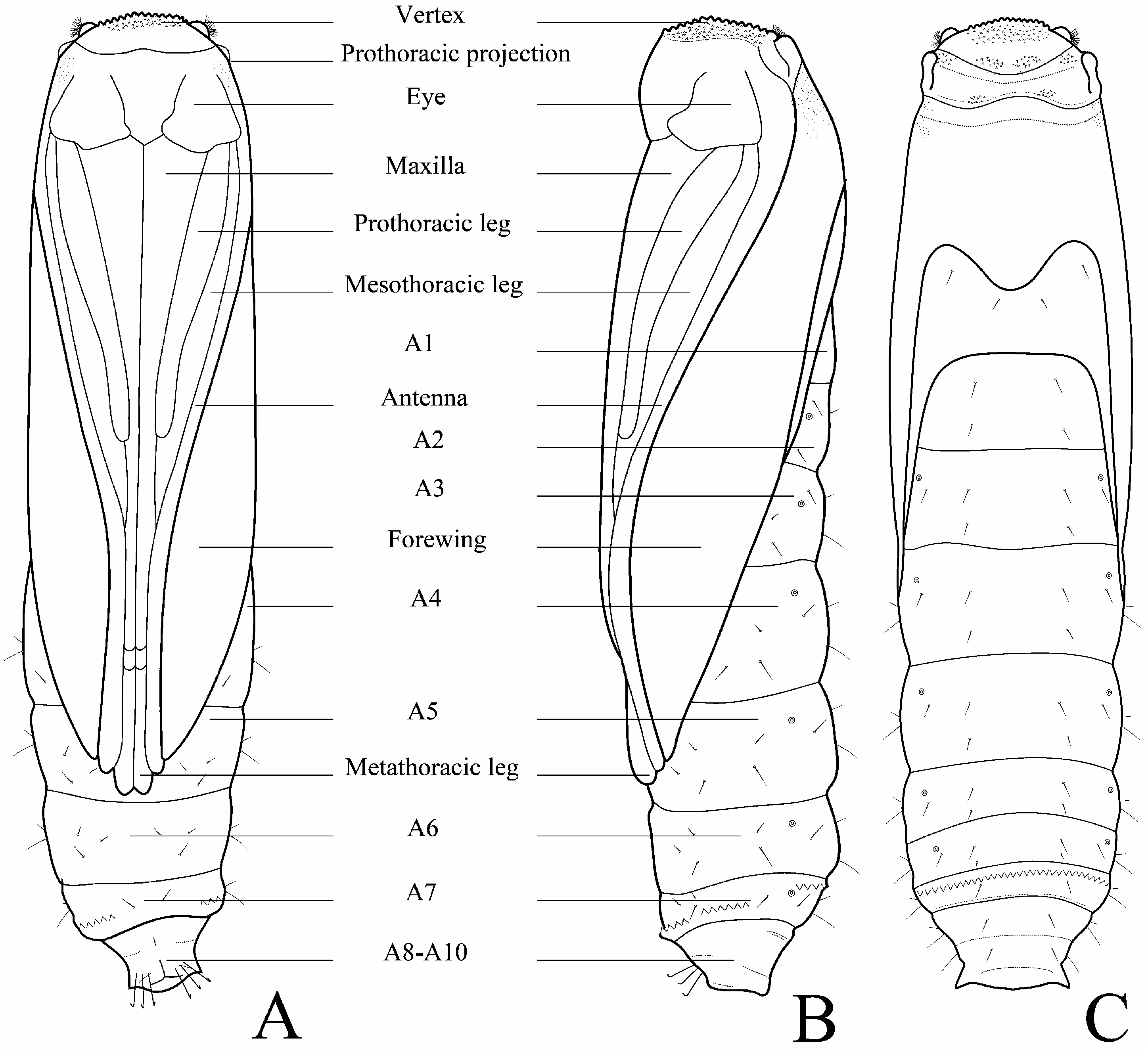
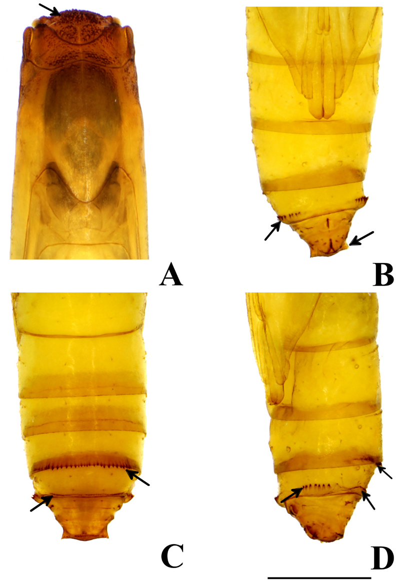
Pupa ( Figs. 11 View FIGURE 11 , 13A–C View FIGURE 13 , 14 View FIGURE 14 ). Length 3.2–4.4 mm (n=13). Yellowish brown, cylindrical. Head slightly flattened, with many granular projections, and a pair of knob-like processes each bearing with hairs. Vertex with many minute granular projections. Prothorax with a pair of sub-triangular projections on dorsolateral corners of tergite. Antennae and forewings reaching to A5. Maxilla (galea) basally broad, gradually narrowing and extending to near posterior end of A4. Prothoracic legs extending to near posterior margin of A2; mesothoracic legs extending to middle of A4; metathoracic legs extending to near posterior margin of A5 or anterior margin of A6. Proleg scars indistinct. A7 with a transverse row of tergal spinules on anterior margin directed posteriorly and with a transverse row of minute spinules on caudal margin. Sternite A7 with a pair of oval pads equipped with a row of spines directed anteriorly. A10 with a pair of triangular tergal projections on posterolateral, apically with three pairs of hooked setae on ventral surfaces of A9 and A10, no true cremaster present.
Remarks. Immature stages of this species were already reported and tentatively assigned to Polyhymno sp. and Polyhymno sp. 4 , respectively ( Ueda 2011, 2013). However, after this publication, Thiotricha was separated from Polyhymno ( Karsholt et al. 2013) . Thus, based on the larval mode of feeding, with 4 Radial veins (R5 absent, or coincident with R4), and the similarity of the male genitalia, we treat this species as Thiotricha .

| T |
Tavera, Department of Geology and Geophysics |
| KM |
Kotel'nich Museum |
| KGU |
Geology and Mineralogy Museum |
| TU |
Tulane University, Museum of Natural History |
No known copyright restrictions apply. See Agosti, D., Egloff, W., 2009. Taxonomic information exchange and copyright: the Plazi approach. BMC Research Notes 2009, 2:53 for further explanation.
|
Kingdom |
|
|
Phylum |
|
|
Class |
|
|
Order |
|
|
Family |
|
|
Genus |
Thiotricha lumnitzeriella Kyaw, Ueda, & Hirowatari
| Kyaw, Khine Mon Mon, Ueda, Tatsuya, Yagi, Sadahisa, Okamoto, Tomoko, Wang, Min & Hirowatari, Toshiya 2021 |
Polyhymno sp. 4: Ueda, 2013: 298
| Ueda, T. 2013: 298 |
Polyhymno sp.: Ueda, 2011: 645
| Ueda, T. 2011: 645 |
Thiotricha sp.1: Murphy, 1990: 162
| Murphy, D. H. 1990: 162 |