
Quedius przewalskii Rеittеr, 1887
|
publication ID |
https://doi.org/ 10.11646/zootaxa.4394.1.5 |
|
publication LSID |
lsid:zoobank.org:pub:B37194D0-F2E7-46DE-B057-D6CDDA997B8B |
|
DOI |
https://doi.org/10.5281/zenodo.5990947 |
|
persistent identifier |
https://treatment.plazi.org/id/18024119-C43A-4D30-52C1-FD14920C5145 |
|
treatment provided by |
Plazi |
|
scientific name |
Quedius przewalskii Rеittеr, 1887 |
| status |
|
Quedius przewalskii Rеittеr, 1887 View in CoL
Quedius lamus Smetana, 1995 , 239
Reitter, 1887, 211 (original description); Bernhauer and Schubert, 1916, 432 (catalog); Gridelli, 1924, 71 (characters); Scheerpeltz, 1933, 1458 (catalog); Boháč, 1988, 554 (cited as Q. przewalskyi ; redescription based on holotype; first illustration of aedeagus; records); Smetana, 1999, 521, 524 (revision of the holotype; redescription; characters; notes; records; synonymy with Q. lamus ); Solodovnikov & Hansen, 2016 (notes; discussion on the composition of Q. przewalskii View in CoL species group); Smetana, 2014, 13 (notes; distribution records).
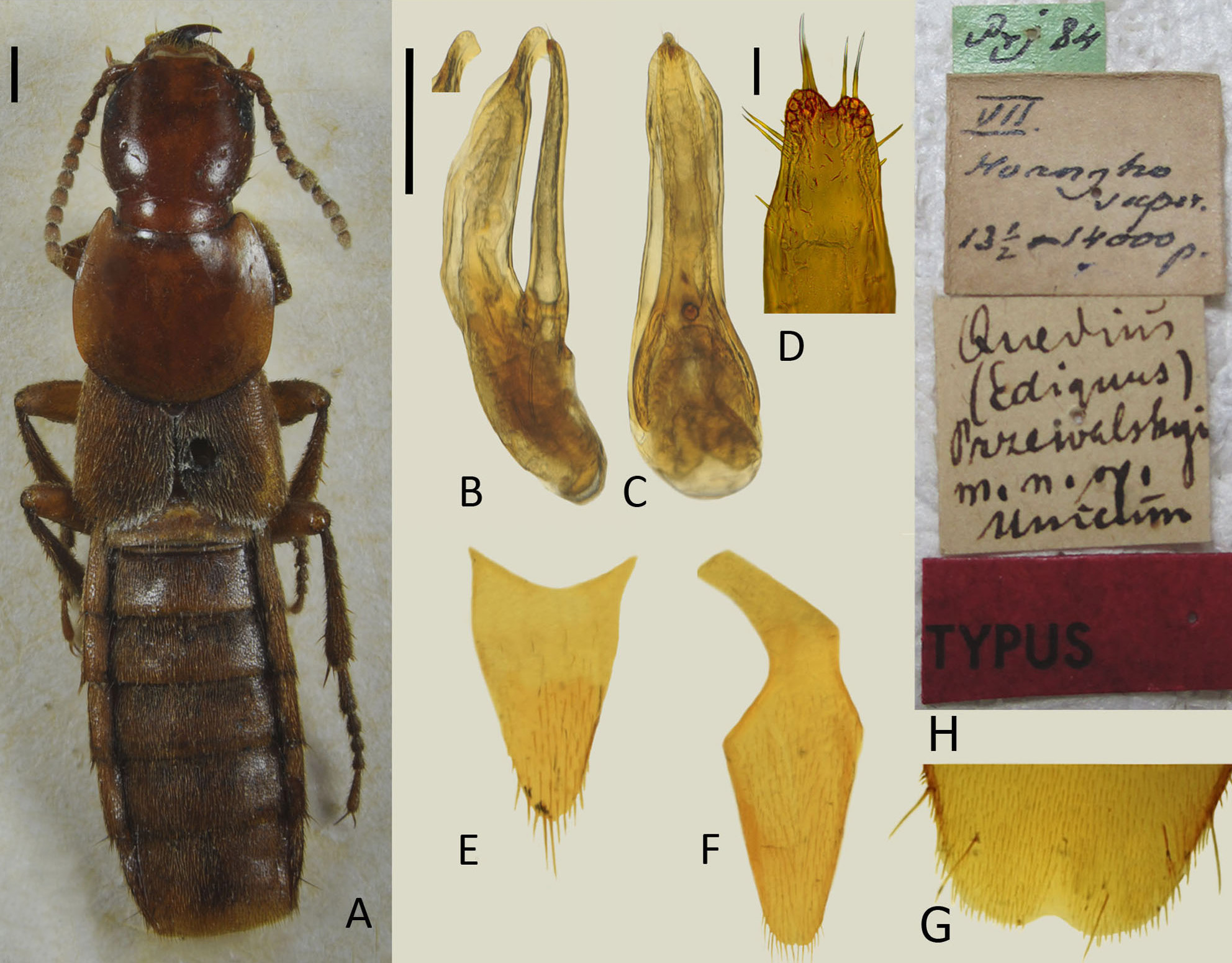
The taxonomic history of Q. przewalskii and the Q. przewalskii species group was recently summarized by Solodovnikov & Hansen (2016). As follows from this paper, the diagnosis and composition of the Q. przewalskii group remains ambiguous. Moreover, in view of the similar Q. roma described in that paper, and the similar Q. mutilatus group newly defined in Salnitska & Solodovnikov (2018), it became apparent that the morphology of the parameral side of the median lobe (often best observed in lateral view, or when the paramere is dissected) is important for the identification of these species and species groups. In that respect the aedeagi of Q. przewalskii and other species assigned to this group were not sufficiently described or illustrated, even though the former species was subject to several publications. Boháč (1988) provided somewhat distorted illustrations not fully matching the real structure, while Smetana (1999) did not provide the lateral view of the aedeagus at all. To facilitate informative comparisons for the new species described in this paper and in the future, here we redescribe the holotype of Q. przewalskii and provide such missing illustrations ( Fig. 2 View FIGURE 2 ).
Type material examined: China: Holotype (♂): ”Prj 84 [green label]/ VII Huangho super. [superior] 13 1/2 – 14000 p. [handwritten label]/ Quedius (Ediquus) przewalskyi [sic!] m. n. sp. unicum [handwritten label]/ TYPUS [red printed label]” ( Fig. 2F View FIGURE 2 ) ( ZIN).
Redescription. Measurements and ratios: HL: 1.93; HW: 1.89; PL: 2.25; PW: 2.59; EL: 1.98; EW: 2.66; FB: 6.16; TL: 15.7; HL/HW: 1.02; PL/PW: 0.87; EL/EW: 0.92; PL/EL: 1.24; PW/EW: 0.97.
Body light brown (somewhat reddish, possibly because of the age of the specimen according to Smetana (1995), rather unicolorous, appendages only slightly lighter, anterior margins of abdominal tergites dark brown; elytra and abdomen with dense yellowish pubescence; legs rather long, body flattened dorso-ventrally. Head barely longer than wide with distinctly rounded posterior angles; with dense and shallow microsculpture of transverse waves uniformly covering the whole head. Eyes small and flat, slightly protruding from lateral contour of head; temples about 2.7 as long as longitudinal diameter of eye.
Head disk on each side with the following setiferous punctures: anterior frontal puncture situated very close to inner margin of eye, posterior frontal puncture at about the same distance from posterior margin of eye and nuchal ridge, and a pair of small barely visible vertical punctures between nuchal ridge and posterior frontal puncture. Each temple with two temporal punctures of which the posterior temporal puncture is closer to posterior margin of head than to posterior margin of eye; additionally, temples with fine setiferous punctures bearing short yellowish setae.
Antennae moderately long, with antennal segments: third distinctly longer than second, fourth and fifth as long as wide, sixth to tenth wider than long gradually widening towards apex of antenna, last segment longer than preceding segments.
Pronotum barely wider than long, widest at about middle, more strongly narrowing anteriad than posteriad; posterior lateral portions distinctly explanate. Dorsal rows of setae each with three equal sized small punctures, the basalmost puncture located at a level slightly before large lateral puncture; sublateral group with two punctures; with three shallow hardly visible punctures along basal margin at each side of pronotum; microsculpture slightly shallower than on head.
Scutellum impunctate, with faint transversal microsculpture.
Elytra widening posteriad, very short and wider than long; slightly wider and notably shorter than pronotum; punctation fine and dense with interspaces between setiferous punctures as large as diameter of punctures; interspaces shiny, with minute irregularities; pubescence yellowish. Wings vestigial.
Abdomen: punctation fine and dense, becoming sparser toward posterior part of each tergite and in general toward apex of abdomen; interspaces with minute irregularities; posterior margin of tergite VII without palisade fringe.
First four segments of front tarsus dilated; second segment as wide as apex of tibia. Sternite VIII with three long setae on each side (on the left side one is broken off), with shallow subangulate triangular medio-apical emargination, area before emargination slightly depressed ( Fig. 2G View FIGURE 2 ). Sternite IХ asymmetrical, elongate, with wide and glabrous basal portion, and with sparsely setose and weakly emarginated apical portion ( Fig. 2F View FIGURE 2 ). Tergite Х triangular, gradually narrowed apicad, with numerous variably long setae apically ( Fig. 2E View FIGURE 2 ). Aedeagus ( Figs. 2B– D View FIGURE 2 ): median lobe (in parameral or anteparameral view) narrowed into distinctly asymmetrical, pointed apex ( Fig. 2C View FIGURE 2 ); (in lateral view) with slightly arcuate and elongated narrow apical portion with subacutely triangular apex and minute tooth situated close to apex ( Fig. 2B View FIGURE 2 ). Paramere (in parameral view) much narrower than median lobe, asymmetrical, gradually narrowed apicad, with distinct apical emargination ( Figs. 2C, D View FIGURE 2 ); with one pair of apical setae on each side of emargination and two pairs of lateral setae ( Fig. 2D View FIGURE 2 ); (underside) with 13 sensory peg arranged in two dense groups on each side of apical emargination; (in lateral view) nearly reaching apex of median lobe ( Fig. 2B View FIGURE 2 ). Internal sac without larger sclerotized structures.
| ZIN |
Russian Academy of Sciences, Zoological Institute, Zoological Museum |
No known copyright restrictions apply. See Agosti, D., Egloff, W., 2009. Taxonomic information exchange and copyright: the Plazi approach. BMC Research Notes 2009, 2:53 for further explanation.
|
Kingdom |
|
|
Phylum |
|
|
Class |
|
|
Order |
|
|
Family |
|
|
Genus |
Quedius przewalskii Rеittеr, 1887
| Salnitska, Maria & Solodovnikov, Aleхey 2018 |
Quedius lamus
| Smetana 1995 |
Q. lamus
| Smetana 1995 |
Q. przewalskii
| Riittir 1887 |