
Strombidium tropicum, Liu & Yi & Lin & Li & Al-Farraj & Al-Rasheid & Song, 2015
|
publication ID |
https://doi.org/ 10.1111/zoj.12257 |
|
persistent identifier |
https://treatment.plazi.org/id/18298784-FFF8-9E74-8F3D-F9D8FC2804C6 |
|
treatment provided by |
Felipe |
|
scientific name |
Strombidium tropicum |
| status |
sp. nov. |
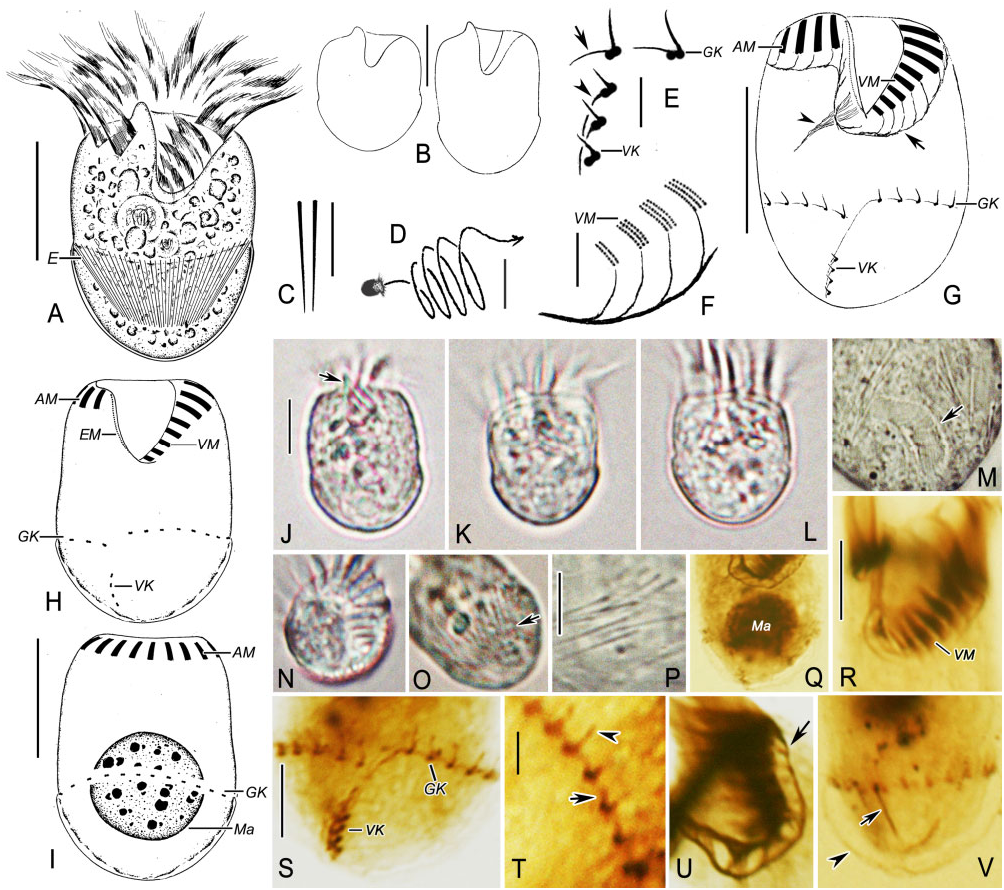
STROMBIDIUM TROPICUM SP. NOV. ( FIG. 3 View Figure 3 , TABLE 1)
Diagnosis: Small Strombidium , cell size usually ∼25 × 20 μm in vivo; body broadly oblong to doliform, anterior end transversely truncated with little apical protrusion; macronucleus globular; ∼12 anterior and five ventral membranelles; girdle kinety comprising ∼20 widely spaced dikinetids with a little gap in the ventral centre, slightly spiralled with the right end shifted posteriad; ventral kinety comprising ∼four dikinetids, separated from the girdle one. Marine form.
Type locality: Coastal waters off Daya Bay (22°43′N, 114°32′E), Guangdong, China, on 27 May 2009. Water temperature was 27.8 °C, salinity 31.0‰, and pH 8.6 GoogleMaps .
Etymology: The Latin adjective tropicum [neuter gender] (tropical) refers to the tropical biotope where the species was discovered.
Deposition of slides: A protargol slide containing the holotype specimen (marked with a black circle) has been deposited at the Natural History Museum, London, with registration number NHMUK 2014.5 View Materials .7.3. Two protargol slides with paratype specimens have been deposited in the Laboratory of Protozoology, Ocean University of China, with registration numbers LWW 09052702 -1 and LWW 09052702 -2.
Deposition of SSU rRNA gene sequence data: The SSU rRNA gene sequence was deposited in GenBank with accession number KJ609050 View Materials . Its length and G + C content are 1774 bp and 48.8 mol %, respectively
Description: Size 15–30 × 15–25 μm in vivo and 16– 25 × 12–19 μm after protargol impregnation. Shape broadly oblong to doliform ( Fig. 3A, B, J–L View Figure 3 ); length: width ratio variable, about 1–1.5: 1 in vivo ( Fig. 3B View Figure 3 ). Anterior cell end transversely truncated and posterior end broadly rounded. Right wall of buccal cavity domed to form an apical protrusion about 2 μm high in vivo, which disappeared after protargol impregnation ( Fig. 3A, B, J View Figure 3 , arrow).
Cytoplasm colourless, containing abundant lipid droplets, 2–4 μm across, and food vacuoles, 3–5 μm across, probably with remnants of bacteria ( Fig. 3A, J View Figure 3 ). Pellicle thin and transparent. Hemitheca covering the cell from the posterior end to the girdle kinety, usually distended in protargol-impregnated cells ( Fig. 3A, L, V View Figure 3 , arrowhead). Polygonal platelets not recognizable. Extrusomes attached to cortex in a shallow bulge above the girdle kinety and forming an equatorial funnel, indistinctly clustered ( Fig. 3A, O, V View Figure 3 , arrows). Extrusomes thin acicular with a sharply pointed posterior end ( Fig. 3C, P View Figure 3 ), about 8 × 0.3 μm in vivo. Macronucleus located in posterior centre of cell, with a globular shape and containing numerous nucleoli 0.5–2 μm across ( Fig. 3I, Q View Figure 3 ). Micronucleus, cytopyge, and contractile vacuole not observed. In Petri dish with in situ water at room temperature, cells keep swimming in spirals (about 60 μm across) by rotating about main cell axis ( Fig. 3F View Figure 3 ).
Oral apparatus occupying anterior end of cell. Oral cavity extending posteriad to about 30% of cell length ( Fig. 3A, G, J View Figure 3 ). Anterior zone of membranelles surrounding the peristomal collar, composed of about 12 membranelles with cilia up to 14 μm long in vivo, which typically extend anteriorly like a crown ( Fig. 3A, K View Figure 3 ). Bases of anterior membranelles about 6 μm long, each composed of three rows of basal bodies. Ventral zone of membranelles closely connected with anterior zone and composed of about five membranelles ( Fig. 3G, H, R View Figure 3 ). Cilia of ventral membranelles about 5–8 μm long in vivo. Bases of ventral membranelles about 3–5 μm long, decreasing in length towards the cytostome: each composed of three parallel rows of basal bodies in the distal portion of zone, but only two rows in the proximal one ( Fig. 3F View Figure 3 ). Endoral membrane on the inner wall of the buccal lip, 7 μm long, probably composed of bare basal bodies as no cilia recognizable ( Fig. 3H View Figure 3 ). System of argyrophilic fibres developed and associated with the adoral zone of membranelles and endoral membrane ( Fig. 3G, U View Figure 3 , arrows). The fibres ‘J’-shaped and about 3–5 μm long. Their anterior ends connecting with the distal membranelle ends or endoral membrane whereas the posterior portions bundled closely together to form a distinct stripe under the adoral zone of membranelles ( Fig. 3F, G, U View Figure 3 ). Pharyngeal fibres prominent, extending obliquely rightwards, up to 7 μm long ( Fig. 3G View Figure 3 , arrowhead).
Somatic cilia arranged in a girdle and a ventral kinety ( Fig. 3G View Figure 3 ). Girdle kinety located in the posterior third of the cell length, with a gap (about 4 μm wide) in the ventral centre. The left end of the girdle kinety positioned higher than the right one by about 1–2 μm, and thus the girdle kinety tending to spiral slightly sinistrally ( Fig. 3G, H, S View Figure 3 ). Girdle kinety composed of about 21 widely spaced dikinetids; within each dikinetid, the left basal body bearing a short cilium about 2 μm long in vivo, whereas the right one associated with a conspicuous argentophilic fibre that is about 2 μm long and curved rightwards ( Fig. 3E, T View Figure 3 , arrows). Ventral kinety in posterior fifth of cell, anterior end below and about 4 μm away from the right end of the girdle kinety, extending posteriad obliquely to the posterior end of the cell ( Fig. 3G, H, S View Figure 3 ). The ventral kinety consisting of about four dikinetids, each bearing a rod-shaped cilium on the anterior basal body, 1 μm long in vivo ( Fig. 3E, G, S View Figure 3 ). An argentophilic fibre originated from the posterior basal body of each ventral dikinetid and extended posteriad to associate with their neighbouring ones, forming a stripe on the right side of the ventral kinety, which tends to connect with the left end of the girdle kinety ( Fig. 3E View Figure 3 , arrow; Fig. 3G, S View Figure 3 ).
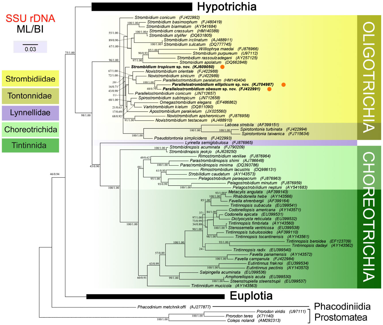
SSU rRNA GENE SEQUENCE ANALYSES ( FIG. 4 View Figure 4 )
The SSU rRNA gene sequences of P. obesum showed a similarity of 98.6% when compared with that of Parallelostrombidium paralatum , which is higher than those obtained with Parallelostrombidium conicum (96.9%) and P. ellipticum (98.2%). Parallelostrombidium ellipticum showed the highest similarity with P. obesum (98.2%) and lower similarity with P. paralatum (97.6%) as well as P. conicum (96.5%). The pairwise sequence similarities between Strombidium tropicum and other sequenced Strombidium species ranged from 93.8 to 97.4%, with Strombidium conicum exhibiting the highest similarity.
In the trees, our new species P. obesum and P. ellipticum were sister to each other, and then clustered with P. paralatum with fully supported values (ML 100%, BI 1.00). This clade grouped with two Novistrombidium species , and then form a larger clade with P. conicum which was moderately supported by the ML tree (43%) and strongly supported by the BI tree (0.93) ( Fig. 4 View Figure 4 ). Strombidium tropicum was related to the non- Strombidium clade comprising Parallelostrombidium , Novistrombidium , Spirostrombidium , Omegastrombidium , Varistrombidium , and Apostrombidium species in the ML tree (11%; Fig. 4 View Figure 4 ), whereas their relationships were not resolved in the BI tree. Strombidium conicum represented the earliest clade of oligotrichs (ML 96% and BI 1.00; Fig. 4 View Figure 4 ). All the nine other Strombidium species composed a moderately wellresolved assemblage into which Williophrya maedai nested ( Fig. 4 View Figure 4 ). The monophylies of the genera Strombidium and Parallelostrombidium were both rejected by the AU test (P = 0.00002 and 0.0001, respectively).
No known copyright restrictions apply. See Agosti, D., Egloff, W., 2009. Taxonomic information exchange and copyright: the Plazi approach. BMC Research Notes 2009, 2:53 for further explanation.