
Parallelostrombidium obesum, Liu & Yi & Lin & Li & Al-Farraj & Al-Rasheid & Song, 2015
|
publication ID |
https://doi.org/ 10.1111/zoj.12257 |
|
persistent identifier |
https://treatment.plazi.org/id/18298784-FFFF-9E78-8F60-F911FB5807C6 |
|
treatment provided by |
Felipe |
|
scientific name |
Parallelostrombidium obesum |
| status |
sp. nov. |
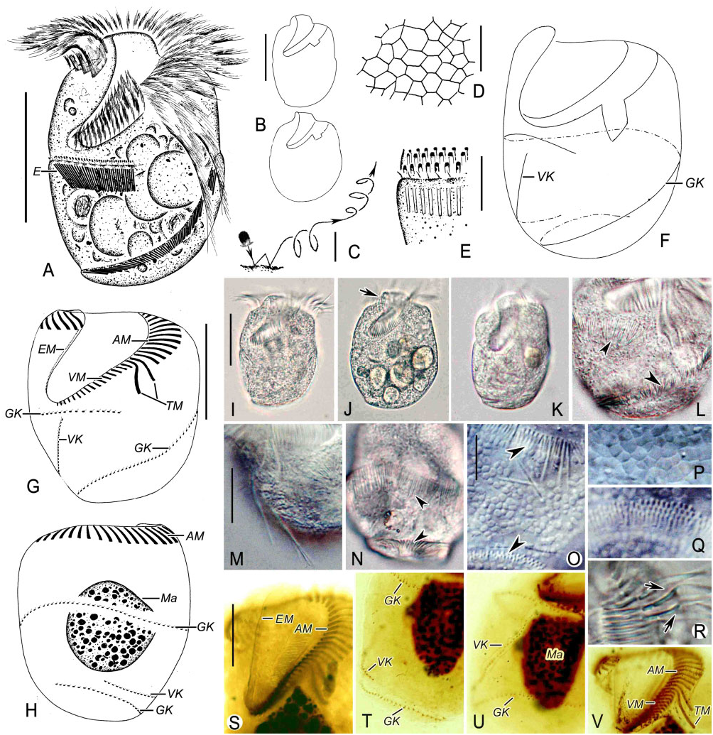
PARALLELOSTROMBIDIUM OBESUM SP. NOV.
( FIG. 1 View Figure 1 , TABLE 1)
Diagnosis: Large-sized Parallelostrombidium ; cell size ∼90 × 65 μm in vivo, body doliform, anterior and posterior ends transversely truncated with conspicuous apical protrusion; macronucleus ovoid; ∼30 anteri- or, ∼17 ventral, and consistently two conspicuous thigmotactic membranelles; girdle kinety comprising ∼130 dikinetids, spiralling around cell with one and a half whorls and the anterior end of the girdle kinety extending to the left of the ventral kinety and located far away from the ventral kinety; ventral kinety with ∼35 dikinetids; the posterior portions of girdle and ventral kineties crossing the right margin of cell and extending transversely on the posterior dorsal side. Marine form.
Type locality: A mangrove wetland near Shenzhen (22°37′N, 114°04′E), Guangdong, China. Water temperature was 27.0 °C, salinity 17.0‰, and pH 8.2 GoogleMaps .
Etymology: The Latin adjective obesum [neuter gender] (fat) refers to the fat body of the species.
Deposition of slides: A protargol slide containing the holotype specimen (marked with a black circle) has been deposited at the Natural History Museum, London, with registration number NHMUK 2014.5 View Materials .7.1. One protargol slide with paratype specimens has been deposited in
*Anterior end of girdle kinety in P. obesum and P. ellipticum , left end of girdle kinety in S. tropicum .
†Posterior end of girdle kinety in P. obesum and P. ellipticum , right end of girdle kinety in S. tropicum .
Data based on protargol-impregnated and randomly selected specimens.
Measurements in μm.
Max, maximum; Mean, arithmetic mean; Min, minimum; N, number of specimens measured; SD, standard deviation; –, data unavailable.
the Laboratory of Protozoology, Ocean University of China, with registration number LWW08040807.
Deposition of SSU rRNA gene sequence data: The SSU rRNA gene sequence has been deposited in GenBank with accession number FJ422991 View Materials , which was previously published by Gao et al. (2009) as misidentification Spirostrombidium sp. 3 .
Description: Cell in vivo 65–110 × 55–70 μm and 69– 103 × 54–72 μm after protargol impregnation. Body shape generally variable, slightly asymmetric and doliform with anterior and posterior ends transversely truncated ( Fig. 1A, B, I, J View Figure 1 ). Collar region domed to form a conspicuous apical protrusion about 10 μm high in vivo, which is undetectable after protargol impregnation ( Fig. 1A, B, J View Figure 1 , arrow). Cell length: width ratio about 5:4 and dorsoventrally flattened with a thickness: width ratio of about 2: 3 in vivo ( Fig. 1A, I, K View Figure 1 ).
Pellicle relatively rigid. Hemitheca composed of polygonal platelets, about 3 μm across in vivo ( Fig. 1D, P View Figure 1 ), covering the posterior dorsal and left ventral portion below the girdle kinety. Distended cell surface not recognizable in protargol-impregnated cells. Cytoplasm colourless, containing many ingested food globules (about 15 μm across) clustered in the posterior half of the cell, and some food vacuoles filled with some small protozoa (about 10 μm in diameter) ( Fig. 1A, J View Figure 1 ). Extrusomes prominent and rod-shaped, about 10 × 0.5 μm in vivo ( Fig. 1E, O, Q View Figure 1 ). Extrusome attachment sites anteriorly positioned to the girdle kinety, producing a stripe surrounding the body with 1.5 whorls ( Fig. 1A, L, N View Figure 1 ). These attachment sites are evenly arranged in three rows in the first whorl and in two rows in the last half whorl ( Fig. 1E, Q View Figure 1 ). Macronucleus oblong to ovoid, about 30 × 25 μm after protargol impregnation, centrally located and containing numerous chromatin granules ( Fig. 1H, U View Figure 1 ). Contractile vacuole, cytopyge, and micronucleus not recognized. In Petri dish with in situ water at room temperature, cells usually swimming in spirals (about 120 μm across) by rotating about the main cell axis, and occasionally crawling over debris on the ventral side using thigmotactic membranelles for attachment ( Fig. 1C View Figure 1 ).
Buccal cavity prominent, extending obliquely to the right and terminating in approximately the anterior 40% of the cell ( Fig. 1A, G, I View Figure 1 ). Adoral zone of membranelles consisting of anterior, ventral, and thigmotactic membranelles ( Fig. 1G, H, S, V View Figure 1 ), all of which are composed of three parallel rows of kinetosomes, except for the two posterior-most membranelles, which probably comprise only two rows. Anterior membranelles composed of 24–34 membranelles with cilia up to about 25 μm long in vivo, stretching perpendicularly to the main cell axis when swimming ( Fig. 1A, I View Figure 1 ). Bases of the anterior membranelles about 11–13 μm long. Ventral membranelles com- posed of 12–20 membranelles with their cilia 6–11 μm long in vivo and their bases about 4–8 μm long, decreasing in length towards the cytostome ( Fig. 1G, V View Figure 1 ). Two dominant thigmotactic membranelles located between the anterior and ventral membranelles, but clearly distinct from these owing to their long cilia (about 55–60 μm in length) pointing posteriorly in vivo ( Fig. 1A, M View Figure 1 ). Bases of the thigmotactic membranelles strikingly long, with the proximal one being about 20 μm long and the distal one about 18 μm long, and with their outside parts curved towards the cytostome ( Fig. 1G, R View Figure 1 , arrows, S, V). Endoral membrane arranged obliquely on the inner wall of the right buccal lip, about 30 μm long, probably composed of a single row of kinetosomes ( Fig. 1G, S View Figure 1 ); no cilia observed. Pharyngeal fibres not observed.
Girdle kinety comprising 101–164 dikinetids, each dikinetid bearing a fusiform cilium, about 2 μm long in vivo, associated with the anterior basal body ( Fig. 1G View Figure 1 ). Girdle kinety beginning below the ventral membranelles, with about seven to ten dikinetids on left of ventral kinety, extending transversely across right ventral and dorsal sides, and obliquely posteriad, crossing the left ventral side of the cell, and curving to the dorsal side from the right margin in the posterior quarter of the cell ( Fig. 1F–H View Figure 1 ). Posterior portion of the girdle kinety running transversely on the posterior dorsal side and terminating in the middle of the posterior dorsal side in 70% of individuals, or in the left margin of the posterior dorsal side in the remaining 30%. Girdle kinety, therefore, spiralling dextrally around the cell in one and a half whorls ( Fig. 1F–H, T View Figure 1 ). Ventral kinety composed of 23–51 densely arranged dikinetids, with the anterior basal body of each bearing a cilium about 2 μm long in vivo ( Fig. 1G View Figure 1 ). The ventral kinety commencing just below the buccal vertex, extending posteriad on the right ventral side, crossing the right margin of the posterior cell, extending above and parallel to the posterior part of the girdle kinety with seven to ten dikinetids, and terminating in the centre of the posterior dorsal area ( Fig. 1F, H, T View Figure 1 ).
No known copyright restrictions apply. See Agosti, D., Egloff, W., 2009. Taxonomic information exchange and copyright: the Plazi approach. BMC Research Notes 2009, 2:53 for further explanation.