
Chrissia acuminata, Zhai & Fan & Wang, 2023
|
publication ID |
https://doi.org/ 10.11646/zootaxa.5323.2.2 |
|
publication LSID |
lsid:zoobank.org:pub:998911D1-EF22-4100-8648-EC4B0AEF24C1 |
|
DOI |
https://doi.org/10.5281/zenodo.8221776 |
|
persistent identifier |
https://treatment.plazi.org/id/186FFE5E-FFF7-5C4A-0D9E-0E7CFBA0F883 |
|
treatment provided by |
Plazi |
|
scientific name |
Chrissia acuminata |
| status |
sp. nov. |
Chrissia acuminata n. sp.
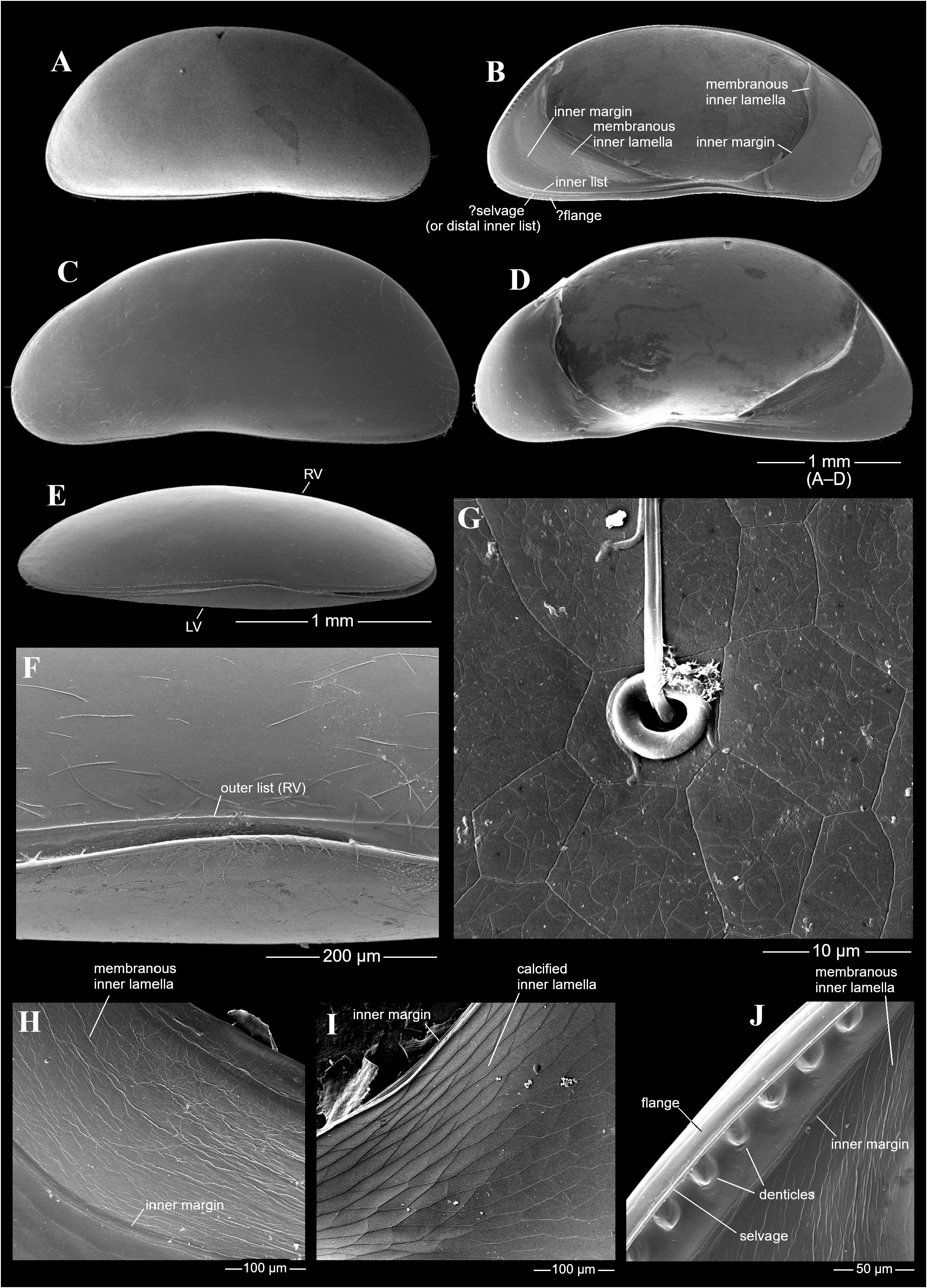
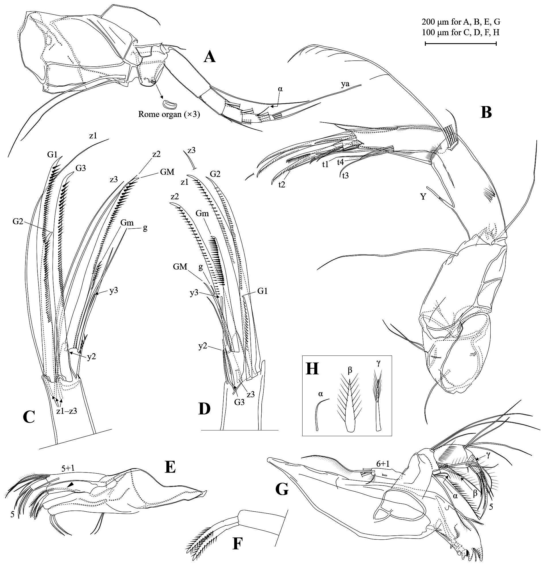
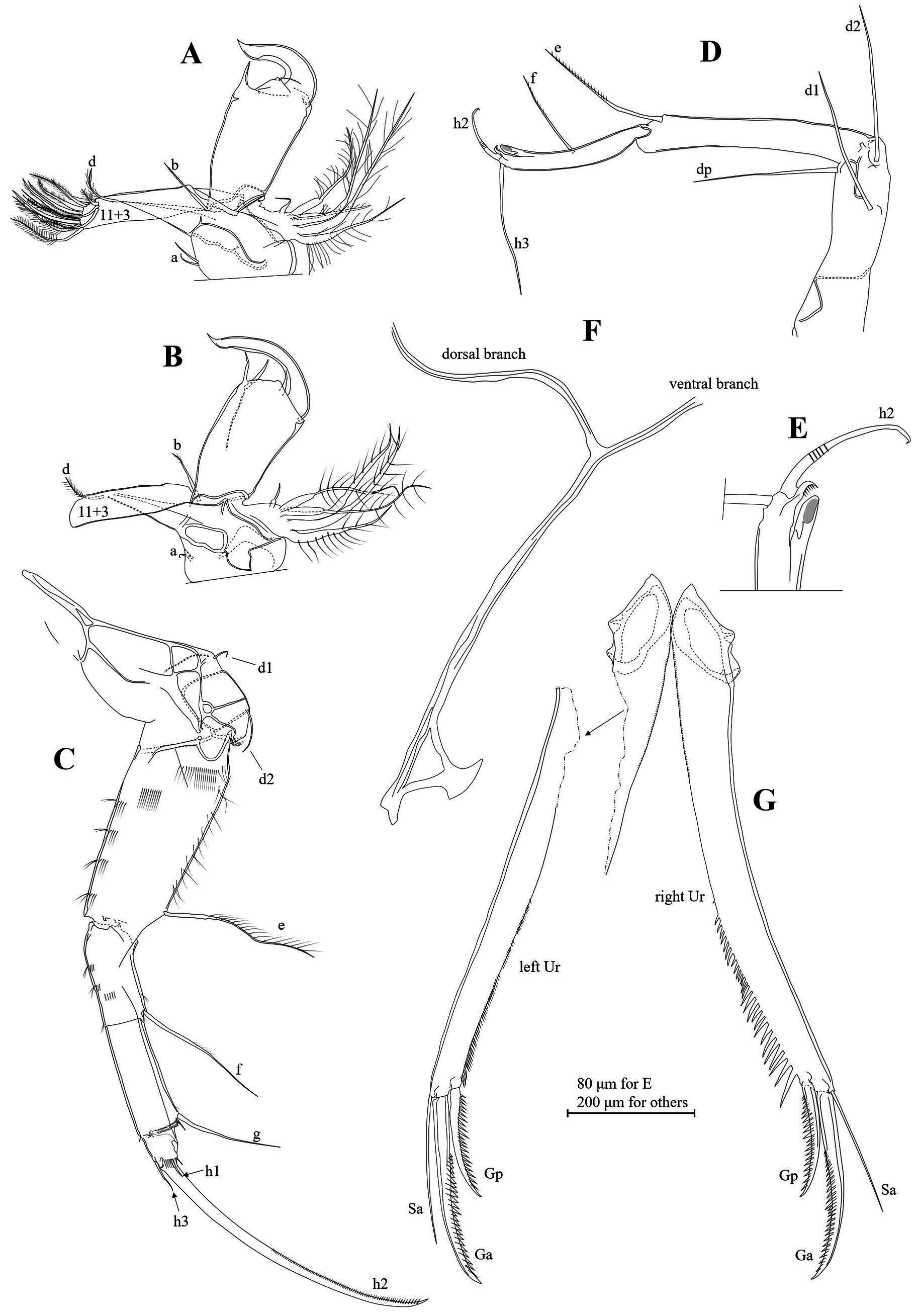
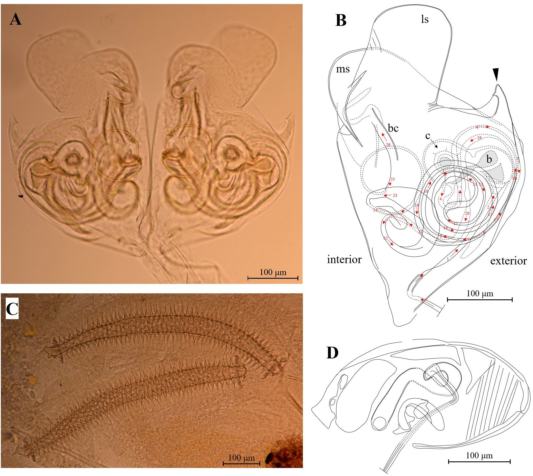
( Figs 8‒11 View FIGURE 8 View FIGURE 9 View FIGURE 10 View FIGURE 11 )
Type locality. Site Y 167 (25°42’18.0”N, 103°06’31.1”E, 1889 m a.s.l.; Table 1 View TABLE 1 ), a small pond (diameter c. 1 m, depth c. 0.5 m) in a valley a few hundred meters away from Xiaohuangtian Village GoogleMaps .
Type material. Holotype: dissected male ( WOC72 ) . Allotype: dissected female ( WOC71 ). Both collected from type locality (site Y167; Table 1 View TABLE 1 ) . Paratype: dissected female ( WOC145 ), from site Y134 (rice field, 25°11’08”N, 102°11’27”E, 1821 m, sampled on August 5th, 2017) ( Note : The site Y134 is not listed in Table 1 View TABLE 1 , and its ostracod assemblage is not discussed in the present paper, because it is situated outside the Kunming area) GoogleMaps .
Other material examined. A- 1 juvenile ( WOC92 ), dissected with valve articulation retained. Dissected A- 2 juvenile ( WOC79 ). Both from type locality (site Y167; Table 1 View TABLE 1 ) .
Derivation of name. From Latin acuminatus, meaning pointed or sharpened, referring to the acuminate lappet at the exterior side of the Hp.
Dimensions of adults. Male (n = 1) RV length 2.69 mm, LV length 2.74 mm. Female (n = 2) RV length 2.78‒ 2.98 mm, LV length 2.86‒3.03 mm. H/L ratio of LV (both sexes, n = 3) 0.45‒0.48.
Diagnosis. Intermediate-sized Chrissia with postero-dorsal valve margin more convex in female than male. Postero-ventral corner narrowly rounded, not angular. Shell surface with polygonal patterns (visible with SEM). Septa absent on valve margin. Postero-dorsal margin of LV with small rounded denticles in interior view. A2 swimming setae extending to about tips of claws. Mx tooth bristles serrated. Male L5 palp sub-rectangular, with left clasper longer and thinner than right. L6 with short d1 and d2 setae. Ur asymmetrically developed, right ramus thicker and with more pronounced denticles. Triangular loop well developed in proximal end of Ur attachment. Hp with subrectangular lateral shield (ls), shorter, distally rounded medial shield, and with acuminate lappet at exterior side. Postlabyrinth spermiductus extending beyond structure ‘c’. Female GL subovate, with dense striations on one end.
Description. (Features for both sexes unless otherwise noted.) Comparatively large cypridid but intermediate-sized for Chrissia (species in genus range from 1 to 4 mm in length), valves moderately elongate, sub-reniform in lateral view ( Fig. 8A‒D View FIGURE 8 ). Dorsal margin evenly rounded (more distinctly arched in female, Fig. 8C & D View FIGURE 8 ), with greatest height more or less posterior to mid-length. Antero-dorsal margin with slight depression anterior to eye. Anterior end rounded. Postero-dorsal margin descending sharply, nearly straight. Postero-ventral corner narrow, but not angular. Ventral margin concave somewhat anterior to mid-length. Shell surface covered with fine setae and polygonal patterns ( Fig. 8F & G View FIGURE 8 ). In interior view, anterior calcified inner lamella much wider than posterior ( Fig. 8B & D View FIGURE 8 ). Calcified inner lamella with shallow reticulation ( Fig. 8I View FIGURE 8 ). Membranous inner lamella with irregular wrinkles ( Fig. 8H View FIGURE 8 ). Selvage (or distal inner list) of LV more or less displaced inwards along free valve margins ( Fig. 8B View FIGURE 8 ). One inner list running close to selvage along ventral margin. About 12 small rounded denticles present on postero-dorsal part of LV ( Fig. 8B & J View FIGURE 8 ), not in RV. LV slightly overlapping RV in ventral view ( Fig. 8E & F View FIGURE 8 ).
A1 ( Fig. 9A View FIGURE 9 ) with eight segments, first two of which fused into large base, supporting one short dorsal seta and two long ventral setae. Third segment short and wider than more distal ones, bearing one short dorso-apical seta. RO small, with fine longitudinal striations. Fourth through eighth segments progressively smaller. Fourth segment elongate, with one long dorso-apical seta and one very short ventro-apical seta. Fourth and fifth segments each carrying two very long dorso-apical setae and two shorter ventro-apical setae, and with dorso-apical sclerotized extensions resembling setae. Seventh segment with four very long apical setae on interior surface, and with fine, short “α” seta on exterior surface. Eighth segment with four apical setae. Aesthetasc seta ya conspicuously longer than ventral seta. Two intermediate setae very long.
A2 ( Fig. 9B‒D View FIGURE 9 ) with two protopodal segments, one exopodal segment, and three endopodal segments (penultimate segment undivided in both sexes). Coxa segment with one extero-posterior seta and two sub-equal ventro-medial setae. Basis segment with one long ventro-apical seta. Exopodal plate carrying three setae, longest one almost reaching to distal end of first endopodal segment. First endopodal segment carrying aesthetasc Y, six dorso-apical setae (swimming setae and accompanying setae), and one ventro-apical seta. Dorso-medial and apical parts of this segment with well-developed pseudochaetae. Aesthetasc Y 2-segmented, not reaching to end of this segment. Swimming setae extending almost to tips of terminal claws. Accompanying seta (6th seta) short. Ventro-apical seta of first endopodal segment reaching to about end of terminal segment. Penultimate segment dorsomedially with two setae and ventro-medially with four t-setae: t2 longest, extending beyond mid-way of terminal claws; t3, t1, and t4 progressively shorter. Aethestasc y1 only observed in allotype female, situated slightly proximal to t setae, difficult to be distinguished from accompanying pseudochaetae. Distal chaetotaxy of last two segments sexually dimorphic. In male ( Fig. 9D View FIGURE 9 ), G2 normally developed. G1 less than half length of G2. G3 small, reaching to only about basal part of GM. z1 and z2 transformed to thick claws with z1 being longest terminal claw. z3 not transformed, being seta somewhat longer than z1. G1–G3, z1, and z2 serrated. y2 slender, extending to about end of terminal segment. Terminal segment small, ventro-apically with seta-like sclerotized extension, and dorsally with lobe-like extension. Gm (more exterior claw of this segment) strongly serrated, being comb-like, extending beyond half way of z1. GM (more interior claw of this segment) about 2/3 of Gm in length, slightly serrated.Aesthetasc part of y3 extending to about half way of GM. Seta g somewhat longer than setal part of y3. In female ( Fig. 9C View FIGURE 9 ), G1 and G3 not reduced, with G1 being longest claw. G2 significantly shorter than G1 and G3. z1 and z2 not transformed into claws, being setae extending beyond G1 claw. GM (more interior claw of terminal segment) thick, extending to tip of G3, serrated. Gm (more exterior claw) slender, being about half length of GM.
Md ( Fig. 9G & H View FIGURE 9 ) coxa elongate, distally with tooth-like masticatory processes, with one and two small plumose setae on antero-exterior and postero-interior edges respectively. Palp 4-segmented. First segment (basis) elongate, dorsally bearing exopod with six unequal plumose apical setae and one sub-apical seta, and ventrally with slender seta α and three long plumose setae. Second segment dorsally with three unequal setae and ventrally with thick plumose seta β, four plumose grouped setae, and one shorter accompanying seta. Third segment dorso-subapically with four long setae, and apically with six setae, including thick plumose seta γ, four other lateral setae, and one minute ventro-apical seta. Fourth segment with five claws/setae.
Mx palp ( Fig. 9E View FIGURE 9 ) 2-segmented. First segment elongate, with one smooth sub-apical seta and five dorso-subapical setae, two of which being plumose. Second segment with five setae. Tooth bristles on distal gnathobasic endite ( Fig. 9F View FIGURE 9 ) distinctly serrated or with thick pseudochaetae. Distal endite also with one plumose lateral seta ( Fig. 9E View FIGURE 9 ).
Male L5 ( Fig. 10A & B View FIGURE 10 ) protopod with short setae a, slightly plumose seta b and distinctly plumose seta d. Gnathoobasic endite apically with 11 setae most of which being plumose, and sub-apically with three shorter plumose setae. Exopod with one short and five long plumose rays. Right ( Fig. 10A View FIGURE 10 ) and left ( Fig. 10B View FIGURE 10 ) palps sub-equal in size and shape, thick and elongate. Right palp ventro-apically with one sharp projection, while left palp with two slenderer projections. Right clasper shorter and thicker than left.
L6 ( Fig. 10C View FIGURE 10 ) well developed, with five segments. First segment (protopod) with small setae d1 and d2. Second segment (first endopodal segment) with long plumose seta e, and with clusters of pseudochaetae in anterior, posterior, and proximal areas. Third segment comparatively short, with faintly plumose seta f, and with clusters of pseudochaetae in posterior area. Fourth segment elongate, with smooth seta g, minute accompanying seta, and distally with one row of pseudochaetae. Terminal segment small, distally with one row of pseudochaetae. Seta h1 small. Claw h2 slender, slightly serrated along distal half. Seta h3 somewhat longer than h1.
L7 ( Fig. 10D View FIGURE 10 ) typical of family Cyprididae , with well-developed setae d1, d2, dp, e, and f. Seta h3 comparatively long, anteriorly directed. Pincer organ ( Fig. 10E View FIGURE 10 ) with h2 nearly smooth, only with serrations medially. CL structure serrated.
Ur ( Fig. 10G View FIGURE 10 ) asymmetrically developed, with right Ur thicker than left and slightly bent (left ramus straight). Both rami with serrations in distal half but those on right Ur more pronounced. Claws and setae identical in both sides. Ga and Gp distinctly serrated, with Gp reaching slightly beyond mid-length of Ga. Sa slender, shorter than Ga and longer than Gp. Sp absent.
Ur attachment ( Fig. 10F View FIGURE 10 ) characteristic of subfamily, with triangular structure developed in basal part. Dorsal branch longer than ventral branch.
Hp ( Fig. 11A & B View FIGURE 11 ) lateral shield (ls) subrectangular, with parallel sides and rounded distal end. Medial shield (ms) shorter than lateral shield, asymmetrically oriented. Prominent acuminate lappet present at exterior side of Hp. Internal anatomy with part ‘c’ globular. Spermiductus entering Hp first passing through structures ‘b’ and ‘c’, then going in several sets of coils in different directions (red arrows 3‒20 in Fig. 11B View FIGURE 11 ) that extend beyond ‘c’, then into three descending spiral coils with tubes of varying thicknesses (red arrows 21‒26 in Fig. 11B View FIGURE 11 ) into structure ‘bc’.
ZO ( Fig. 11C View FIGURE 11 ) elongate, with 48‒49 rosettes. Central tube widest medially, narrowest proximally.
GL ( Fig. 11D View FIGURE 11 ) sub-ovate, with transverse striations on one end, and tube-like structures and ovate structures on other end and central part.
Remarks. Within the genus, Chrissia acuminata n. sp. resembles only Chrissia hodgsoni ( Sars 1924a) and Chrissia smaragdina ( Sars 1924a) , which were originally reported from Cape Province of the Republic of South Africa. These three species are unique among congeners in having a sloping postero-dorsal valve margin, narrowly rounded postero-ventral corner, and more importantly, in having an acuminate lappet at the exterior side of the Hp. For distinction, the postero-dorsal margin of female C. smaragdina is less inflated than the female of Chrissia acuminata n. sp., the ls and ms of the Hp are wider than those of the new species, and there is a tiny angular obtrusion on the exterior margin of ms, which is absent in the new species. In addition, the claw/ramus ratio or Ur is about 15% longer in C. smaragdina than that of the new species. The postero-dorsal part of the female valve of C. hodgsoni is also less inflated than the new species, ms being longer and more ventrally directed, and the male L5 palps are shorter and distally dilating.
The small denticles on the LV postero-dorsal margin of Chrissia acuminata n. sp. are hitherto not known from any other species of the genus. Similar structures were also observed in Chrissia achandii (George & Martens, in George et al. 1993), where they were present in the postero-dorsal margin of the RV.
No known copyright restrictions apply. See Agosti, D., Egloff, W., 2009. Taxonomic information exchange and copyright: the Plazi approach. BMC Research Notes 2009, 2:53 for further explanation.
|
Kingdom |
|
|
Phylum |
|
|
Class |
|
|
Order |
|
|
Family |
|
|
Genus |