
Simulium (Simulium) cavum Takaoka
|
publication ID |
https://doi.org/ 10.11646/zootaxa.3961.1.1 |
|
publication LSID |
lsid:zoobank.org:pub:EFA2C0F4-35FC-47D3-91F9-5D8B5C68624D |
|
DOI |
https://doi.org/10.5281/zenodo.6108829 |
|
persistent identifier |
https://treatment.plazi.org/id/190987B3-1346-7B40-5AF6-F939F267F8A9 |
|
treatment provided by |
Plazi |
|
scientific name |
Simulium (Simulium) cavum Takaoka |
| status |
|
Simulium (Simulium) cavum Takaoka View in CoL & Ya’cob sp. nov.
( Figs. 32 View FIGURE 32 A–34D)
Female. Unknown.
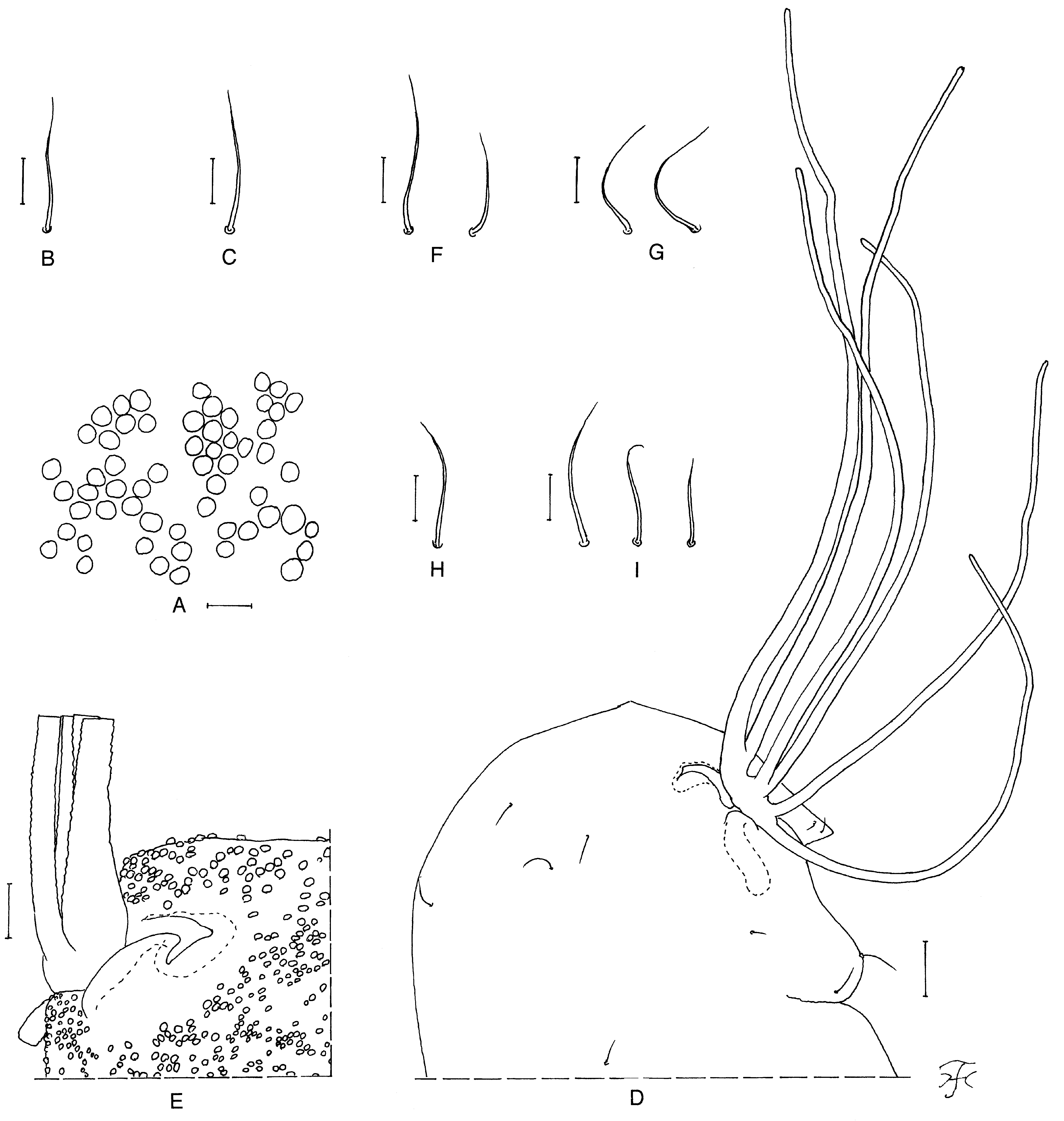
Male. Body length 3.0 mm. Head. Slightly wider than thorax. Upper eye medium brown, consisting of large facets in 21 vertical columns and in 21 horizontal rows. Clypeus black, thickly white pruinose, silvery or bluish, shiny when illuminated at certain angles, covered with dark brown hairs along and near lateral margins (central portion widely bare). Antenna composed of scape, pedicel and nine flagellomeres, brownish-black except base of first flagellomere yellow; first flagellomere elongate, 1.8 times as long as second one. Maxillary palp grayish to dark brown, composed of five segments with proportional lengths of third, fourth, and fifth segments 1.0:1.1:2.4; third segment ( Fig. 32 View FIGURE 32 A) of moderate size; sensory vesicle ( Fig. 32 View FIGURE 32 A) small (0.16–0.18 times as long as third segment), ellipsoidal, and with opening of moderate size. Thorax. Scutum black, with white pruinose pattern, i.e., anterior pair of elliptical spots (not sharply pointed posteriorly) on shoulders extending posteriorly along lateral margins but disconnected to large transverse spot entirely covering prescutellar area; these pruinose areas silvery or bluish iridescent when illuminated at certain angles; scutum uniformly and moderately covered with brassy recumbent short hairs interspersed with dark-brown long upright hairs on prescutellar area. Scutellum black, with several dark-brown long upright hairs. Postnotum black, white pruinose when illuminated at certain angles and bare. Pleural membrane bare. Katepisternum longer than deep, black, white pruinose when illuminated at certain angles, and bare. Legs. Foreleg: coxa yellow to dark yellow with anterior surface darkened; trochanter light brown; femur light brown with apical cap dark brown; tibia dark brown except median large portion on inner surface white on outer surface, and with bright white sheen when illuminated at certain angles; tarsus brownish-black to black, with moderate dorsal hair crest; basitarsus greatly dilated, 5.4 times as long as its greatest width. Midleg: coxa brownish-black; trochanter dark brown; femur medium brown with apical cap dark brown; tibia medium brown except apical cap dark brown and extreme base white; tibia with white sheen on posterior surface when illuminated at certain angles; tarsus light brown except basal two-thirds of basitarsus white. Hind leg: coxa brownish-black; trochanter light brown; femur medium brown except apical cap dark brown and extreme base yellow; tibia medium brown except apical cap dark brown and basal tip dark yellow; tarsus ( Fig. 32 View FIGURE 32 B) medium brown except basal half of basitarsus yellowish white (though base darkened) and basal half of second tarsomere whitish; basitarsus ( Fig. 32 View FIGURE 32 B) much enlarged, spindle-shaped, gradually widened from base to middle, then gradually narrowed to apex, 3.13 times as long as its greatest width, and 1.07 and 1.19 times as wide as greatest widths of hind tibia and femur, respectively; calcipala ( Fig. 32 View FIGURE 32 B) small, nearly as long as wide, 0.22 times as wide as greatest width of basitarsus; pedisulcus ( Fig. 32 View FIGURE 32 B) well developed. Wing. Length 2.0 mm. Costa with dark spinules and hairs; subcosta bare; basal section of radial vein bare; R1 with dark spinules and hairs; R2 with hairs; hair tuft on base of radial vein dark brown; basal cell absent. Halter . White except basal portion darkened. Abdomen. Basal scale brownish-black, with fringe of dark long hairs. Dorsal surface of abdomen brownish-black to black, with dark short hairs; segments 2, 6 and 7 each with pair of silvery or bluish iridescent spots dorsolaterally, those on segment 2 connected broadly to each other in middle. Genitalia. Coxite in ventral view ( Fig. 32 View FIGURE 32 C) nearly quadrate, covered with stout hairs on posterior half. Style in ventral view ( Fig. 32 View FIGURE 32 C) elongate, gradually tapered to apex, with apical spine; style in ventrolateral view ( Fig. 32 View FIGURE 32 D) elongate, 2.8 times as long as its greatest width at basal one-third, nearly parallelsided from base to basal one-third, then tapered toward middle, and nearly parallel-sided to apex; style in medial view ( Fig. 32 View FIGURE 32 E) 1.5 times as long as coxite, moderately produced dorsally near base forming protuberance having several small cone-like spines on its apex. Ventral plate in ventral view ( Fig. 32 View FIGURE 32 C) well sclerotized, with body subquadrate (though slightly narrowed basally, and rounded on each posterolateral corner), 0.6 times as long as wide, posterior margin somewhat concave, and with round projection directed ventrally which is covered with many minute setae; arms divergent from each other; ventral plate in lateral view ( Fig. 32 View FIGURE 32 F) with posterior margin weakly serrated along basal two-thirds; ventral plate in end view ( Fig. 32 View FIGURE 32 G) rounded ventrally, with lateral margin serrated along basal two-thirds, with numerous setae on posterior surface except most of basal portion bare. Median sclerite ( Fig. 32 View FIGURE 32 F, H) plate-like, gradually widened from base toward apex, with round apex, brown basally, but not so well sclerotized apically. Paramere ( Fig. 32 View FIGURE 32 I) with several distinct hooks and several smaller ones. Aedeagal membrane ( Fig. 32 View FIGURE 32 I) densely covered with minute setae, and with moderately sclerotized dorsal plate in form of horizontal bar ( Fig. 32 View FIGURE 32 J). Abdominal segment 10 ( Fig. 32 View FIGURE 32 K, L) with three or four hairs on ventral surface and four to seven hairs on lateral surface on each side. Cercus ( Fig. 32 View FIGURE 32 K, L) small, rounded, with eight distinct hairs.
Pupa. Body length 3.0– 3.2 mm. Head. Integument including antennal sheaths ochreous, moderately covered with relatively larger tubercles (most of them lack minute secondary projections) on frons ( Fig. 33 View FIGURE 33 A), and relatively smaller tubercles on antennal sheaths and face; frons with two pairs of unbranched slender medium-long trichomes ( Fig. 33 View FIGURE 33 B), arising close together; face with pair of unbranched medium-long trichomes ( Fig. 33 View FIGURE 33 C). Thorax. Integument ochreous, moderately covered with relatively larger tubercles (similar to those on frons though somewhat smaller) on anterior half, and relatively smaller tubercles on posterior half, and with pit-like organ near base of each gill ( Fig. 33 View FIGURE 33 D, E); thorax on each side with two anterodorsal trichomes ( Fig. 33 View FIGURE 33 F), two anterolateral trichomes ( Fig. 33 View FIGURE 33 G), one mediolateral trichome ( Fig. 33 View FIGURE 33 H), and three ventrolateral trichomes (one trichome longer than two others) ( Fig. 33 View FIGURE 33 I); all trichomes medium-long and unbranched. Gill ( Fig. 33 View FIGURE 33 D) with six slender thread-like filaments in three pairs (one dorsal, one middle and one ventral) arising directly from thorax and lacking common basal stalk; all pairs short-stalked (slightly longer stalk for dorsal pair in one pupa), and stalks of dorsal and ventral pairs lying at angle of 90 degrees or little more when viewed laterally; ventral filament of ventral pair directed downward, then curved forward (or backward in one pupa); upper filament of dorsal pair longest (1.5 mm long), other filaments gradually shortened from dorsal to ventral, and lower filament of ventral pair shortest (0.9–1.0 mm long); relative thickness of six filaments from dorsal to ventral when compared basally 1.00:0.69:0.63:0.56:0.56:0.56; upper filament of dorsal pair gradually tapered from base toward apex, other filaments nearly subequal in thickness from base to one-half or one-third, then tapered toward apex; all filaments dark brown, with distinct annular ridges and furrows forming definite reticulate surface patterns, and densely covered with minute tubercles. Abdomen. Dorsally, all segments unpigmented except segment 1 dark yellow and posterior part of segment 9 yellow; segment 1 with one unbranched medium-long seta on each side; segment 2 with one unbranched seta and five spinous short setae on each side; segments 3 and 4 each with four distinct hooked spines and one unbranched spinous short seta on each side; segment 5 and 6 lacking spine-combs; segments 7–9 with distinct spine-combs in transverse row (though those on segments 7 and 9 fewer in number and smaller than those on segment 8); segments 6–9 each with comb-like groups of minute spines on each side; segment 9 without terminal hooks. Ventrally, segments 4–8 unpigmented, each with comb-like groups of minute spines; segment 4 with one unbranched hook and few slender minute setae on each side; segment 5 with pair of bifid hooks submedially and few unbranched short setae on each side; segments 6 and 7 each with pair of bifid inner and unbranched outer hooks somewhat separated from each other, and few unbranched short setae on each side. Grapnel-shaped hooklets absent on each side of segment 9. Cocoon. Wall-pocket-shaped, thickly woven (in particular anterior margin), medium-brown, not extended ventrolaterally, and with thick anterodorsal margin; individual threads invisible; 2.5–2.7 mm long by 1.2–1.3 mm wide.
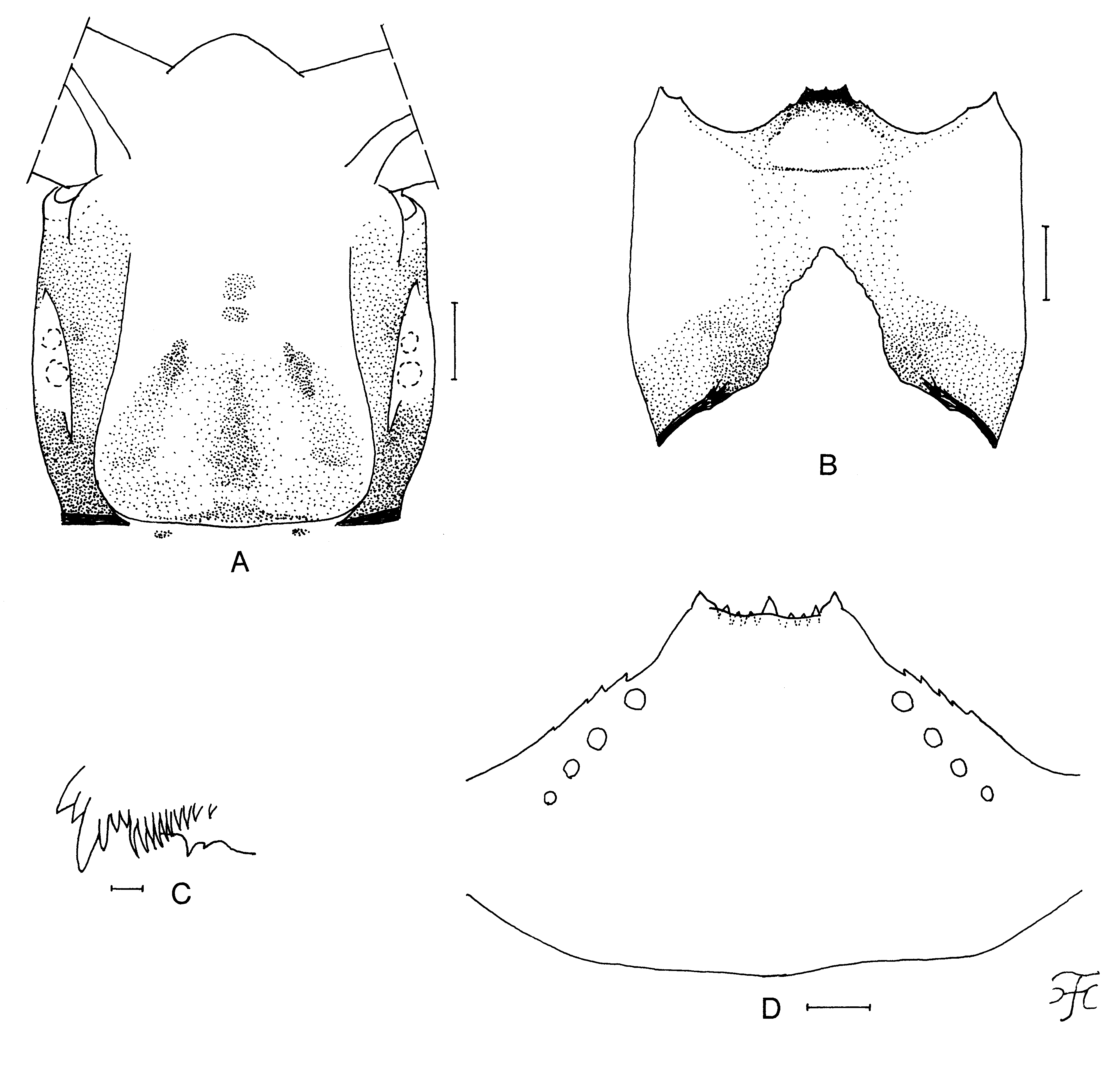
Mature larva. Body length 4.8–5.2 mm. Body grayish black to brownish black, except ventral surface of abdominal segments 4–9 white though there is dark line mediolongitudinally on ventral surface of segments 4–7. Cephalic apotome ( Fig. 34 View FIGURE 34 A) whitish on anterior half and light brown on posterior half with narrow portion along posterior margin dark brown medially, moderately covered with minute setae; head spots light to dark brown, usually surrounded by dark areas and mediolateral spots and posterolateral spots connected to each other by dark areas. Lateral surface of head capsule medium brown except eye-spot region whitish and anterior two-thirds of lower half yellowish; eyebrow distinct; few spots before posterior margin indistinct or faintly negative. Ventral surface of head capsule ( Fig. 34 View FIGURE 34 B) yellowish except posterior one-third and medial wide portion medium to dark brown though mediolongitudinal portion narrowly paler; long spot on each side of postgenal cleft faintly positive or negative. Antenna composed of three segments and apical sensillum, much longer than stem of labral fan; length ratio of three segments (from base to tip) 1.0:1.0 – 1.1:0.7 – 0.8. Labral fan with 34–44 main rays. Mandible ( Fig. 34 View FIGURE 34 C) with serrations composed of two teeth (one medium-sized, one small); main tooth at obtuse angle against mandible on apical side; supernumerary serrations absent; comb-teeth decreasing in length from first to third. Hypostoma ( Fig. 34 View FIGURE 34 D) with nine anterior teeth, of which medin tooth and corner teeth most prominent, subequal in length to each other, and longer than intermediate teeth on each side; lateral margins weakly serrate apically; four or five hypostomal bristles per side slightly divergent posteriorly from lateral margin. Postgenal cleft ( Fig. 34 View FIGURE 34 B) triangular, pointed apically, 2.2 times length of postgenal bridge; sheath of subesophageal ganglion not pigmented. Histoblast of pharate pupal gill with six short filaments. Thoracic and abdominal cuticle almost bare except last segment of abdomen moderately covered with short colorless setae on each side of anal sclerite. Rectal scales present. Rectal organ compound, each lobe with 11 – 17 finger-like secondary lobules. Anal sclerite X-shaped, with short broad anterior arms 0.8 times length of posterior ones; 10 – 12 sensilla posterior to posterior arms. Last abdominal segment somewhat swollen laterally but lacking ventral papillae. Posterior circlet with 80 rows of hooklets with up to 14 hooklets per row.
Type material. HOLOTYPE: Male, reared from a pupa collected from a small stream (width 0.5–1.0 m, depth 5–30 cm, water temperature 15.0˚C, shaded, altitude 1,186 m, 16˚11’45.123” N/107˚50’55.600” E), moderately flowing from a natural forest, near the top of Bach Ma National Park, Phu Loc, Thua Thien Hue Province, central Vietnam, 23-II-2014, by H. Takaoka, M. Sofian-Azirun, Z. Ya’cob, C.D. Chen & K.W. Lau. PARATYPES: One pupa, one pupal exuviae collected from a small stream (width 1–2 cm, water temperature 14.0˚C, shaded, altitude 1,273 m, 16˚11’42.061” N/107˚51’27.955” E), moderately flowing from a natural forest, near the top of Bach Ma National Park, Phu Loc, Thua Thien Hue Province, central Vietnam, 23-II-2014, by H. Takaoka, M. Sofian-Azirun, Z. Ya’cob, C.D. Chen & K.W. Lau; three mature larvae, same data as those of the holotype.
Biological notes. The pupae and larvae of this new species were collected from dead tree leaves in two small streams. Associated species were S. (G.) phulocense sp. nov., S. (G.) thuathienense sp. nov., S. (N.) bachmaense , S. (S.) atipornae and S. (S.) rufibasis .
Etymology. The species name cavum refers to the hollow on the thoracic integument near the base of the pupal gill. The Latin adjective ‘ cavus ’ means ‘hollow’.
Remarks. Simulium (S.) cavum sp. nov. is assigned to the S. tuberosum species-group of the subgenus Simulium , redefined by Takaoka and Davies (1996), based on the male genitalia ( Fig. 32 View FIGURE 32 C). This new species is characterized by having the pit-like organ on the thoracic integument near the base of the gill ( Fig. 33 View FIGURE 33 D, E). Five species of the S. tuberosum species-group, i.e., S. (S.) brevipar Takaoka & Davies , S. (S.) tiomanense Takaoka, Sofian-Azirun & Belabut , both from Peninsular Malaysia, S. (S.) tianchi Chen, Zhang & Yang from China, S. (S.) sigiti Takaoka & Hadi from Java and S. (S.) yuphae Takaoka & Choochote from Thailand, have a similar pit-like organ ( Takaoka and Davies 1995; Takaoka et al. 2012b; Takaoka and Choochote 2005b; Takaoka and Hadi 1991; Chen et al. 2003). This new species is distinguished in the male from S. (S.) brevipar , S. (S.) tiomanense , S. (S.) tianchi and S. (S.) yuphae by having the upper-eye facets in 21 vertical columns (cf., in 17 vertical columns in S. (S.) brevipar , 16 vertical columns in S. (S.) tiomanense , 15 vertical columns in S. (S.) tianchi and 17 or 18 vertical columns in S. (S.) yuphae ), and in the pupa from S. (S.) brevipar , S. (S.) sigiti and S. (S.) yuphae by the relatively large tubercles on the pupal frons without minute secondary projections ( Fig. 33 View FIGURE 33 A) (cf., large tubercles with minute secondary projections in the latter three known species), and also from S. (S.) tianchi and S. (S.) sigiti by having six gill filaments of different length and thickness (cf., six gill filaments are subequal in length and thickness to one another in the latter two known species).

No known copyright restrictions apply. See Agosti, D., Egloff, W., 2009. Taxonomic information exchange and copyright: the Plazi approach. BMC Research Notes 2009, 2:53 for further explanation.
|
Kingdom |
|
|
Phylum |
|
|
Class |
|
|
Order |
|
|
Family |
|
|
Genus |