
Rhynchotermes Holmgren
|
publication ID |
https://doi.org/ 10.11646/zootaxa.4109.5.1 |
|
publication LSID |
lsid:zoobank.org:pub:6690022A-AE4C-4E45-8A22-161FA9784E47 |
|
DOI |
https://doi.org/10.5281/zenodo.5316456 |
|
persistent identifier |
https://treatment.plazi.org/id/1918971F-FFEF-FFD1-FF33-FBD6FB7DFB01 |
|
treatment provided by |
Plazi |
|
scientific name |
Rhynchotermes Holmgren |
| status |
|
Genus Rhynchotermes Holmgren
Armitermes (Rhynchotermes) Holmgren 1912: 55 ; Snyder 1925b: 181, 188–190; Rhynchotermes ; Snyder 1949: 264; Mathews 1977: 154 –159, redescription; Mill 1983: 183, 188; Fontes 1985: 9; Nickle & Collins 1992: 226, 227, 234; Constantino 1999: 403, 416; 2002: 22; Krishna et al. 2013: 1471 (catalog)
Type-species. Armitermes nasutissimus Silvestri, 1901 , by monotypy.
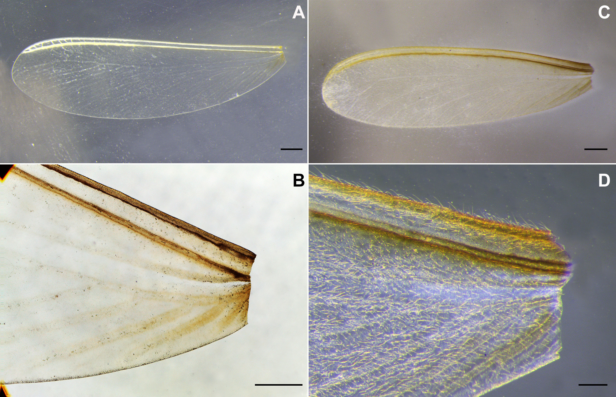
Imago. Head capsule rounded with large eyes near lower margin. Ocelli near eyes, major diameter less than half the diameter of eyes, protruding. Fontanelle rounded to sub-oval, medium sized, paler than rest of head capsule. Mandibles as in worker (described below, Fig. 8 View FIGURE 8 D) or with second marginal tooth of right mandible very small. Antenna with 14 articles, first and third longer than second, fourth shorter than second and remaining articles progressively increasing in length. Postclypeus inflated with dark line. Pronotum with anterolateral margin projected slightly forward or slightly downward; anterocentral margin projected upward ( Fig. 1 View FIGURE 1 D) or not ( Fig. 1 View FIGURE 1 B). Labrum subpentagonal with rounded hyaline tip. Margin of forecoxa slightly elevated ( Fig. 7 View FIGURE 7 A). Wings membranous with costal margin and radial sector conspicuous. Tibial spur formula 2:2:2. Bristles with lighter base spread all over the head capsule. Postclypeus with four long bristles highlighted and several other shorter bristles. Labrum with varying number of bristles. Pronotum with some medium size bristles on anterior margin and group of long bristles on anterolateral angles. Wings densely covered by hair ( Fig. 2 View FIGURE 2 D) or not ( Fig. 2 View FIGURE 2 B). Abdomen and legs with numerous bristles. In female, pleural region has many dark hairs while the male has only a few inconspicuous hairs. Head capsule and pronotum brown, eyes black. Wings with blackish brown subcosta and radial veins. Body yellowish.
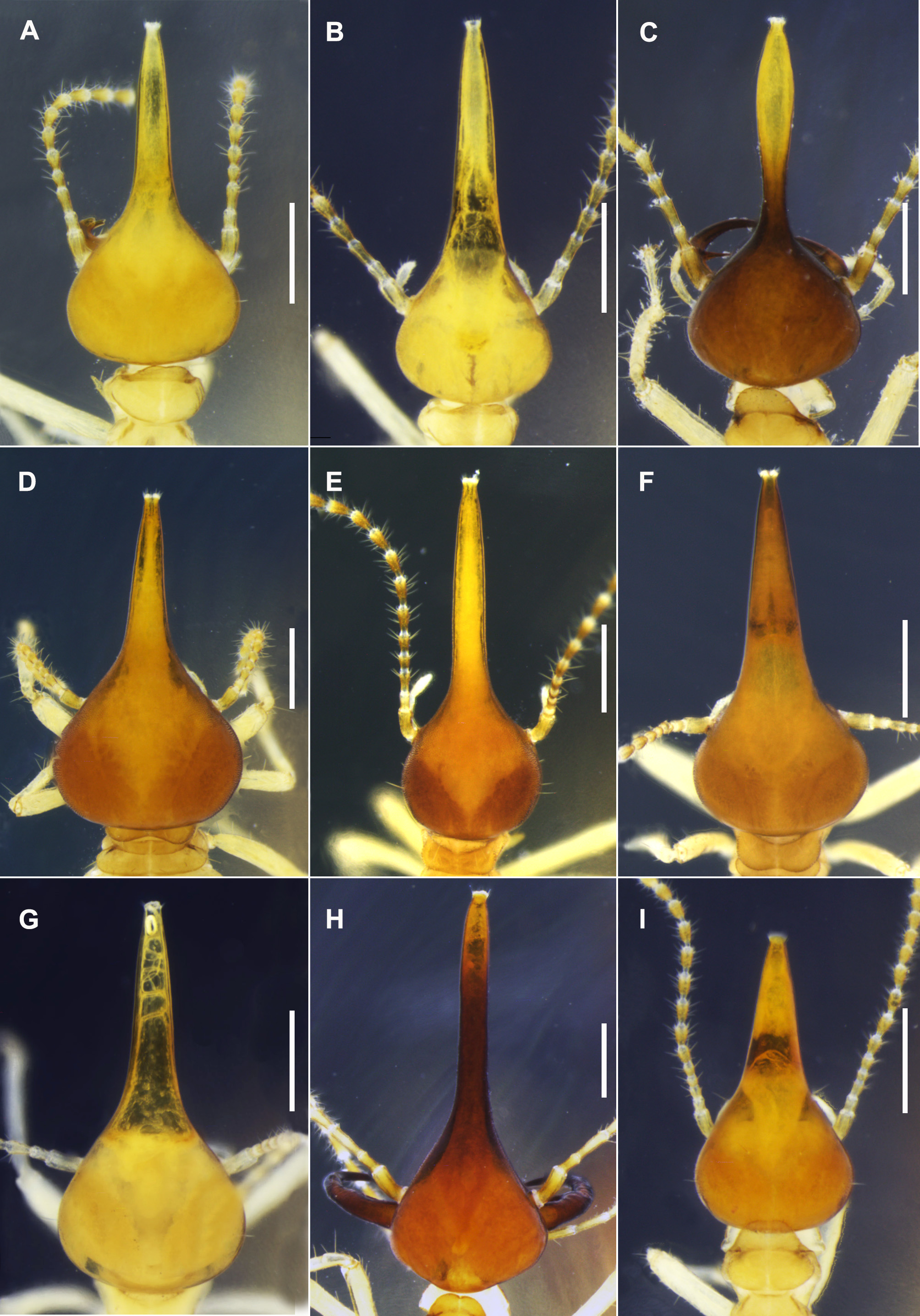
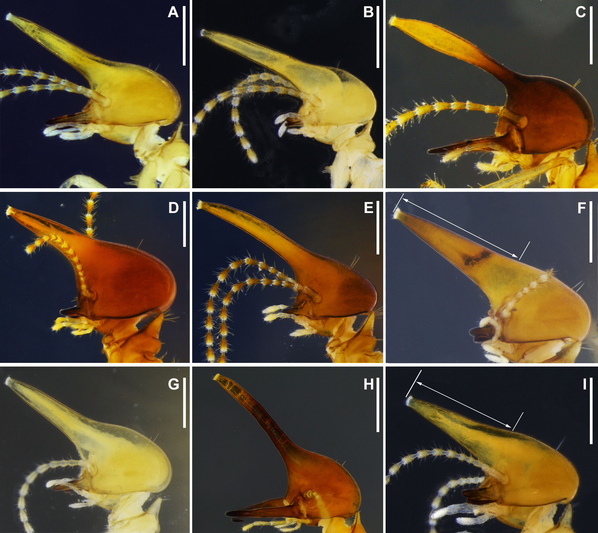
Soldier. Monomorphic or dimorphic. Head capsule rounded to pear-shape in dorsal view. Antenna with 14 articles, first and third articles longer than second, fourth article shorter than second and remaining articles progressively increasing in length, long ( Fig. 4 View FIGURE 4 A, B, C) or short articles ( Fig. 4 View FIGURE 4 D, F, G, I). Labrum subrectangular, subpentagonal or tongue-shaped. Frontal tube about same length of head or longer and always covering the postclypeus in dorsal view. Frontal tube with bulbous shape ( Fig. 3 View FIGURE 3 C), conical ( Fig. 3 View FIGURE 3 A, B, D, F, G, I) or subcylindrical ( Fig. 3 View FIGURE 3 E, H) forming an angle of about 45 degree in lateral view with dorsal line of head. Dorsal margin of head plus margin of frontal tube concave ( Fig. 4 View FIGURE 4 A, B, C, D, E, G, H) or straight ( Fig. 4 View FIGURE 4 F, I) in profile.
Mandibles evenly ( Fig. 5 View FIGURE 5 B, C, D, E, H) or strongly curved ( Fig. 5 View FIGURE 5 A, F, G, I), apical portion sharp with inner margin serrated ( Fig. 5 View FIGURE 5 A, F, G, I) or not ( Fig. 5 View FIGURE 5 B, C, D, E, H), and marginal tooth conical ( Fig. 5 View FIGURE 5 C, H) or subrectangular ( Fig. 5 View FIGURE 5 A, B, D, E, F, G, I). Apical region of each mandible aligns to the proximal region of the opposite mandible ( Fig. 6 View FIGURE 6 A) or apical region of each mandible extending well beyond the opposite mandible when closed ( Fig. 6 View FIGURE 6 B). Postmentum small, slightly projected, subquadrangular to subrectangular. Pronotum with two rounded projections close to posterolateral margins, length of anterior lobe variable. Meso- and metanotum with rounded lateral margins. Forecoxa process conical ( Fig. 7 View FIGURE 7 B, C) or subcylindrical ( Fig. 7 View FIGURE 7 D). Tibial spur formula 2:2:2. Digestive tube visible by transparency. Head capsule, postmentum, pronotum and forecoxa process with cuticular microsculpture (points). Head capsule with two to ten bristles (none short) on surface. Labrum with two to four bristles. Articles of antennae with whorls of bristles that vary in size on posterior portion. Frontal pore surrounded by a couple of short bristles. Pronotum without bristles or with one to six bristles on the anterior lobe. Meso- and metanotum without bristles or with one bristle on each anterolateral angle. Forecoxa process glabrous or with a single erect bristle. Tergites with a variable number of bristles on posterior margin. Sternites with more numerous bristles than tergites, projected obliquely in anteroposterior direction and also on posterior edge of each plate. Legs with variable number of long bristles distributed throughout extension. Head capsule orange to reddish-brown. Mandibles with base yellowish and distal portion orange to reddish-brown. Body yellow to pale yellow.
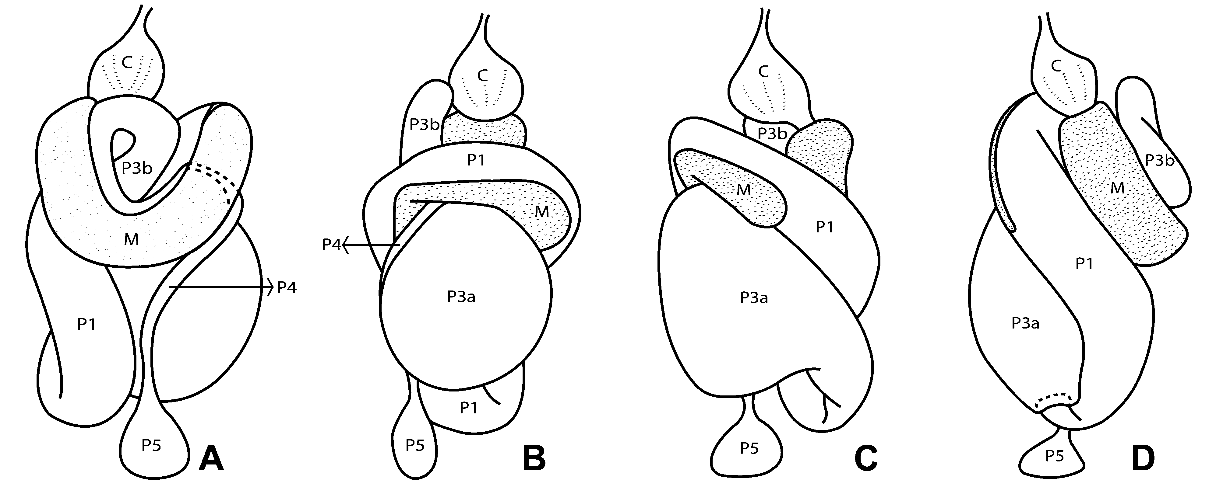
Worker. Head capsule rounded in dorsal view, flattened dorso-ventrally ( Fig. 8 View FIGURE 8 C), highly sclerotized. Fontanelle rounded, conspicuous ( Fig. 8 View FIGURE 8 B) or inconspicuous. Antenna with 14 articles, first and third article larger than second, fourth article shorter than second and remaining articles progressively increasing in length. Postclypeus inflated with or without dark line ( Fig. 8 View FIGURE 8 A, B). Labrum subpentagonal with rounded and hyaline margins. Left mandible with small apical tooth (A) slightly shorter than M1+2, cutting margin between tip of M1+2 and third marginal tooth (M3) and molar prominence with 5–6 developed ridges ( Fig. 9 View FIGURE 9 D). Right mandible with apical tooth almost equal in size to M1, acute angle between them, M1 and M2 robust and molar plate with 5–6 ridges. Meso- and metanotum as described for soldier. Posterior margin of tergites darkened or not. Forecoxa process inconspicuous. Digestive tract ( Fig. 9 View FIGURE 9 A–C, 10A–D) with gizzard armature complete, columnar belt with 24 visible folds, 6 first-, 6 second- and 12 third order ( Fig. 9 View FIGURE 9 A). Pulvilli without ornamentation but with scales tips distinguishable ( Fig. 9 View FIGURE 9 B). Mesenteric tongue external to mesenteric arc with short proximal constriction and extending toward first proctodeal segment (P1), ending in oval shape ( Fig. 10 View FIGURE 10 B, C). Two pairs of Malpighian tubules, closely inserted, each pair joined at their base and inserted at the midgut-hindgut junction. P1 fusiform, positioned to left and almost parallel to body axis in dorsal view ( Fig. 10 View FIGURE 10 A). Internally, distal portion of P1 ornamented with scales, each bearing a row of 15 short wide spines ( Rocha & Constantini 2015, Fig. 21). Enteric valve (EV) oriented at 180° relative to the axis of body, starting at P1 and extending as far as the paunch or proctodeal third segment (P3) on right, in left side view. Symmetrical EV with three conspicuous fingerlike cushions ( Fig. 9 View FIGURE 9 C). Cushions and inter-cushion spaces ornamented with triangular spines that become gradually sparser towards transition with P3. Proximal portion of enteric valve with more elongated thin spines. P3 divided into two distinct regions: P3a globular ( Fig. 10 View FIGURE 10 B, C) on right side of dorsal view, and P3b "S" shaped ( Fig. 10 View FIGURE 10 A) in dorsal view. Colon or fourth proctodeal segment (P4) tubular after U-turn on right, in dorsal view. Head capsule with about ten long bristles and various shorter bristles. Two long bristles adjacent to fontanelle. Postclypeus with two to four bristles. Labrum with six bristles. Antenna articles with whorl of bristles, varying in size, on distal portion. Pronotum, mesonotum and metanotum with variable number of bristles on anterior margin. Digestive tube visible by transparency. Tergites and sternites follow the description of soldiers but with more numerous bristles.
Pilosity of legs as described for soldier. Head capsule orange to reddish brown with yellowish mandibles base and brown teeth. Body from yellow to pale yellow.
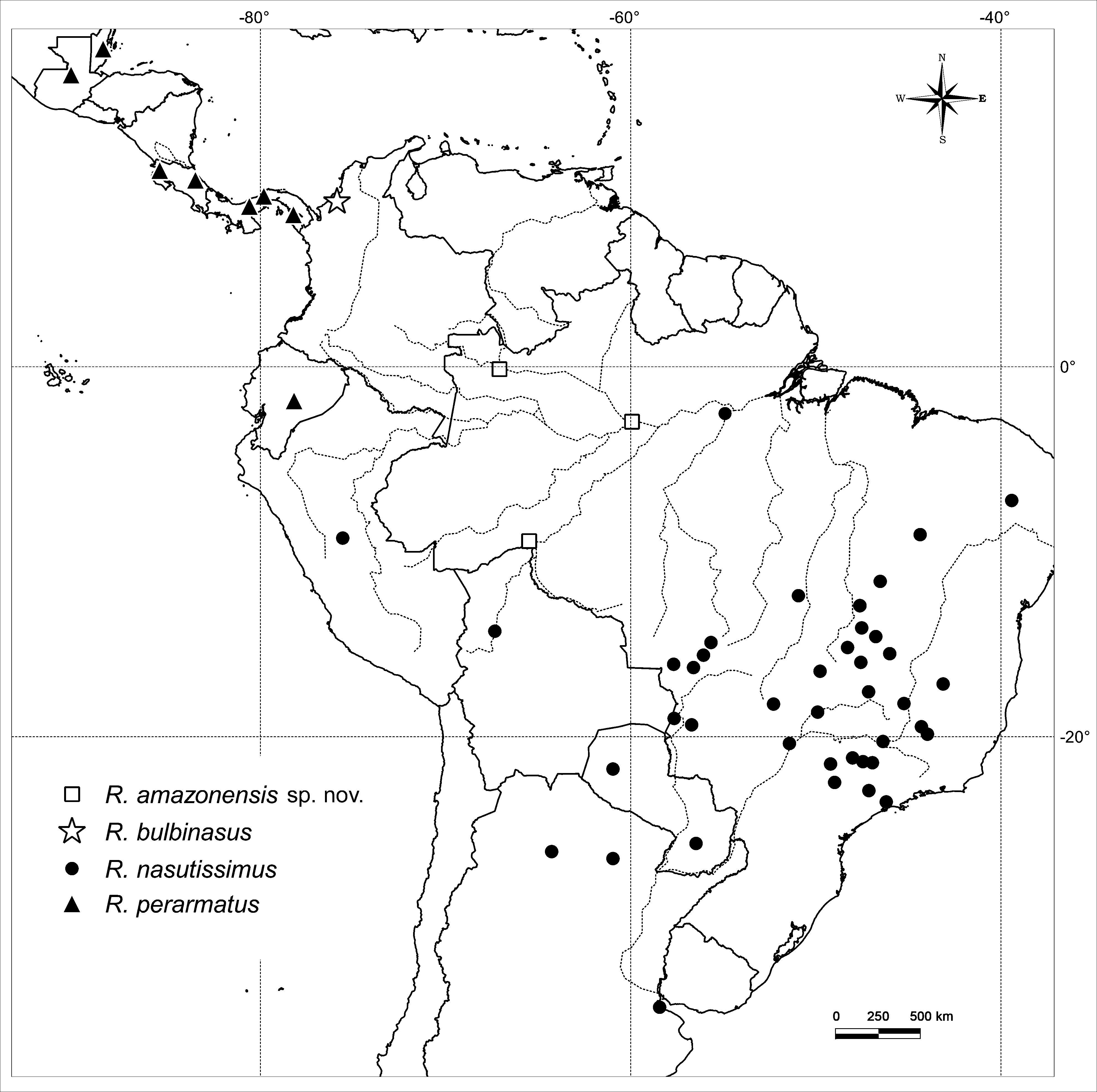
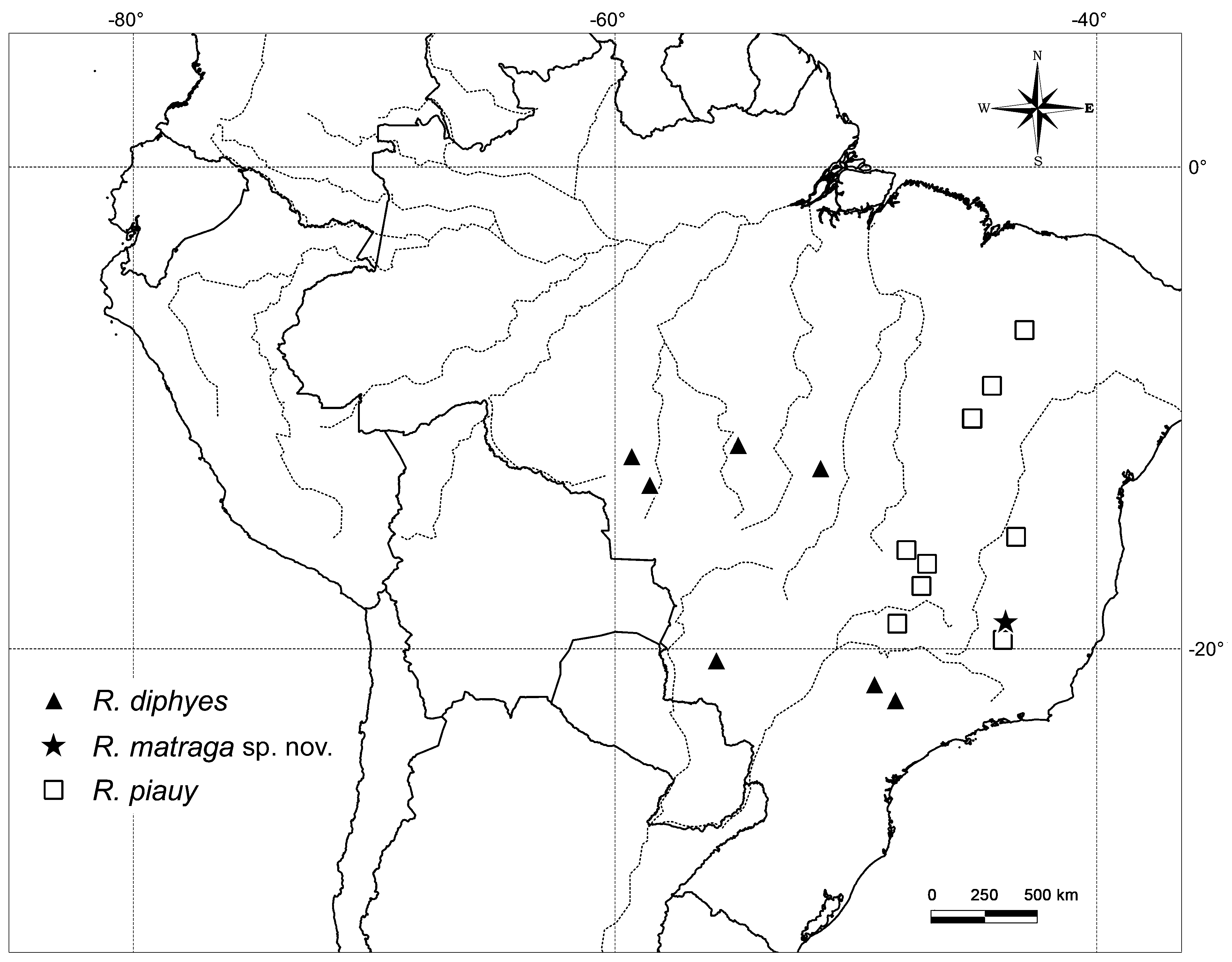
Distribution ( Fig. 12 View FIGURE 12 , 13 View FIGURE 13 ). Throughout the Neotropics in Argentina, Belize, Bolivia, Brazil, Colombia, Costa Rica, Ecuador, Guatemala, Panama, Paraguay and Peru.
Comparisons. Among the Syntermitinae genera, soldiers of Uncitermes Rocha & Cancello (Rocha et al. 2012) are morphologically most similar to Rhynchotermes by their large curved mandible with conical marginal teeth and long frontal tube. Uncitermes teevani has been recorded in northern Brazil, eastern Venezuela, Guyana and Suriname. The main differences between the workers of these species include: apical teeth of left and right mandibles that are respectively smaller than M1 +2 and M1, larger molar prominence and concave molar plate; a mesenteric tongue with initial portion filiform, extending to P1 dilated region, which is almost circular in shape, in U. teevani relative to R. nasutissimus . In the Syntermitinae P1 is usually voluminous, but in U. teevani it is more swollen with a globular shape, whilst in R. nasutissimus it is distinctly fusiform. The enteric valve is asymmetric, consisting of four cushions well ornamented with slender and curved spines in U. teevani.


No known copyright restrictions apply. See Agosti, D., Egloff, W., 2009. Taxonomic information exchange and copyright: the Plazi approach. BMC Research Notes 2009, 2:53 for further explanation.
|
Kingdom |
|
|
Phylum |
|
|
Class |
|
|
Order |
|
|
Family |
Rhynchotermes Holmgren
| Constantini, Joice P. & Cancello, Eliana M. 2016 |
Armitermes (Rhynchotermes)
| Krishna 2013: 1471 |
| Constantino 1999: 403 |
| Nickle 1992: 226 |
| Fontes 1985: 9 |
| Mill 1983: 183 |
| Mathews 1977: 154 |
| Snyder 1949: 264 |
| Snyder 1925: 181 |
| Holmgren 1912: 55 |