
Pelecinobaccha Shannon
|
publication ID |
https://doi.org/ 10.11646/zootaxa.3819.1.1 |
|
publication LSID |
lsid:zoobank.org:pub:355CBCD4-AB75-4F9F-A476-4B300143F8D6 |
|
DOI |
https://doi.org/10.5281/zenodo.5674357 |
|
persistent identifier |
https://treatment.plazi.org/id/191A7A05-0044-1242-FF7E-FDECD16FE9BF |
|
treatment provided by |
Plazi |
|
scientific name |
Pelecinobaccha Shannon |
| status |
|
Genus Pelecinobaccha Shannon View in CoL View at ENA
Pelecinobaccha Shannon, 1927: 10 View in CoL [type species Baccha peruviana Shannon, 1927 View in CoL (original designation)]. Proposed as a subgenus of Baccha View in CoL .
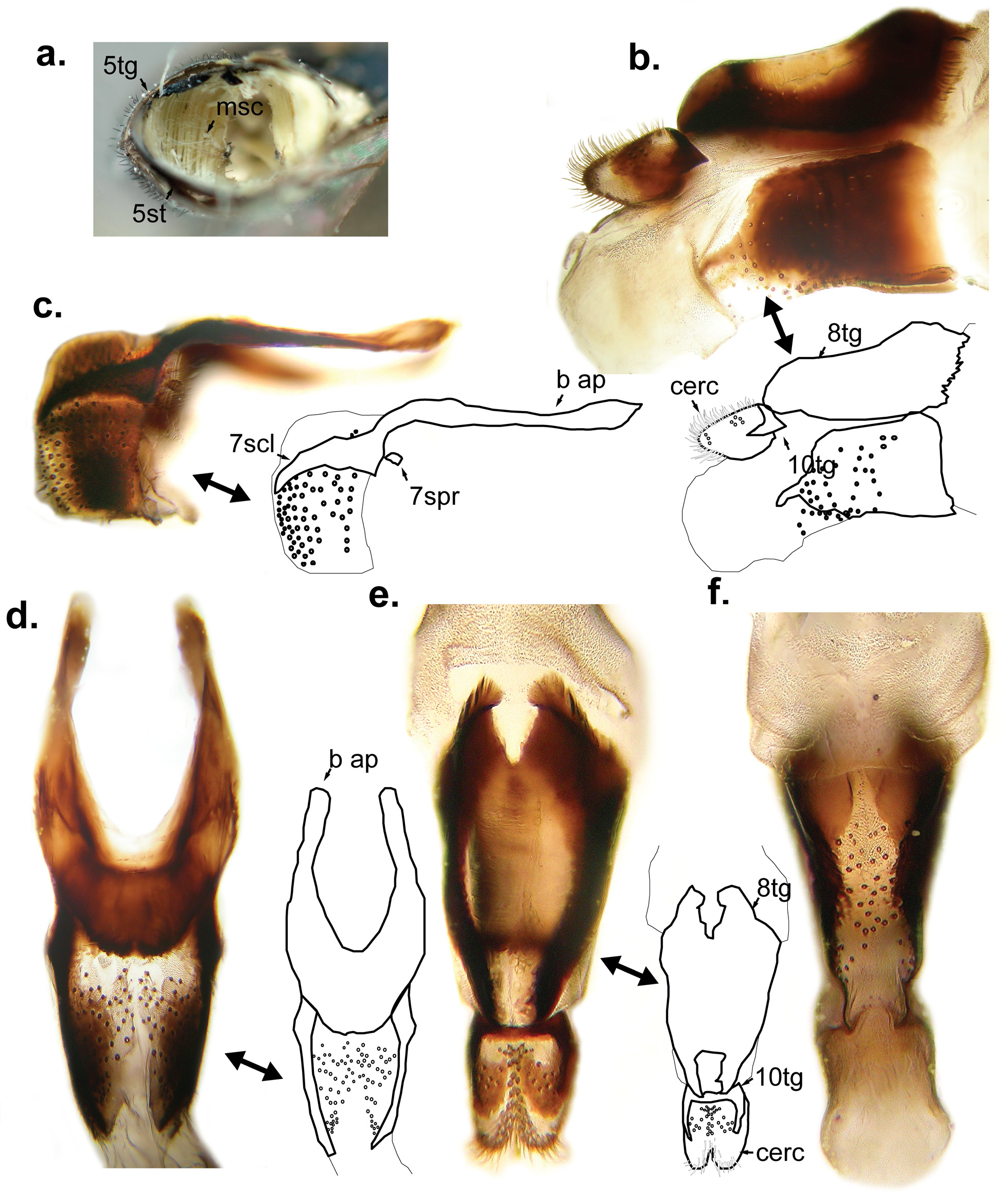
Diagnosis. Head. Face narrow to normal (between ¼ to ⅓ of head width) and mostly dark; tubercle distinct and ventrally positioned ( Fig. 37 View FIGURE 37 e). Frons normal (~⅓ of head width). Antennal insertions usually confluent. Female ocellar triangle usually ~2 ocellus-width from lateral eye margin. Dorsal occiput with 2 rows of pile ( Fig. 16 View FIGURE 16 a). Thorax. Scutum with anterior row of longer pile, anterior row with shorter pile medially, usually entirely dark and without any discernible pattern of pollinosity. Scutellum usually entirely dark. Anterior anepisternum pilose. Katatergite with short to long microtrichia. Metaepisternum pilose. Metasternum bare. Dorsal lobe of calypter with normal pile on margin (but shorter than ventral lobe pile). Male metafemur with normal pile. Metabasitarsomere usually bicoloured, at least basal ½ dark. Wing. Alula width usually 2–3 times as broad as c cell, rarely reduced or absent. Wing usually with extensive dark markings and entirely microtrichose. Abdomen. Abdomen petiolate ( Fig. 25 View FIGURE 25 g) or very narrow and very long ( Fig. 9 View FIGURE 9 i); abdominal tergites with pattern variable. Genitalia. Female 7th tergite with basal pair of long apodemes ( Fig. 10 View FIGURE 10 d); 8th tergite triangular in dorsal view and notched posteriorly, with basal crest flanking posterior notch ( Fig. 10 View FIGURE 10 e); 10th tergite usually reduced and fused to dorsal surface of cercus. Male subepandrial sclerite usually trapezoidal and with slightly extended posterior corners; surstylus usually oval and elongated ( Fig. 3 View FIGURE 3 e) if sub-quadrate, then without extended antero-apical corner ( Fig. 15 View FIGURE 15. a – d e); basiphallus teardropshaped, distiphallus membranous but with dorsal sclerotized triangular region.
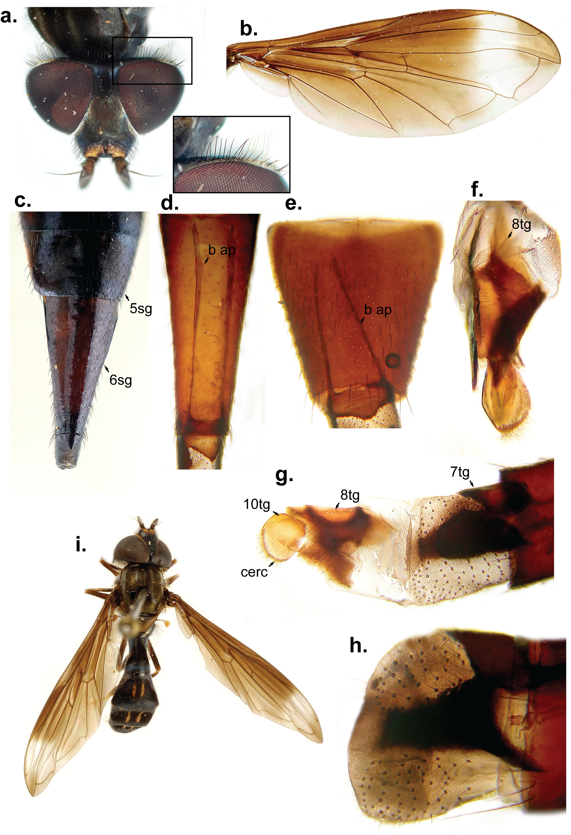
Description. Male. Head: Dark red, dark brown, bluish-black or black. Face narrowing slightly ventral to tubercle, slightly concave dorsal to tubercle, sometimes pale laterally, rarely mostly pale [e.g. P. aster ( Curran, 1941) ], with white pollen laterally, pollen sparse to absent on tubercle apex, usually with white pile laterally except black ventral to antenna, with distinct sub-ventral median tubercle, anterior oral margin sparsely white pilose; gena narrow (appears as an elongated triangle from ventral view). Lunule usually black but sometimes distinctly pale above antennal insertion, bare, shiny. Frontal triangle sometimes pale laterally, with dull brown pollen medially and silvery-white pollen laterally that sometimes is restricted to oval/triangular maculae, usually with long, erect, black pile; ocellar triangle 1–2 times its length from posterior eye margin. Eye contiguity slightly shorter to 2 times as long as vertical triangle length; antenna dark, basoflagellomere oval and at least slightly longer than wide, black pilose, arista dark red. Occiput usually with sparse dark pollen on dorsal ¼ and dense silvery-white pollen on ventral ¾, rarely homogeneously covered by white pollen [e.g. P. ovipositoria ( Hull, 1943a) ], dorsal ¼ usually with 2 regular rows of simple black pile, pile usually shorter on anterior row, posterior row sometimes with white and scale-like pile, middle ½ with 2–4 regular rows of pile, posterior row always with longer, scale-like white pile, anterior rows ranging from scale-like white pile to simple black pile, ventral ¼ with 2–3 irregular rows of white, scale-like pile, usually with shorter pile on anterior row.
Thorax: Prothorax dark with sparse, dull pollinosity, without pile. Scutum entirely dark red, brown, bluishblack or black, dull pollinose, with a rectangular area of concentrated pollen anterior to scutellum, markings, if present, formed by vittae of pale pollen, pilosity usually erect, with anterior row of longer white pile that sometimes has shorter pile in the middle, notopleuron with pile longer anterior to transverse suture, pile slightly thicker and densely arranged latero-posteriorly to transverse suture. Scutellum usually as dark as scutum and covered by dull pollen. Pleuron dark, sometimes with pale markings, with sparse white microtrichia, longer on anterior ½ of katatergite (‘velvet’-like); with pile on anterior anepisternum, posterior ½ of posterior anepisternum, anterior anepimeron, ventro- and dorso-posterior katepisternum, katepimeron and sub-appressed on metaepisternum, pile on katepimeron and metaepisternum sometimes inconspicuous; metasternum dark, bare and very narrow dorso-laterally to metacoxa; metaepimeron flared laterally on posterior ½ being connected to body by a membrane; post metacoxal bridge incomplete, usually metathoracic epimera widely separated. Calypter dorsal lobe narrow, with marginal fringe of short pile, ventral lobe with long branching pile.
Wing: Usually with dark brown markings basally and rarely with bare areas; alula absent (rare, e.g. P. invisibilis sp. nov.) to ~4 times wider than c cell, rarely with bare areas.
Legs: Procoxa with only 1 row of pile antero-apically, protrochanter bare ventrally, profemur with sparse pile on basal ½ and slightly longer pile on baso-ventral ½, densely arranged pale pile ventro-laterally on apex of protibia and ventrally on protarsus. Mesofemur bare to sparsely pilose ventrally and with posterior row of longer black pile, mesotibia with ventro-apical short, thick, black pile, 1st and 2nd mesotarsomeres with densely arranged pale pile and short, thick, black pile intermixed ventrally, mesobasitarsus thinner than mesotibia and slightly thinner than the other mesotarsomeres; metacoxa with distinctly longer pile, basal ½ of metafemur with only sparse pile ventrally, metatarsus usually bicoloured but in some species entirely dark [e.g. P. adspersa ( Fabricius, 1805) ], metabasitarsomere usually with no more than apical ½ pale ( P. susio ( Hull, 1941) usually with ⅔ to ¾ pale); pile usually the same color as background.
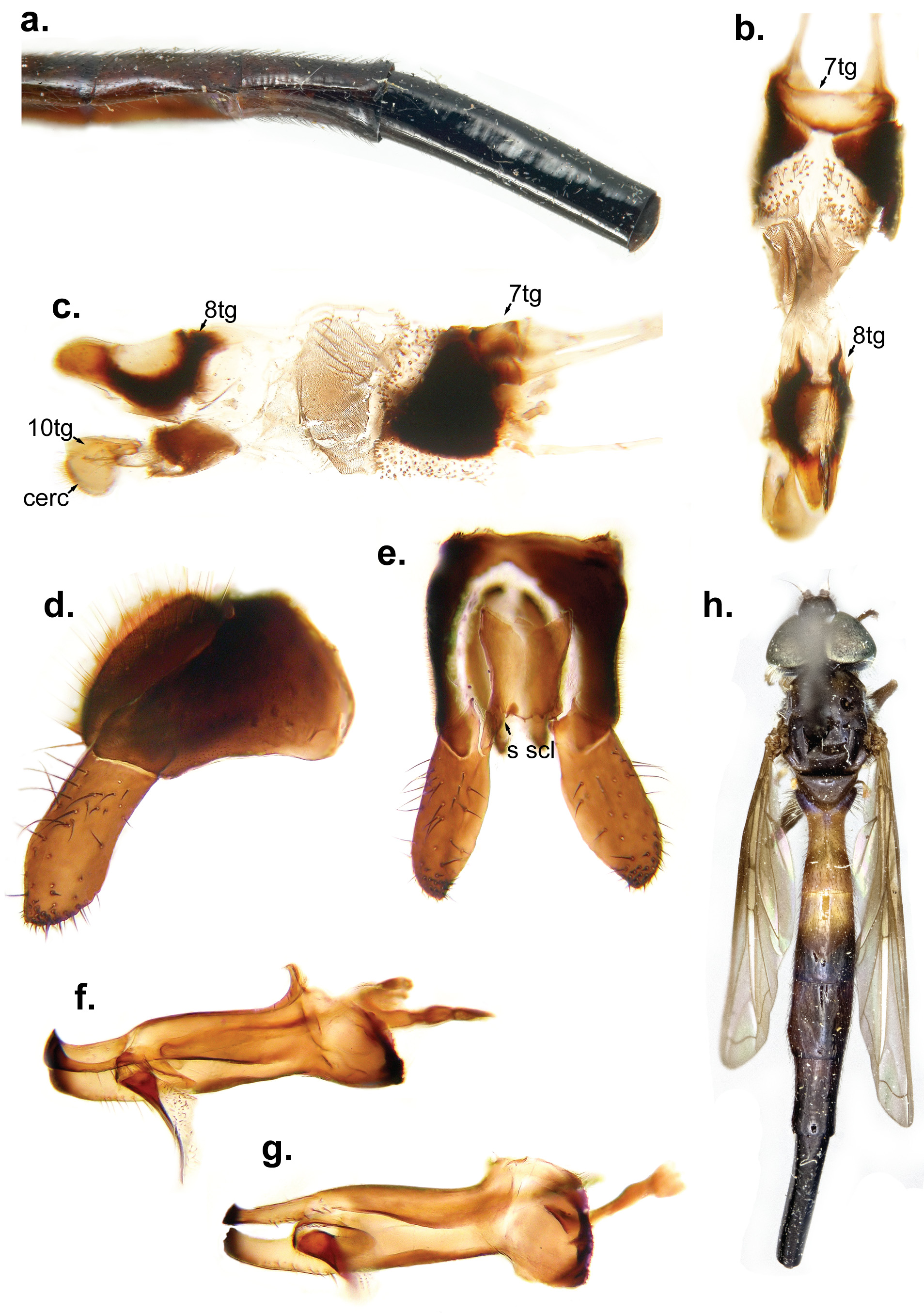
Abdomen: Usually petiolate but sometimes parallel-sided or narrow, 1st abdominal segment crescent shaped with lateral extremities directed laterally or posteriorly, with long and erect pile laterally, usually bare or with shorter and sparse pile dorso-medially; 2nd abdominal segment longer than wide, usually constricted medially and sometimes very narrow and long (e.g. P. vesca sp. nov.); other abdominal segments variable; sterna dark, well sclerotized. Genitalia: Small; epandrium and cercus densely microtrichose; epandrium trapezoidal in lateral view; base of surstylus articulated to epandrium apicoventrally, usually flattened dorso-ventrally. Subepandrial sclerite usually trapezoidal with slightly extended posterior corners and homogeneously sclerotized. Hypandrium usually sub-oval with a narrower quadrate apical ⅓, ventral anterior notch variable, ventral surface bare. Basiphallus dropshaped in dorsal view, tapering on basal ⅓ in lateral view; distiphallus seen as a sclerotized triangular plate in dorsal view, with sclerite involving the circumference of the base, apex slightly curving dorsally, apical membranous portion usually uninformative. Postgonite apex either with acute dorsal and ventral extremities or with only dorsal extremity acute, pilose on baso-ventral surface.
Female: Usually very similar to male except: frons usually of normal width, distinctly widening from vertex. Pro- and mesotarsomeres sometimes enlarged. Wing and abdominal markings sometimes differ from male; 2nd abdominal tergite usually shorter and wider than male but still longer than wide; 5th segment rarely with tergite and sternite fused apically; 6th segment tergite and sternite usually completely fused laterally, entirely dark with short appressed black pile. Genitalia: Strongly sclerotized, 7th tergite bare, with pair of long baso-lateral extensions (apodemes) into 6th segment, apodemes usually as long as the 6th segment; extensive membranous region between 7th and 8th segments, pile restricted to region immediately apical to 7th tergite; 7th sternite modified into separate pair of lateral sclerites, dorsally fused to sides of 7th tergite; 8th tergite triangular in dorsal view, notched posteriorly, sometimes unsclerotized medially, with basal crest flanking posterior notch, usually with a pair of rounded apices; 8th sternite reduced to pair of lateral rectangular sclerites, with pair of apical narrow extensions that fold into the segment; 10th tergite usually reduced and fused to dorsal surface of cercus; 10th sternite, if visible, just a narrow sclerite with sparse pile. Cercus usually with only 1 row of pile on apical margin.
Comments. Pelecinobaccha Shannon was proposed as a subgenus of Baccha to hold B. peruviana , characterized by an elongated abdomen with segments 2–6 of similar length. Hull (1949a) added B. telescopica ( Curran, 1930) to the subgenus.
Although many female specimens seem to have a dorso-ventrally flattened 6th segment ‘ovipositor’ (and sometimes an apical ‘ridge’ sensu Hull, 1949a ), this is an artefact of preservation. The 6th segment can be slightly laterally flattened (natural condition, more easily preserved in females with longer 6th segments) through to dorsally flattened, even within the same series of specimens. This artefact is most likely due to dehydration of the tissues and collapsing of the sclerite walls, a phenomenon even more common in material pinned straight from alcohol. This effect sometimes also hinders the observation of the shape of some of the abdominal tergites, due to ‘curving’ of the lateral margin.
The common conical form of the female 6th segment ( Fig. 16 View FIGURE 16 c) probably results from the lateral fusion of its tergite and sternite; as suggested by the incomplete fusion of these sclerites in the Pelecinobaccha susio group (see below) and by evidence of incomplete lateral sclerite fusion of the female 5th segment on other species (e.g. P. telescopica , Fig. 39 View FIGURE 39 a).
As mentioned above, the female terminalia of Pelecinobaccha are very different from other syrphids ( Fig. 10 View FIGURE 10 c–f). The 7th tergite is somewhat shortened and possesses a pair of very long baso-lateral apodemes. The 7th sternite is modified into a pair of separate lateral rods, partially fused to the tergite laterally and with short ventral apodemes into the 6th segment, leaving the segment membranous ventrally. The 8th tergite is modified into a triangular sclerite that is naturally folded longitudinally. The 8th sternite is usually divided into a pair of lateral lightly sclerotized regions connected by a basal narrow sclerotized bridge and with apicoventral extensions that fold into the segment. It is assumed that the 10th tergite (epiproct) is either a narrow transverse strip or a pair of sclerites, in either case usually fused to the dorsal margin of the cerci as demonstrated by a distinct thickening of that region on the cercus. The cercus is usually bare with only a single row of pile along its apicoventral margin. The resting condition of the female terminalia is always laterally flattened, and the segments are usually easily extended by light dorso-ventral compression of the 6th segment in dissections.
It is assumed that the female terminalia is extended by hydraulic pressure of the haemolymph due to dorsoventral compression of the pre-abdominal segments. This is suggested by the presence of massive muscles running from tergite to sternite of the 5th segment of P. adspersa ( Fig. 10 View FIGURE 10 a). The uniquely long baso-lateral apodemes of the 7th tergite and the basal crest of the 8th tergite are most likely used to pull the modified terminalia back into the abdominal cavity.
The mostly membranous ventral region of the female terminalia, and the acute (while at rest) abdominal tip probably serve to ease the insertion of the terminalia beneath the ‘scale’ of scale insects ( Coccidae ). After insertion, the female probably extends and expands the terminalia, as described above, raising the scale enough to place an egg.
Species (e.g. P. telescopica ) with an elongate female 6th segment also have a relatively elongate male surstylus and hypandrium. In these species the female 2nd segment is shorter than the same segment in males.
Four distinct groups of Pelecinobaccha are here treated as species groups: P. adspersa , P. brevipennis ( Schiner, 1868) , P. peruviana and P. susio .


No known copyright restrictions apply. See Agosti, D., Egloff, W., 2009. Taxonomic information exchange and copyright: the Plazi approach. BMC Research Notes 2009, 2:53 for further explanation.
|
Kingdom |
|
|
Phylum |
|
|
Class |
|
|
Order |
|
|
Family |
Pelecinobaccha Shannon
| Miranda, Gil Felipe Gonçalves, Marshall, Stephen A. & Skevington, Jeffrey H. 2014 |
Pelecinobaccha
| Shannon 1927: 10 |