
Abyssocladia, LEVI, 1964
|
publication ID |
https://doi.org/ 10.1111/j.1096-3642.2006.00234.x |
|
persistent identifier |
https://treatment.plazi.org/id/191D4B17-FFC5-FF9E-0764-FEA8FC78A6F7 |
|
treatment provided by |
Felipe |
|
scientific name |
Abyssocladia |
| status |
|
ABYSSOCLADIA LÉVI, 1964 View in CoL
Type species: Abyssocladia bruuni Lévi, 1964 (by monotypy).
Diagnosis, modified from Lévi (1964): Cladorhizidae with abyssochelae and sigmancistras, most often pedunculate and disciform with a radial skeleton.
Remarks
The genus Abyssocladia Lévi, 1964 has been synonymized with Phelloderma Ridley & Dendy, 1886 by van Soest & Hajdu (2002b) based on a single shared character, i.e. a peculiar isochela with the two frontal alae nearly in contact for which they proposed the term ‘abyssochela’. This measure appears unsound and will not be followed here. van Soest & Hajdu (2002b) mistakenly indicated that Abyssocladia was monotypic. It actually includes three species: A. bruuni Lévi, 1964 , known by the type specimen and found again by Koltun (1970); A. claviformis Koltun, 1970 ; and A. oxeata Koltun, 1970 . These three species, from the deep Pacific, have a thin stalk to which is attached a spherical or disc-shaped body with a radiate skeleton. They share a similar spicule complement, including styles, abyssochelae and sigmancistras to which may be added double bent microxea and subtylostyles. [In the type species, A. bruuni, Lévi (1964) reported sigmas, but from his description they are more likely sigmancistras.) By contrast, Phelloderma radiatum Ridley & Dendy, 1886 , from the deep south-west Atlantic, while having rather similar spiculation, differs considerably in its sessile shape, the presence of oscules on small papillae and a cork-like cortex, suggesting to them (p. 113) that ‘In the presence of a distinct cortex, and in the radiate arrangement of its skeleton, this genus approaches the Suberitidae ...’. The organization of the three species previously described in Abyssocladia and which is also found in the four new species described below certainly cannot be assimilated to that of the suberitids. The shape of the isochelae alone cannot justify the merging of Abyssocladia with the poorly known, monotypic Phelloderma . As presently construed, with seven species including the four new species described below, the genus is well defined and homogeneous, the only exception being A. naudur sp. nov., which does not display the disciform pedunculate shape and possibly belongs to an undescribed genus or subgenus.
Abyssocladia View in CoL was first assigned to Mycalidae View in CoL by Lévi (1964). This assignment is not well justified, as the skeletal organization is different from that of the Mycalidae ( van Soest & Hajdu, 2002a) View in CoL , and the isochelae are rather of the arcuate type. Furthermore, the presence of sigmancistras in all representatives of the genus argues for affinities with Cladorhizidae View in CoL (see Discussion). It seems more sound temporarily to classify the genus in the Cladorhizidae View in CoL , although the family appears to be polyphyletic, pending more information before a general assignment of the various genera of the family can be made. The general organization, absence of a visible aquiferous system and microsclere spicule arrangement suggest a carnivorous habit, which is confirmed by the presence of crustacean debris in A. huitzilopochtli View in CoL sp. nov.
ABYSSOCLADIA HUITZILOPOCHTLI View in CoL SP. NOV.
( FIGS 9–11 View Figure 9 View Figure 10 View Figure 11 )
Type material
Holotype: NAUTIMATE NM 03-875-15, 21/01/1994, Middle America Trench (off Mexico), 18°17′N, 104°31′W, 3325 m, Muséum National d’Histoire Naturelle, Paris, no. MNHN D JV 85. GoogleMaps
Etymology
From ‘Huitzilopochtli’, the Aztecs’ bloodthirsty god of the sun. Referring to the sun-like body shape of the sponge suggesting Aztec feather ornaments, and to Mexico off which this carnivorous sponge was collected.
Locality and habitat
Middle America Trench (off Mexico), 18°17′N, 104°31′W, 3325 m. Collected during NAUTIMATE cruise, dive NM 03 of the manned submersible Nautile, on brown-green siltstone. No hydrothermal fluid emission was observed during dive NM 03. However, fluid emissions with associated fauna ( Calyptogena sp. , Escarpia sp. ) were observed 16 km from the collection site, at 2600–3000 m depth.
Description
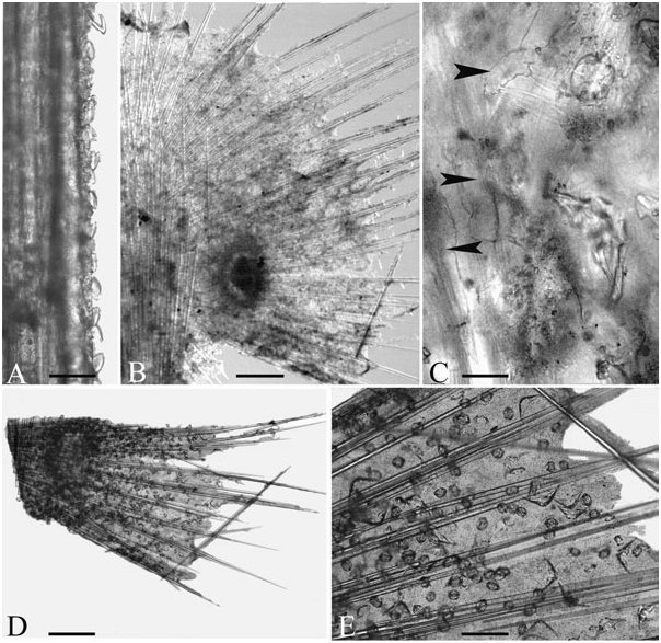
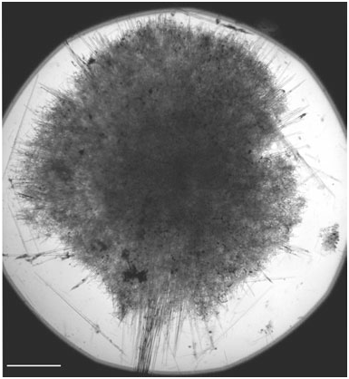
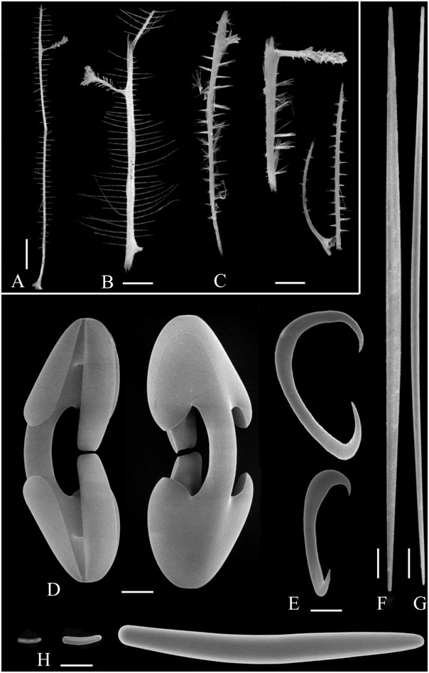
A single specimen of a pedunculate sponge consisting of a long thin peduncle and a flattened semicircular body ( Fig. 9A View Figure 9 ). Peduncle 35 mm long, 1 mm thick near the base then varying from 0.3 to 0.6 mm, attached to a small rocky fragment by an enlarged base. Body in shape of a regular flattened disc, 7 mm in diameter and 0.3–0.5 mm thick, with free radiating spicule fascicles protruding from 2 to 4 mm and surrounded by a small amount of living tissue. Two extensions with flesh and spicule fascicles approximately 15 mm long on the lower part of the disc. A denser area, 0.5 mm in diameter, near the centre of the disc, comprising an unidentified clear mass surrounded by a dense concentration of microscleres, most likely the remains of prey ( Fig. 11B, D View Figure 11 ). Several crustacean appendages included in the disc ( Fig. 11C View Figure 11 ). No cortex, no visible aperture. Colour pure white.
Skeleton: peduncle made of an axis of tightly packed, long substrongyles longitudinally arranged, reaching the centre of the disciform body and slightly diverging after the centre. Peduncle with a small amount of living material, entirely covered on the basal twothirds by isochelae regularly attached by the dorsal surface with the teeth protruding outward ( Fig. 11A View Figure 11 ). On the last third of the peduncle, cover of isochelae progressively replaced by a less dense cover of abyssochelae similarly arranged but more numerous on the two lateral sides. In the body, skeleton of radiating large substrongyles similar to those of the peduncle. Tissue of the body containing rare smaller substrongyles irregularly dispersed, a few isochelae, numerous orthancistras, numerous abyssochelae and extremely abundant sigmancistras ( Fig. 11E View Figure 11 ). Protruding fascicles made of 5–10 megascleres with a few abyssochelae and orthancistras often dorsally attached to the fascicles.
Spicules:
1. Styles or substrongyles ( Fig. 9C View Figure 9 ) of the peduncle and of the fascicles. Straight, with the two ends nearly equal, fusiform (for instance for a spicule 2450 µm long, diameter 30 µm in the middle, 10 µm at the head and 8 µm at the rounded tip). Size 1050–2500 × 15–30 µm. 2. Substrongyles ( Fig. 9D View Figure 9 ) dispersed in the flesh and included in the fascicles. Straight, the longer slightly fusiform, with the two ends rounded and nearly equal. Size 260–660 × 5–10 µm.
3. Substrongyles ( Fig. 9G View Figure 9 ) of the base. Ends nearly equal, often slightly flexuous, with a few intermediaries with the large fusiform substrongyles. Size 560– 750 × 21–30 µm.
4. Isochelae 1 ( Fig. 9B View Figure 9 ), arcuate or subpalmate, with lateral alae linked to the shaft along almost their entire length. Front alae ovoid. Developmental stages present in the body, showing a shaft of size similar to that of the mature spicule and more or less developed alae. Size 67–90 µm.
5. Isochelae 2, found only on the basal part of the shaft. Same shape as isochelae 1 but smaller. Size 40–55 µm.
6. Abyssochelae ( Fig. 9E, F View Figure 9 ) of the terminal part of the shaft and of the body, absent in the distal parts of the filaments. Nearly spherical, with a shaft strongly curved, bearing two large lateral fimbriae roughly triangular, sometimes with a third smaller one in the middle. Lateral alae nearly in contact with the opposite alae, made of a thin lamella folded on the sides. Frontal ala long, nearly in contact with the opposite ala or sometimes bound to it, ovoid or occasionally bifid at the end. Developmental stages present in the body, with more or less developed conical alae and two thickening on the outer side of the shaft. Size 60– 80 µm long and 40–55 µm wide.
7. Orthancistras ( Fig. 10A View Figure 10 ) (new term for a peculiar microsclere spicule, from ‘orth’, Greek, perpendicular and ‘ancistr’, Greek, hook; referring to the general shape of the spicule with a roughly right angle between the two branches), found in the body and on the filaments. Symmetrical spicule arising from a rod curved in the middle at roughly right angles and ending in two conical tips, on which develop lateral lamellae or expansions. Lamellae curved towards each other, increasing in width from the tip to the middle of the rod, cut in two hook-like spines. Two or more rarely three lateral expansions, roughly ovoid, near the middle of the rod. Immature spicules rather common in the body, the younger somewhat similar to an angular toxa, with a smooth shaft of irregular thickness, the later stages with more or less developed lateral lamellae or expansions. Size 150–195 µm.
8. Sigmancistras 1 ( Fig. 10B View Figure 10 ), extremely abundant everywhere. Contorted with the small end in a perpendicular plane, without notch. Size 20–24 µm.
9. Sigmancistras 2 ( Fig. 10B View Figure 10 ), extremely abundant everywhere. Contorted but with a smaller angle than sigmancistras 1, with a well-marked notch. Size 11–12 µm.
10. Microxeas, probably foreign, rare and found on a single slide, straight or slightly curved, the thicker sometimes fusiform. Size 30–95 × 0.3–1 µm.
Remarks
The sponge is more complete than the other species of the genus described below. It remains uncertain, however, if the radiating processes around the disc are complete or if they were similar in the live specimen to the two longer filaments of the base of the disc. The species is remarkable for its sophisticated organization, with a precise arrangement of the microscleres in the diverse parts of the body. The new type of spicule for which the term ‘orthancistra’ is proposed probably does not derive from chelae, as the tips of the juvenile spicules do not show enlargement, and the spicule seems to derive from a smooth angulate rod on which appear lateral expansions. Although their angulate shape is reminiscent of that of toxas, they do not appear to derive from this type of microsclere, which is rare in Mycalina .
The presence of crustacean debris in the body clearly indicates a carnivorous habit, which in the other species of Abyssocladia could be only inferred from the organization. The presence of a cover of isochelae along the peduncle, not found in the other Abyssocladia and more generally in other pedunculate carnivorous sponges, suggests that the peduncle plays a role in the capture of prey.
ABYSSOCLADIA INFLATA SP. NOV.
( FIGS 12 View Figure 12 , 13 View Figure 13 )
Type material
Holotype: PITO PI 17 , 02 /1993, East Pacific Rise , Easter microplate near Easter Island, 24°14.85′S, 115°39.70′W, 3142 m, Muséum National d’Histoire Naturelle, Paris, no. MNHN D JV 86. Spicule and skeleton slides only. GoogleMaps
Etymology
From ‘ inflatus ’, Latin, adj. swollen. Referring to the shape of the shaft of isochelae 1.
Locality and habitat
East Pacific Rise, Easter microplate, west of Eastern Island on the Terevaka fault, 24°14.85′S, 115°39.70′W, 3142 m. Collected during PITO cruise, dive PI 17 of the manned submersible Nautile, 02/1993 ( Segonzac et al., 1997; Naar et al., 2004). Attached to a piece of rock from a vertical cliff, with hanging sediment and very poor macrofauna, 423 km distant from the main hydrothermal venting of Pito Seamount.
Description
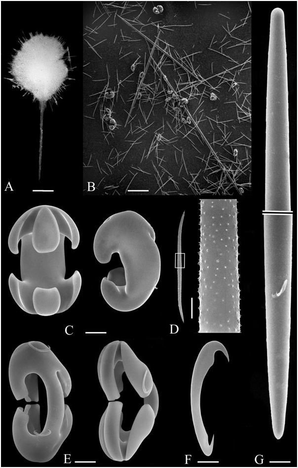
A single specimen of a small sponge ( Figs 12A View Figure 12 , 13 View Figure 13 ) consisting of a thin peduncle 5 mm long, bearing a disciform body, 3.5 mm in diameter. Peduncle very thin (0.2–0.3 µm in diameter), devoid of fixation base, made of longitudinally arranged fusiform styles, with extremely reduced living tissue. Body circular, flattened, with a skeleton of radiating styles protruding from the surface by 0.2–0.4 µm ( Fig. 13 View Figure 13 ). No cortex present. Acanthomicroxeas and isochelae irregularly dispersed in the body, mostly near the surface. Consistency soft, highly fragile after preservation. Colour white. No visible aperture and aquiferous system.
Spicules:
1. Styles of the peduncle and the radiating skeleton of the body ( Fig. 12B, G View Figure 12 ), straight, with a short acerate tip, strongly fusiform (for instance, for a spicule 1250 µm long, head 7 µm and middle 22 µm, or for a spicule 1500 µm long, head 7 µm and middle 26 µm). Size 1075– 1800 × 21–33 µm in the middle.
2. Acanthomicroxeas ( Fig. 12B, D View Figure 12 ), slightly fusiform, most often with a marked curve at both ends, some of the larger ones nearly straight. Surface finely rugose, with tiny conical spines 0.15 µm high. Size 130– 350 × 3–5 µm.
3. Arcuate isochelae ( Fig. 12C View Figure 12 ), very stout. Shaft strongly curved, very thick, with a thin axial canal. Front alae ovoid, curved around the edges. Lateral alae large, flattened, partially attached to the shaft. Size 140–150 µm, shaft 25–50 µm in diameter.
4. Abyssochelae ( Fig. 12E View Figure 12 ). Shaft strongly curved. Front alae long, curved, nearly in contact with the opposite ala, made of a thin lamella curved on the lateral sides and in the shape of a hollow chisel, abruptly cut at the end. Lateral alae partially attached to the shaft, more or less curved around half the free length. Immature spicules with a curved shaft and poorly developed alae. Size 80–100 µm, shaft 6–11 µm in diameter.
5. Sigmancistra ( Fig. 12F View Figure 12 ), very numerous at the surface of the body, with a well-marked notch and acerate points. Size 15–18 µm.
Remarks
This tiny sponge, very fragile, was entirely used in spicule and skeleton mounts.
The acanthomicroxeas are of relatively large size for microscleres. Although it has not been possible to observe their arrangement thoroughly owing to the
The two type specimens, small and fragile, have been permanently mounted in Araldite on a microscopic slide.
Etymology
From ‘ domina ’, Latin, noun fem. lady, sovereign, and album, Latin, adj. white. Referring to the active hydrothermal site ‘White Lady’ in the vicinity of which the sponge was living.
Locality and habitat
North-Fijian back-arc Basin, site White Lady, 16°59.50′S, 173°55.47′W, 1997 m. Collected during STARMER II cruise ( Auzende et al., 1989; Desbruyères et al., 1994), dive PL 13 (08/07/1989) of the manned submersible Nautile, on a dead smoker near of the active site White Lady. Water temperature 2.6 °C at the site of collection.
size and the fragility of the specimen, they appear to have no apparent function in the main skeleton and are thus considered here as microscleres.
The new species resembles A. oxeata Koltun, 1970 in the presence of double bent microxeas of similar size. Koltun did not indicate a spination of these spicules, but this is visible only under SEM. Abyssocladia oxeata differs, however, by larger styles, absence of stout isochelae, and sigma 35 µm long instead of small sigmancistra. The other species of the genus, A. bruuni Lévi, 1964 , A. claviformis Koltun, 1970 , A. huitzilopochtli sp. nov., A. dominalba sp. nov. and A. naudur sp. nov., do not have microxeas.
ABYSSOCLADIA DOMINALBA SP. NOV.
( FIG. 14 View Figure 14 )
Type material
Holotype: STARMER II, PL13 , 08/07/1989, 16°59.50′S, 173°55.47′W, North-Fijian back-arc Basin, site White Lady, 1997 m. Muséum National d’Histoire Naturelle, Paris, no. MNHN D JV 87. GoogleMaps
Paratype: STARMER II, PL 13 , 08/07/1989, 16°59.50′S, 173°55.47′W, North-Fijian back-arc Basin, site White Lady, 1997 m. Muséum National d’Histoire Naturelle, Paris, no. MNHN D JV 88 GoogleMaps .
Description
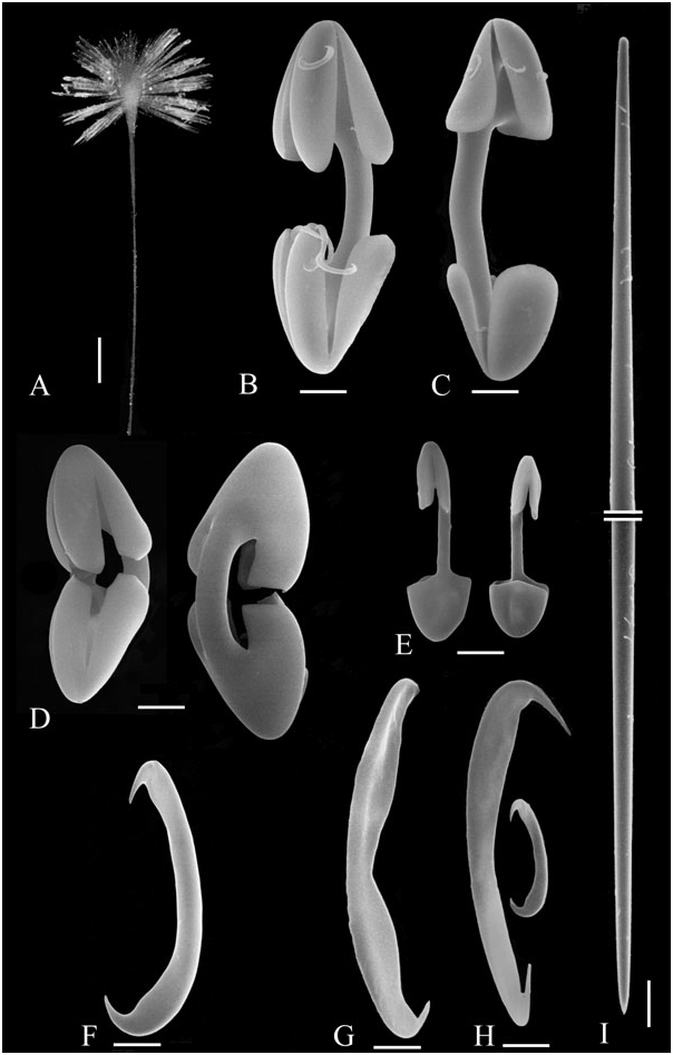
Two small pedunculate sponges ( Fig. 14A View Figure 14 ), consisting of a thin peduncle bearing an ovoid or subspherical body with dense spicular fascicles laterally radiating. Body 3.5 × 1.5 mm, with radiating fascicles up to 5 mm long. Peduncle 25–28 mm long, without attachment base, 0.3–0.5 mm in diameter, made of longitudinally arranged long fusiform styles. Living tissue poorly preserved, nearly absent in the paratype, containing numerous microscleres. Radiating fascicles made of fusiform styles and of smaller styles with the tip outwardly directed. Consistency very fragile after preservation. Colour white. No visible aperture or aquiferous system, no cortex.
Spicules:
1. Styles of the radiating fascicles and of the peduncle ( Fig. 14I View Figure 14 ), straight, with two ill-defined categories. In the peduncle, spicules large and fusiform, with a rather blunt tip. In the body and radiating fascicles, smaller spicules, less fusiform and with an acerate short tip, in addition to styles of the peduncle type. Size 620–2500 × 7–35 µm.
2. Arcuate isochelae ( Fig. 14B, C View Figure 14 ). Shaft curved. Alae ovoid, rather short. Size 80–170 µm, shaft 8 µm in diameter.
3. Abyssochelae ( Fig. 14D View Figure 14 ). Shaft strongly curved. Front alae long, nearly in contact with the opposite ala, laterally curved, abruptly cut at the end. Lateral alae long, laterally curved. Size 40–45 µm.
4. Anisochelae ( Fig. 14E View Figure 14 ), moderately abundant, generally twisted, the plane of the two ends varying from a few degrees to 180°. Shaft straight. One end tridentate with the lateral alae cut away from the shaft and the frontal ala bifid, the other end with fused alae. Size 9.5–11 µm.
5. Sigmancistra 1 ( Fig. 14G, H View Figure 14 ), moderately abundant, most of them with the shaft laterally thickened in a lamella with a central depression, a few with the same shape as sigmancistra 2 but larger. Size 30–40 µm.
6. Sigmancistra 2 ( Fig. 14F, H View Figure 14 ), very abundant. Shaft slightly contorted, the large end with a notch. Size 9.5–12.5 µm.
Remarks
This sponge has the same morphology and general organization as the representatives of the genus Abyssocladia , and its spiculation includes abyssochelae. It differs from the other species of the genus in the microscleres, especially in the presence of tiny anisochelae. These spicules are remarkable for the difference in the two ends, one being palmate and the other arcuate. Anisochelae are known in a member of the Mycalidae , Anomomycale Topsent, 1924 , but they are arcuate, whereas in the new species the anisochelae are of an unusual type, both arcuate and palmate. The presence of anisochelae in otherwise typical representatives of Abyssocladia is one of the reasons temporarily to classify the genus within the Cladorhizidae , possibly indicating affinities with the Mycalidae as already found in Asbestopluma .
ABYSSOCLADIA NAUDUR SP. NOV.
( FIGS 15 View Figure 15 , 16 View Figure 16 )
Type material
Holotype: NAUDUR ND 5 (7-1B), 10/12/1993, 17°23.11′S, 113°11.60′W, 2581 m. Muséum National d’Histoire Naturelle, Paris, no. MNHN D JV 89. GoogleMaps
Paratypes: NAUDUR ND 5 (7-1B), 10/12/1993, 17°23.11′S, 113°11.60′W, 2581 m, approximately 15 specimens or fragments, Muséum National d’Histoire Naturelle, Paris, no. MNHN D JV 90. NAUDUR ND 15 (4-1B), 20/12/1993, 18°15.90′S, 113°22.08 W, 2689 m, one specimen. Muséum National d’Histoire Naturelle, Paris, no. MNHN D JV 91 GoogleMaps .
Etymology
From the NAUDUR cruise (‘NAUtile Dorsale Ultra Rapide’) of Ifremer.
Locality and habitat
East Pacific Rise, North of Easter Island. NAUDUR ND 5, 17°23.11′S, 113°11.60′W, 10/12/1993, 2581 m, on a dead smoker. The sample was collected from the top of the highest chimney (up to 6 m high) of an inactive site, a few metres from some active black smokers with a relatively dense population of the pink sea anemone Chondrophellia sp. The sponges were attached to the vertical face of the chimney, made of sulfide deposits, which also bears the sponge Cladorhiza segonzaci sp. nov. (see above), some hydroids and about 20 specimens of a small white sea anemone. NAUDUR ND 6, 17°24.85′S, 113°12.15′W, 11/12/1993, 2580 m, in a rich assemblage of Vesicomyidae clams Calyptogena , mytilids Bathymodiolus , stalked barnacles Neolepas , etc. NAUDUR ND 15, 20/12/1993, 18°15.90′S, 113°22.08 W, 2689 m, on a dead chimney.
Description
Approximately 20 specimens or fragments of a small erect sponge ( Fig. 15A–C View Figure 15 ), attached on a minute solid substratum by an enlarged base 2–3 mm in diameter, forming a slender, flattened spicular axis with numerous lateral filaments or processes, frequently with a bud-like larger branching process. Holotype 40 mm in total length, with a lateral bud and filaments 1.4 mm in maximum length. Paratypes more or less complete specimens, some with well-preserved thin filaments ( Fig. 15B View Figure 15 ) up to 6 mm long, 170–200 µm in diameter at the base and one or two spicule thick at the end, others with only the base of the filaments. Filaments regularly arranged perpendicularly in two lateral rows along the axis, with a spacing of 0.3–0.6 mm, alternating on each side. Bud-like processes perpendicular or slightly oblique to the main axis, with a few short lateral processes on their axis, thickened at the end. No visible aquiferous system. Colour yellowish grey to clear brown.
Skeleton: Main axis of large fusiform styles longitudinally arranged, spirally twisted in the basal portion where the axis is lined by substrongyles. Axis of the filaments slightly conical at the base, with the styles anchored by their head and the point outwardly directed, entirely crossing the stem ( Fig. 16 View Figure 16 ), reduced to one or two styles near their end. Living tissue poorly preserved, containing numerous sigmancistra. Best preserved zones of the stem and of the base of the filaments with a continuous lining of isochelae, generally with the alae outwardly directed and the shaft lying parallel on the axis.
Spicules:
1. Styles or substrongyles of the main axis ( Fig. 15F View Figure 15 ), straight, the larger strongly fusiform with a blunt point nearly similar to the head. Size 700–1600 × 10–37 µm. 2. Styles of the lateral branches ( Fig. 15G View Figure 15 ), straight, the larger slightly fusiform, with a short acerate point. Size 330–1000 × 5–15 µm.
3. Styles, substrongyles or strongyles ( Fig. 15H View Figure 15 ) from the base and from the coating of the main axis, very variable in size. The smaller located at the fixation base, slightly curved, sometimes with a double bend, with equal ends. The larger located along the basal part of the axis, often a little flexuous, fusiform, with unequal ends. Size 30–825 × 8–30 µm.
4. Abyssochelae ( Fig. 15D View Figure 15 ) with a curved shaft approximately 5 µm in diameter. Frontal ala roughly parallelepipedal, long, nearly in contact with the opposite frontal ala, with a broadly quadrangular section, abruptly cut at the end. Lateral alae large, linked to the shaft along almost their entire length. Size 48– 72 µm, most between 60 and 65 µm, smaller in one specimen (30–65 µm).
5. Sigmancistra 1 ( Fig. 15E View Figure 15 ), with a slightly enlarged shaft. Size 15–19 µm. Smaller sigma in one specimen, in which the size is only 6.5–9.5 × 1 µm.
6. Sigmancistra 2 ( Fig. 15E View Figure 15 ), very abundant, without notch. Size 5–8 µm.
Remarks
The sponge is very fragile, and several specimens were broken upon examination in the laboratory. Most specimens have the filaments reduced to short lateral processes. It cannot be separated with certainty from Cladorhiza segonzaci sp. nov. on the underwater pictures taken in situ after collection ( Fig. 17F View Figure 17 ).
The sponge closely resembles Cladorhiza segonzaci from the same sample, and the two sponges were confused during the preliminary sorting operation and in the first spicule preparations. There are, however, some differences in gross morphology, which have been given in the description of C. segonzaci .
The spicule characters appear rather constant in the 20 available specimens or fragments. One specimen, however, differs slightly from the holotype and the other paratypes in smaller isochelae and small sigmas, 6.5–9.5 µm, instead of sigmancistras, 1 15–19 µm.
This species is classified in Abyssocladia as here redefined based on its spiculation, including abyssochelae. However, it does not display the typical pedunculate, disciform shape characteristic of the genus, but rather closely resembles the shape of some Cladorhiza or Asbestopluma . Furthermore, the abyssochelae have their lateral alae nearly palmate. Its inclusion in the genus is thus made with reservation and may be temporary.


| MNHN |
Museum National d'Histoire Naturelle |
No known copyright restrictions apply. See Agosti, D., Egloff, W., 2009. Taxonomic information exchange and copyright: the Plazi approach. BMC Research Notes 2009, 2:53 for further explanation.
|
Kingdom |
|
|
Phylum |
|
|
Class |
|
|
Order |
|
|
Family |
Abyssocladia
| Vacelet, Jean 2006 |
A. huitzilopochtli
| Vacelet 2006 |
ABYSSOCLADIA HUITZILOPOCHTLI
| Vacelet 2006 |
Abyssocladia
| Levi 1964 |
Cladorhizidae
| DENDY 1922 |
Cladorhizidae
| DENDY 1922 |