
Atractides cf. tuberipalpis ( K. Viets, 1913 )
|
publication ID |
https://doi.org/ 10.11646/zootaxa.3911.3.2 |
|
publication LSID |
lsid:zoobank.org:pub:76C1CD8D-4C66-441D-97C0-A197C2005D79 |
|
DOI |
https://doi.org/10.5281/zenodo.6095011 |
|
persistent identifier |
https://treatment.plazi.org/id/1A2D87A8-4C7F-920D-67E1-FB60B5861DA8 |
|
treatment provided by |
Plazi |
|
scientific name |
Atractides cf. tuberipalpis ( K. Viets, 1913 ) |
| status |
|
Atractides cf. tuberipalpis ( K. Viets, 1913)
( Figs. 5A–I View FIGURE 5 A – I , 6A–F View FIGURE 6 A – F )
Megapus tuberipalpis K. Viets 1913: 26 .
Material examined. Supon stream near Asiakwa, Atewa Hills, 6º 15.530 N, 0º 30.642 E, alt. 250 m asl., 7.iii.2011, Smit, 2/0/0; Afiaso stream, Kakum NP, 5º 30.087 N, 1º 26.373 W, alt. 114 m asl., 12.ii.2013, Smit 1/0/0; Nubui River, Agumatsa Wildlife Sanctuary, 7º 06.986 N, 0º 35.548 E, alt. 254 m asl., 22.ii.2013, Smit 1/0/0 (mounted); Kue River, hyporheic, Kyaboba NP, 8º 31.087 N, 0º 36.049 E, alt. 208 m asl., 25.ii.2013, Smit 0/1/0; Kue River, Kyaboba NP, 8º 31.087 N, 0º 36.049 E, alt. 208 m asl., 25.ii.2013, Smit 1/2/0 (1/1/0 mounted); Laboun River, downstream of falls, Kyabobo NP, 8º 19.836 N, 0º 35.487 E, alt. 342 m asl, 24.ii.2013, Smit 1/0/0 (mounted); Kuluga River, N of Biakpa, upstream, 6º 51.223 N, 0º 25.141 E, alt. 410 m asl., 20.ii.2013, Smit 0/1/0.
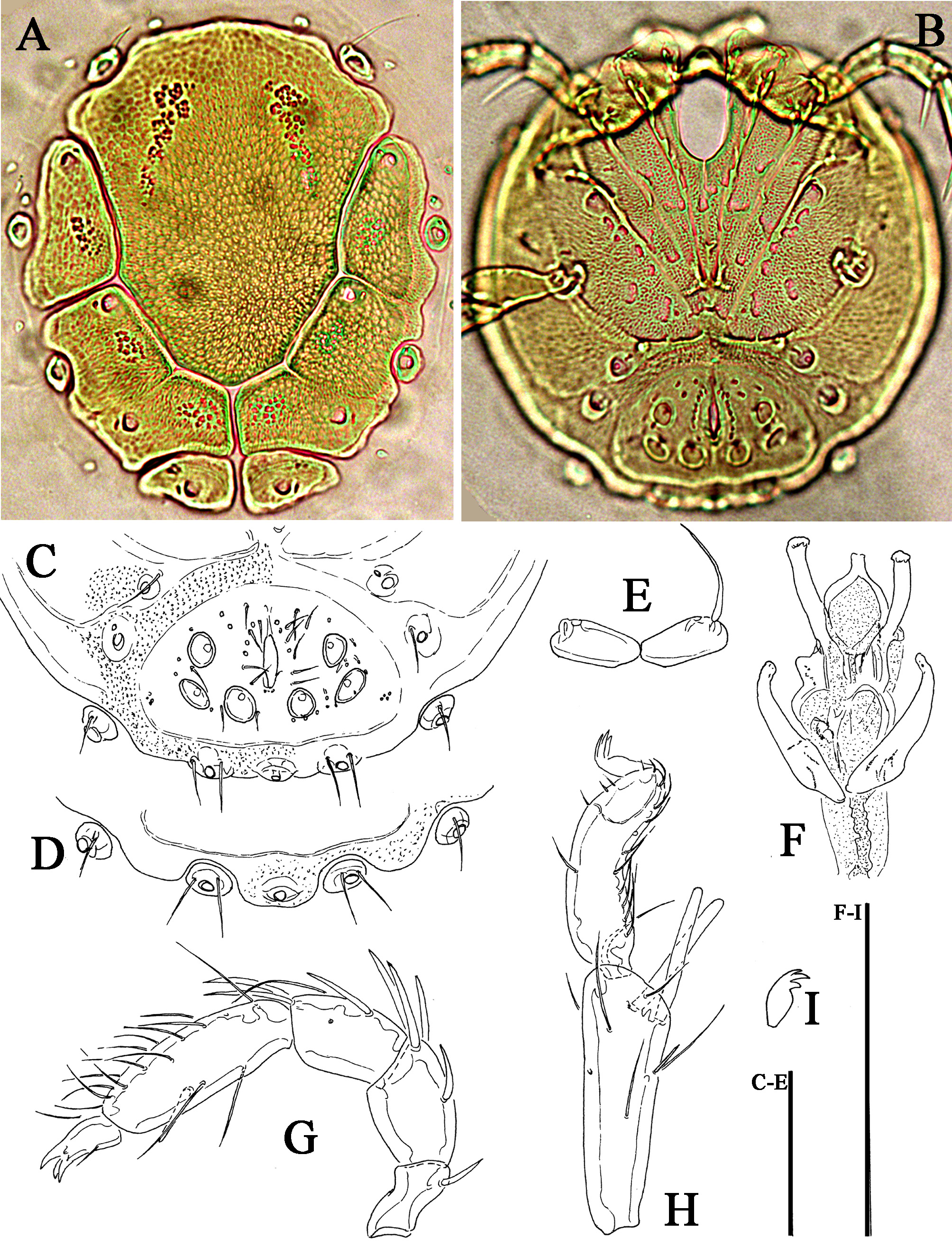
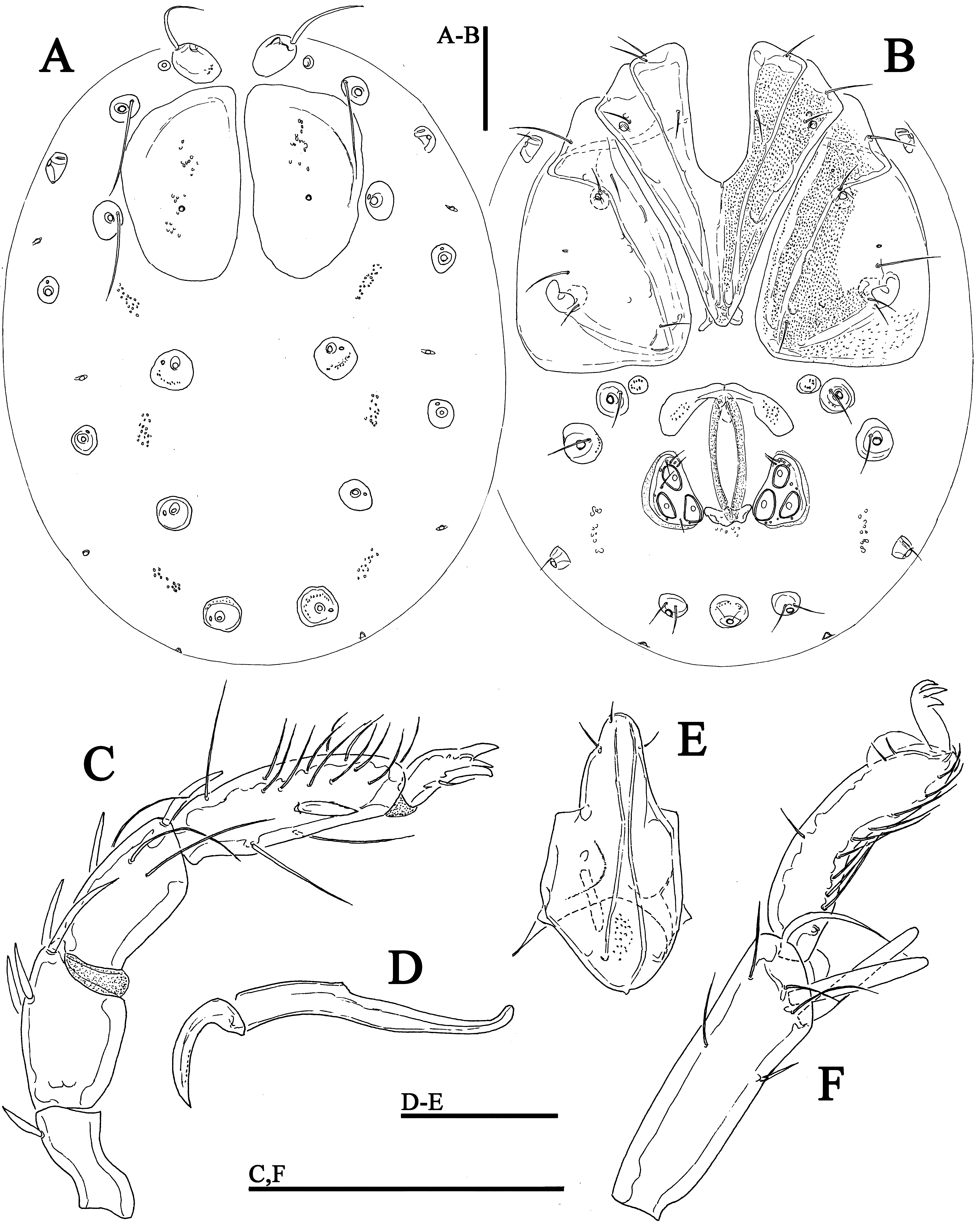
Description. Muscle insertions sclerotized: sexual dimorphism in the dorsal area and coxal field. Palp: ventral margin of P-2 convex, P-4 with straight ventral and equally curved dorsal margins, sword seta pointed, little enlarged, but not hair-like, inserting distal to distoventral seta, P-5 without “cheeks” ( Figs. 5G View FIGURE 5 A – I , 6C View FIGURE 6 A – F ). Legs: I-L-5 S- 1 and -2 closely together; I-L-6 strong and thick, weakly curved, of nearly equal height from the base to the claw furrow; claws with ventral and dorsal clawlets ( Fig. 5I View FIGURE 5 A – I ). Male: Arrangement of dorsal muscle insertions as illustrated in Fig. 5A View FIGURE 5 A – I : postoc on a subtriangular plate covering large part of the dorsum, Dgl-3 fused with D-2, Dgl- 4 fused with D-3, Dgl-5 fused with D4, Dgl-6 fused with D-5, all plates rather large, adjacent to each other. Coxal field: ventral surface covered by an extended shield including coxae, Vgl-3/4, genital field, excretory pore and Vgl- 1+2 ( Fig. 5C View FIGURE 5 A – I ). Female: muscle attachments: postoc on large separate platelets ( Fig. 6A View FIGURE 6 A – F ). Genital field: pregen very robust, with extended border of secondary sclerotization ( Fig. 6B View FIGURE 6 A – F ).
Measurements. Male (from Kue River, in parentheses specimen from Nubui River): Idiosoma length/width 402 (406)/369 (378); large dorsal anteromedial plate length/width 270 (281)/238 (247); ventral shield length/width 381 (373)/350 (363); Cx-III width 253 (256); genital field: width between most lateral pair of Ac 117 (111), length Ac 1–3: 22 (20–21), 23 (22–23), 20–22 (19–21); ejaculatory complex ( Fig. 5F View FIGURE 5 A – I ) length 78.
Palp: Total length 191, dorsal length/height, dorsal length/height ratio: P-1, 25/12, 2.1 (23/11, 2.1); P-2, 40/26, 1.53 (40/25, 1.63); P-3, 39/21, 1.85 (37/23, 1.6); P-4, 61/14, 4.36 (63/15, 4.32); P-5, 26/9, 3.0 (26/10, 2.6); length P-2/P-4 ratio 0.66 (0.63). Capitulum ventral length 115 (120); chelicera total length 142 (155), claw length 49 (43–46), basal segment length 125 (121), length basal segment/claw ratio 2.6 (2.6–2.8).
Legs: I-L-5 dorsal length 79 (81), ventral length 61 (62), dorsal length/ventral length ratio 1.3 (1.3), maximum height 25 (26), dorsal length/maximum height 3.2 (3.1), S-1 length 38 (42), length/width ratio 11.4 (10.6), S-2 length 39 (42), length/width ratio 8.5 (8.6), distance S-1-2, 3 (4-5), length ratio S-1/2, 0.97 (1.0); I-L-6 length 61 (62), central height 15 (17), length/central height ratio 4.0 (3.6); length I-L-5/6 ratio 1.28 (1.31).
Female (from Kue River): Idiosoma length/width 600/484; dorsal anterior plates including postocularia length/ width 188/116; coxal field: length 316; Cx-III width 334; Cx-I+II medial length 137, lateral length 244; genital field length/width 140/163, pregenital sclerite width 131, genital plates length/width 80/51, length Ac 1-3: 25-27, 29, 30-35.
Palp: Total length 253, dorsal length/height, dorsal length/height ratio: P-1, 34/12, 2.8; P-2, 55/34, 1.63; P-3, 55/28, 2.0; P-4, 75/17, 4.4; P-5, 34/12, 2.8; length P-2/P-4 ratio 0.73. Capitulum length 184; chelicera total length 224, claw length 74, basal segment length 178, length basal segment/claw ratio 2.4.
Legs: I-L-5 dorsal length 99, ventral length 77, dorsal length/ventral length ratio 1.28, maximum height 31, dorsal length/maximum height 3.2, S-1 length 48, length/width ratio 8.4, S-2 length 46, length/width ratio 6.6, distance S-1-2, 4, length ratio S-1/2, 1.03; I-L-6 length 82, central height 18, length/central height ratio 4.6; length I-L-5/6 ratio 1.21.
Remarks. This species belongs to the testudo -group (sensu Cook 1966), characterized at the first line by the similar arrangement of the separate dorsal platelets. This group includes seven rather similar African species: A. testudo Cook, 1966 , A. pseudotestudo Cook, 1966 , A. neotestudo Cook, 1966 , A. paratestudo Cook, 1966 and A. subtestudo Cook, 1966 ), all known from Liberia ( Cook 1966), A. scutifer Lundblad, 1951 from East and South Africa (Pešić et al. 2011) and A. tuberipalpis ( K.Viets, 1913) .
Atractides tuberipalpis was described by K. Viets (1913) based on a single male from a stream on the road Kumba-Mundame in Cameroon. No further records were published ever since. In the original description Viets (1913) did not illustrate the dorsum, but he explicitly stated that a dorsal shield is formed by one extended plate resembling the body outline.
A study of populations of A. scutifer Lundblad, 1951 from South Africa by Pešić et al. (2011) shows considerable age-dependent variation in the extension of dorsal sclerotization in males, from separate platelets to a large dorsal shield with an irregular undulating margin. Similarly, we find a variation in the degree of sclerotization in A. tuberipalpis males from our study, from completely separated to partly fused dorsal platelets as showed in Fig. 5A View FIGURE 5 A – I . It is possible that the male on which the original description is based, is a mature specimen with a well developed dorsal shield. Furthermore, the holotype differs from specimens in our study in the medially fused Dgl- 1. In males examined from Ghana these glandularia are medially touching suggesting that this character could be age-depending as well. Small button-like tubercles on the palp and I-L-6, a character not found in any other water mite species, were interpreted in the original description as part of appendages, but shape and location of these structures suggest that Viets probably dealt with small air bubbles, or epibionts.
For the time being we tentatively keep the populations from Ghana assigned to A. tuberipalpis , mainly because of the shape of the ventral shield including the excretory pore and all Vgl, but not incorporating Lgl-4 (incorporated into ventral shield in the closely related A. testudo Cook, 1966 from Liberia). However, in a male from Laboun River the ventral shield includes the excretory pore, but Vgl-1+2 and Lgl-4 are free in the membranous integument ( Fig. 5D View FIGURE 5 A – I ). Understanding the taxonomic position of the species of the A. testudo -group is not possible without additional material from a wider area and/or without the application of molecular techniques.
No known copyright restrictions apply. See Agosti, D., Egloff, W., 2009. Taxonomic information exchange and copyright: the Plazi approach. BMC Research Notes 2009, 2:53 for further explanation.
|
Kingdom |
|
|
Phylum |
|
|
Class |
|
|
Order |
|
|
Family |
|
|
Genus |
Atractides cf. tuberipalpis ( K. Viets, 1913 )
| Pešić, Vladimir & Smit, Harry 2015 |
Megapus tuberipalpis
| Viets 1913: 26 |