
Silutanispermum kvacekiorum, Friis & Crane & Pedersen, 2018
|
publication ID |
https://doi.org/ 10.2478/if-2018-0010 |
|
persistent identifier |
https://treatment.plazi.org/id/1A4787F5-FFA1-E10C-FF0F-6609FB15BE7F |
|
treatment provided by |
Diego |
|
scientific name |
Silutanispermum kvacekiorum |
| status |
gen. et sp. nov. |
Silutanispermum kvacekiorum gen. et sp. nov.
Text-figs 11–12 View Text-fig View Text-fig
H o l o t y p e. Designated here, S170238 (Famalicão sample 025; illustrated here on Text-figs 11a, b, d, e View Text-fig , 12a, b View Text-fig ).
P l a n t F o s s i l N a m e s R e g i s t r y N u m b e r.
PFN000099 (for new species).
P a r a t y p e s. Designated here, S174352 (Famalicão sample 025).
R e p o s i t o r y. Palaeobotanical Collections , Department of Palaeobiology, the Swedish Museum of Natural History, Stockholm, Sweden .
E t y m o l o g y. In recognition of Zlatko and Jiří Kvaček for their many contributions to angiosperm palaeobotany.
T y p e l o c a l i t y. Famalicão , Portugal (39°42′16″N;
8°46′12″W).
T y p e s t r a t u m a n d a g e. Below the Figueira da Foz Formation; Early Cretaceous (late Aptian – early Albian or older).
D i a g n o s i s. As for the genus.
D i m e n s i o n s. Length of seeds: 1.6–2.0 mm; width of seeds: 1.2–1.9 mm.
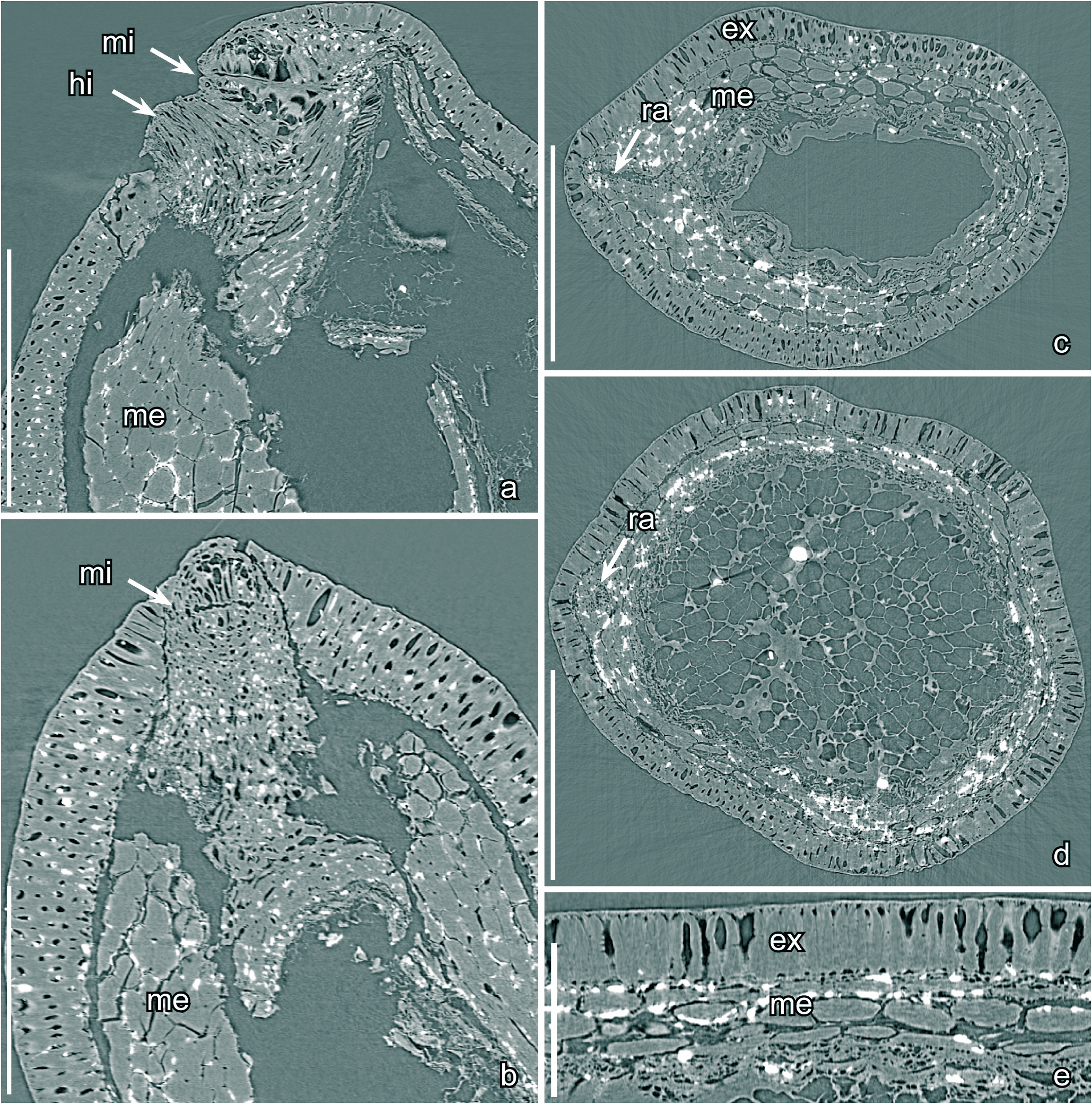
D e s c r i p t i o n a n d r e m a r k s. The species is based on two isolated seeds that were studied using SRXTM (S170238, 174352). There is no information on the fruits in which the seeds were borne. The seeds are small, anatropous, bitegmic and exotestal with bilateral symmetry. They are elliptic in lateral view ( Text-fig. 11a–c View Text-fig ) and elliptical to circular in transverse section ( Text-fig. 12c, d View Text-fig ). The seed surface is smooth with a jigsaw puzzle-shaped pattern formed from the slightly raised undulate anticlinal walls of the exotesta cells.
The micropyle and hilum are very close together and slightly displaced towards the raphal side of the seed (Textfig. 11a–d). The hilum is rounded triangular in outline without a hilar rim ( Text-fig. 11d View Text-fig ). The course of the raphe is seen on the seed surface as a slightly raised ridge that is also distinctive in its transversely aligned cells. The micropyle is formed from the inner integument and is marked on the outside by a transverse, slightly undulate slit in the testa ( Text-figs 11d View Text-fig , 12b View Text-fig ). Internally the micropylar slit is lined by tall cells of the exotesta ( Text-fig. 12a, b View Text-fig ) and there is a tendency towards a Y-shaped branching internally.
The seed coat is composed of a thick exotesta, a thick mesotesta/endotesta, and a thin tegmen. The exotesta consists of a single layer of tall, columnar sclerenchyma cells that are about 65 µm tall over most of the seed (Textfig. 12a, c–e) but taper out in the micropylar region ( Text-fig. 12a View Text-fig ). The anticlinal walls of the exotestal cells are thickened, and of almost even thickness from the outside to the inside, resulting in an almost straight lumen ( Text-fig. 12c–e View Text-fig ). The anticlinal walls are strongly undulate towards the outside and the inside forming stellate-undulate facets and a jigsaw puzzle-like pattern with rounded, undivided lobes on the seed surface; except over the raphe where the cell walls are straight and the facets are polygonal. The mesotesta/ endotesta is as thick as the exotesta, about 65 µm over most of the seed, and consists of up to five layers of large rectangular, tangentially elongated parenchyma cells, (Textfig. 12c–e). The mesotesta/endotesta is thickest around the raphe and close to the hilar region it is up to about 180 µm thick ( Text-fig. 12c View Text-fig ). The tegmen is thin.
Nutritive tissue is well preserved in one of the specimens studied using SRXTM (S174352). It is cellular, consisting of isodiametric cells, about 55 µm in diameter, with thin, straight, cell walls ( Text-fig. 12d View Text-fig ).

| R |
Departamento de Geologia, Universidad de Chile |
| T |
Tavera, Department of Geology and Geophysics |
No known copyright restrictions apply. See Agosti, D., Egloff, W., 2009. Taxonomic information exchange and copyright: the Plazi approach. BMC Research Notes 2009, 2:53 for further explanation.
|
Kingdom |
|
|
Phylum |
|
|
Class |
|
|
Order |
|
|
Family |
|
|
Genus |