
Pazlia hilaris, Friis & Crane & Pedersen, 2018
|
publication ID |
https://doi.org/10.2478/if-2018-0010 |
|
persistent identifier |
https://treatment.plazi.org/id/1A4787F5-FFB4-E114-FC1B-62E5FE17B918 |
|
treatment provided by |
Diego |
|
scientific name |
Pazlia hilaris |
| status |
gen. et sp. nov. |
Pazlia hilaris gen. et sp. nov.
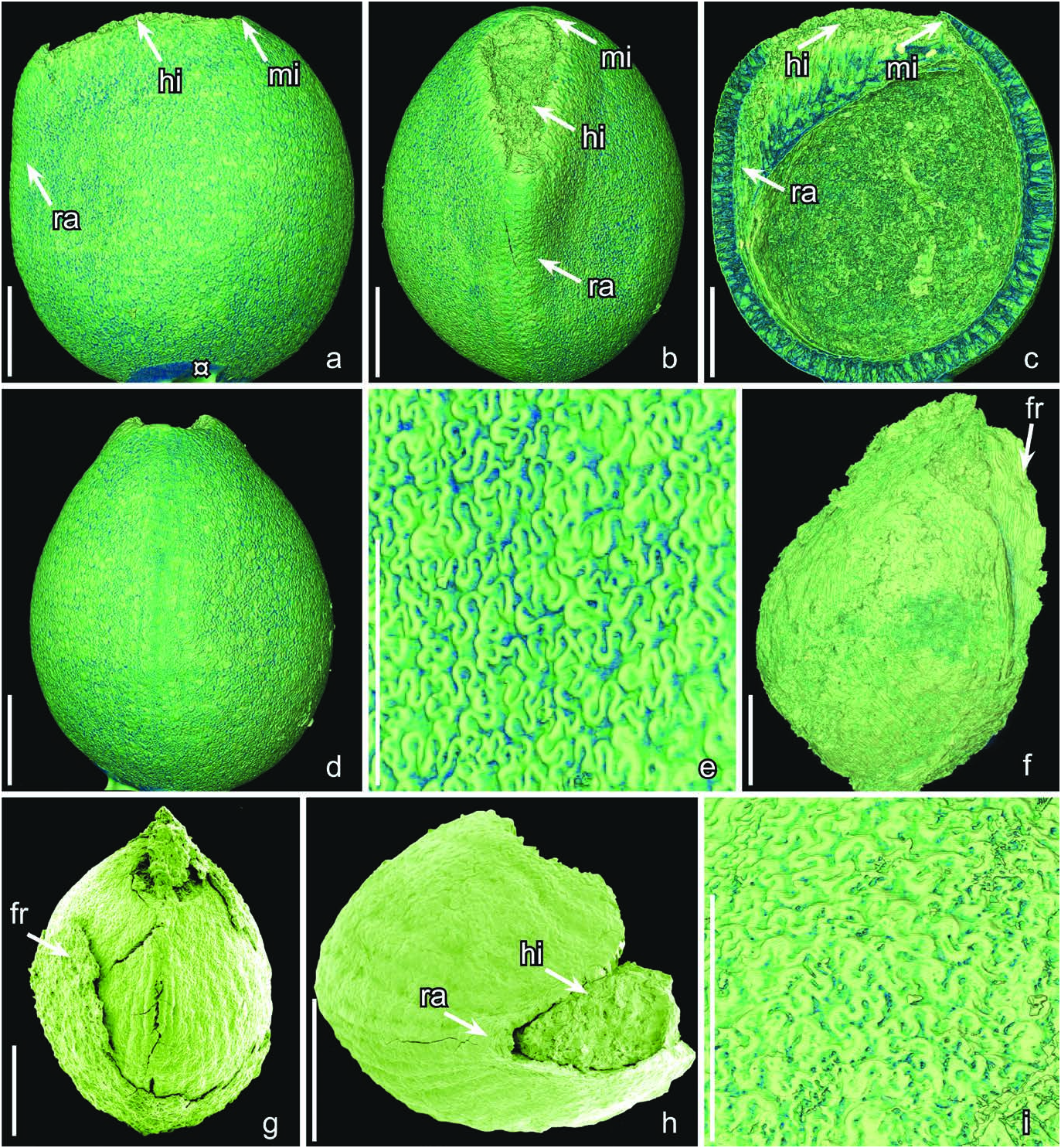
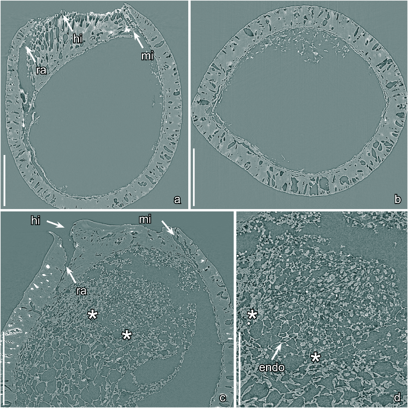
Text-figs 3a–e View Text-fig , 4a, b View Text-fig
H o l o t y p e. Designated here, S175096 (Famalicão sample 025; illustrated here on Text-figs 3a–d View Text-fig , 4a, b View Text-fig ).
P l a n t F o s s i l N a m e s R e g i s t r y N u m b e r.
PFN000091 (for new species).
P a r a t y p e s. Designated here, S174336, S174342, S175083, S175098, S175105, S175106 (Famalicão sample 025).
R e p o s i t o r y. Palaeobotanical Collections , Department of Palaeobiology, the Swedish Museum of Natural History, Stockholm, Sweden .
E t y m o l o g y. From Latin: hilaris, relating to the hilum to emphasis the large hilar scar and strongly developed sclerenchyma tissue under hilum.
T y p e l o c a l i t y. Famalicão , Portugal ( 39°42′16″N;
8°46′12″W).
T y p e s t r a t u m a n d a g e. Below the Figueira da Foz Formation; Early Cretaceous (late Aptian – early Albian or older).
D i a g n o s i s. As for the genus.
D i m e n s i o n s. Length of seeds: 1.1–1.4 mm; width of seeds: 0.9–1.2 mm.
O t h e r m a t e r i a l. S175109 ( Vale de Água 330).
D e s c r i p t i o n a n d r e m a r k s.Thespeciesisbased on about 20 seeds of which five were studied using SRXTM (S174336, S174342, S175083, S175096, S175098). All seeds are isolated and there is no information on the fruit in which they were borne. The seeds are small, anatropous, bitegmic and exotestal with bilateral symmetry. The seed surface is almost smooth with a jigsaw puzzle-like pattern formed from the raised undulate anticlinal walls of the exotesta cells. The seeds are almost circular in lateral view with a truncate hilar-micropylar region ( Text-figs 3a–c View Text-fig , 4a View Text-fig ), and they are broadly elliptical in transverse section (Textfigs 3b, 4b).
The micropyle and hilum are separated by a broad zone of sclerenchyma tissue beneath the very large and distinct hilar scar ( Text-figs 3a–c View Text-fig , 4a View Text-fig ). This hilar sclerenchyma is strongly expanded toward the inside of the seed by cells that are thin-walled, elongate and radiate perpendicular to the hilar scar ( Text-figs 3c View Text-fig , 4a View Text-fig ). The hilum lacks a rim. The course of the raphe is distinct on the seed surface and seen as a slightly raised, rounded ridge extending from hilum to the chalazal end of the seed ( Text-fig. 3b, c View Text-fig ). The micropyle is formed from the inner integument, and seen on the seed surface as a narrow slit. There is a strongly thickened zone of sclerenchyma cells between micropyle and raphe ( Text-figs 3b View Text-fig , 4a View Text-fig ).
Most of the seed coat is composed of exotesta, which is one cell layer deep. The mesotesta/endotesta and tegmen are typically collapsed. The exotesta is one cell layer deep and consists of columnar sclerenchyma cells that are about 105 µm tall over most of the seed, but that gradually become shorter towards the hilar-micropylar region ( Text-fig. 4a, b View Text-fig ). The exotestal cells are arranged in longitudinal rows over the raphe and on the anti-raphal side, but otherwise their arrangement appears irregular. The anticlinal walls of the exotestal cells are thickened and of almost even thickness from the outside to the inside resulting in an almost straight lumen ( Text-fig. 4a, b View Text-fig ). They are strongly undulate towards the outside and inside forming a jigsaw puzzle-like pattern on the surface with rounded, deep, sometimes bifurcate, lobes in the stellate-undulate facets ( Text-fig. 3d View Text-fig ). The cell walls are also undulate over the raphe and in the micropylar region.
Patchy remains of cellular nutritive tissue were observed in one of the specimens studied using SRXTM, but none of the specimens show sufficient internal details to reveal the size or nature of the embryo.
| R |
Departamento de Geologia, Universidad de Chile |
| T |
Tavera, Department of Geology and Geophysics |
No known copyright restrictions apply. See Agosti, D., Egloff, W., 2009. Taxonomic information exchange and copyright: the Plazi approach. BMC Research Notes 2009, 2:53 for further explanation.