
Reyispermum parvum, Friis & Crane & Pedersen, 2018
|
publication ID |
https://doi.org/ 10.2478/if-2018-0010 |
|
persistent identifier |
https://treatment.plazi.org/id/1A4787F5-FFB8-E110-FC6B-64EAFEDABD5C |
|
treatment provided by |
Diego |
|
scientific name |
Reyispermum parvum |
| status |
gen. et sp. nov. |
Reyispermum parvum gen. et sp. nov.
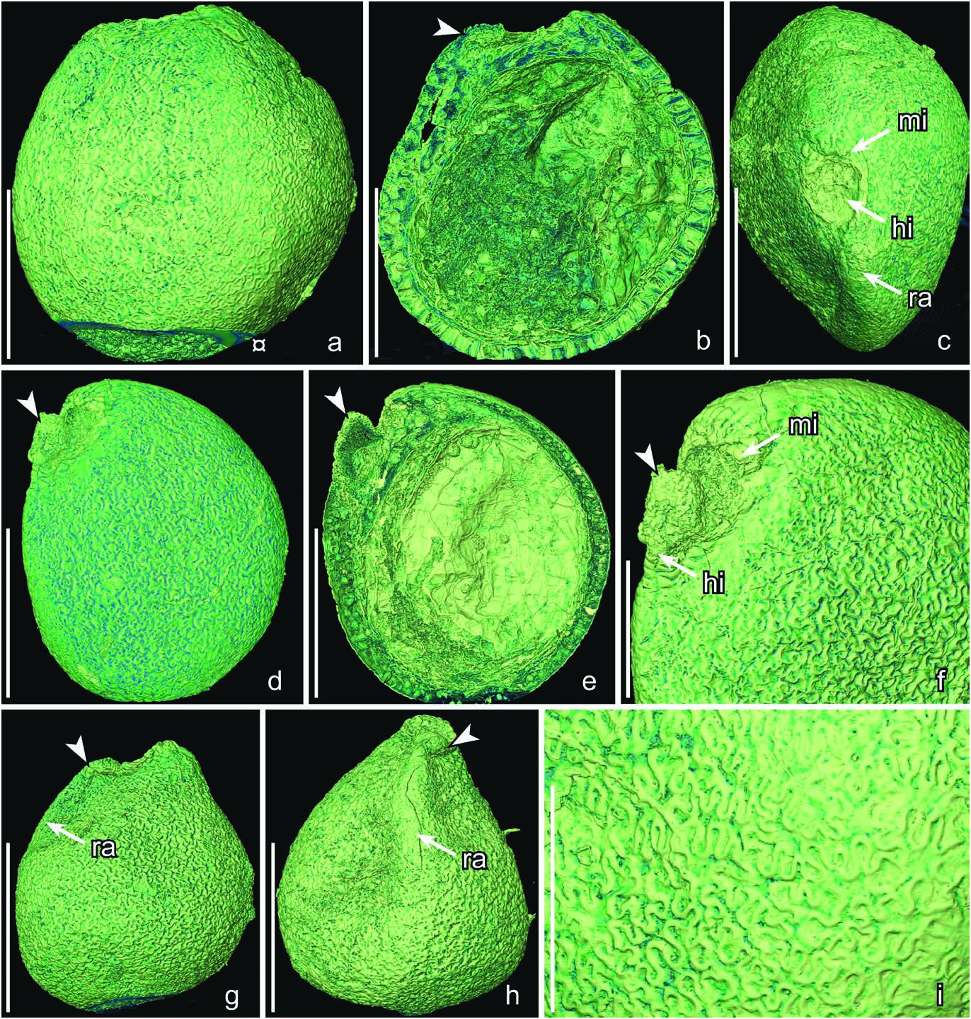
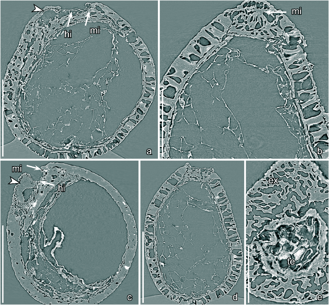
Text-figs 5 View Text-fig , 6 View Text-fig
H o l o t y p e. Designated here, S174178 (Vale de Água sample 141; illustrated here on Text-figs 5a–c, i View Text-fig , 6a, b View Text-fig ).
P l a n t F o s s i l N a m e s R e g i s t r y N u m b e r.
PFN000094 (for new species).
P a r a t y p e s. Designated here, S174179, S175111 (Vale de Água sample 141), S174495 (Vale de Água sample 300).
R e p o s i t o r y. Palaeobotanical Collections , Department of Palaeobiology, the Swedish Museum of Natural History, Stockholm, Sweden .
E t y m o l o g y. From Latin: parvus, small relating to the small size of the seeds.
T y p e l o c a l i t y. Vale de Água , Portugal (39°37′15″N,
08°51′30″W).
T y p e s t r a t u m a n d a g e. Basal part of the Figueira da Foz Formation; Early Cretaceous (late Aptian – early Albian).
D i a g n o s i s. As for the genus.
D i m e n s i o n s. Length of seeds: 0.44–0.52 mm; width of seeds: 0.28–0.5 mm.
D e s c r i p t i o n a n d r e m a r k s. The species is based on about eight isolated seeds of which three (S174178, S174179, S174495) were studied using SRXTM. All seeds are isolated and there is no information on the fruit in which they were borne. The seeds are tiny, anatropous, bitegmic and exotestal. They are broadly ovate in lateral view, bilaterally symmetrical with dorsiventral plane of symmetry, and sometimes slightly compressed laterally ( Text-fig. 5a– h View Text-fig ). The raphal region is seen on the outside as a slightly raised rounded ridge ( Text-fig. 5c, g, h View Text-fig ).
The hilum and micropyle are separated by a zone of testal tissue. The hilum lacks a rim and is seen as a small rounded depression ( Text-fig. 5b, c View Text-fig ). The hilar tissue is slightly expanded inside. The micropyle is formed by the inner integument (tegmen) and marked on the seed surface by a transverse slit through the outer integument (testa) ( Text-figs 5c, f View Text-fig , 6b View Text-fig ). Internally the micropylar slit is lined on all sides by testal cells.
The testa consists of an outer layer of palisade-shaped sclerenchyma cells and an inner layer of thin-walled parenchyma cells ( Text-figs 5b, e View Text-fig , 6a–e View Text-fig ). The palisadeshaped cells of the exotesta are about 45 µm tall over most of the seed but gradually become shorter towards the micropyle and hilum; in the hilar region the thin exotesta is sometimes loosened forming a slightly bulging structure ( Text-figs 5b, d–h View Text-fig , 6a, c View Text-fig ). The anticlinal cell walls of the exotesta cells are evenly thickened walls and the lumen is more or less straight ( Text-fig. 6a, b, d View Text-fig ). The outer and inner parts of the anticlinal walls are strongly undulate and slightly raised resulting in stellate-undulate facets and a jigsaw puzzle-like pattern on the seed surface ( Text-fig. 5a, d, f–i View Text-fig ) except in the micropylar region where the anticlinal walls are straight and the outer facets polygonal ( Text-fig. 5f View Text-fig ). The lobes of the exotestal walls are deep and sometimes bifurcated. The tegmen is thin, but thickened around the micropyle and composed of rectangular epidermal cells with a striate wrinkled surface that are often collapsed ( Text-fig. 6e View Text-fig ).
None of the specimens observed using SRXTM had an embryo preserved, but the irregular cavity in the nutritive tissue, which is in the appropriate position for the embryo, suggests that the embryo was tiny. The nutritive tissue consists of thin-walled and equiaxial cells, about 45 µm in diameter ( Text-fig. 6d View Text-fig ).
| R |
Departamento de Geologia, Universidad de Chile |
| T |
Tavera, Department of Geology and Geophysics |
No known copyright restrictions apply. See Agosti, D., Egloff, W., 2009. Taxonomic information exchange and copyright: the Plazi approach. BMC Research Notes 2009, 2:53 for further explanation.