
Lusitanispermum choffatii, Friis & Crane & Pedersen, 2018
|
publication ID |
https://doi.org/ 10.2478/if-2018-0010 |
|
persistent identifier |
https://treatment.plazi.org/id/1A4787F5-FFBD-E112-FF18-600FFDF7BBA8 |
|
treatment provided by |
Diego |
|
scientific name |
Lusitanispermum choffatii |
| status |
gen. et sp. nov. |
Lusitanispermum choffatii gen. et sp. nov.
Text-figs 7–10 View Text-fig View Text-fig View Text-fig
H o l o t y p e. Designated here, S174345 (Famalicão sample 025; illustrated here on Text-figs 7a, b View Text-fig , 9a, b View Text-fig ).
P l a n t F o s s i l N a m e s R e g i s t r y N u m b e r.
PFN000097 (for new species).
P a r a t y p e s. Designated here, S105097, S105098, S170239, S174035, S174353, S174467 – S174470, S174472, S174474, S174811, S175113 – S175118 (Famalicão sample 025).
R e p o s i t o r y. Palaeobotanical Collections , Department of Palaeobiology, the Swedish Museum of Natural History, Stockholm, Sweden .
E t y m o l o g y. In recognition of Léon Paul Choffat (*1849, †1919) for his geological studies of Cretaceous deposits in Portugal.
T y p e l o c a l i t y. Famalicão , Portugal (39°42′16″N;
8°46′12″W).
T y p e s t r a t u m a n d a g e. Below the Figueira da Foz Formation; Early Cretaceous (late Aptian – early Albian or older).
D i a g n o s i s. As for the genus.
D i m e n s i o n s. Length of seeds: 1.7–2.8 mm; width of seeds: 0.8–1.9 mm.
O t h e r m a t e r i a l. S175046 (Vale de Água sample
333).
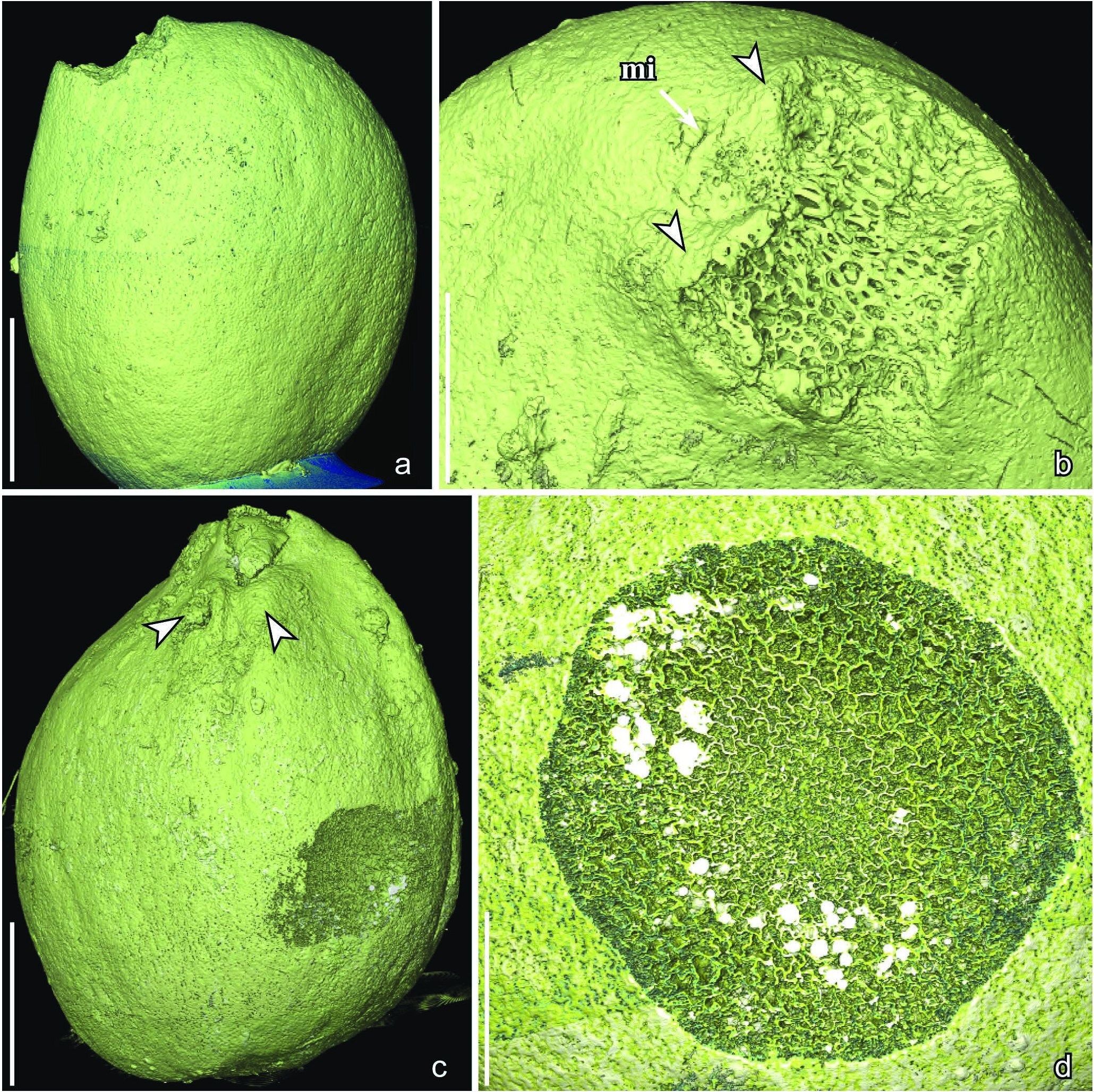
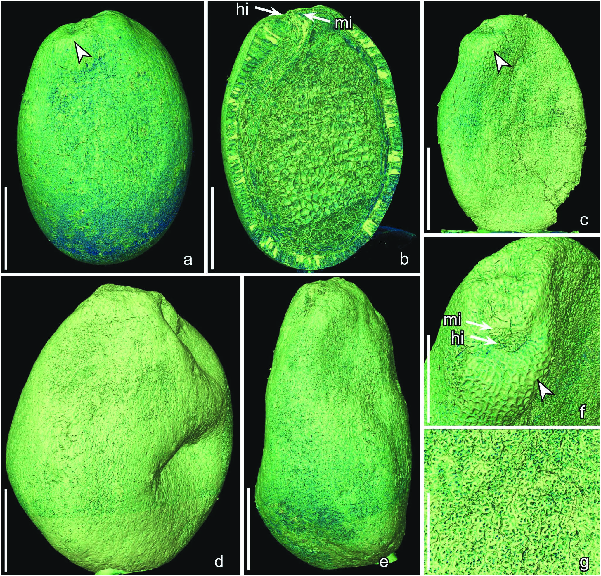
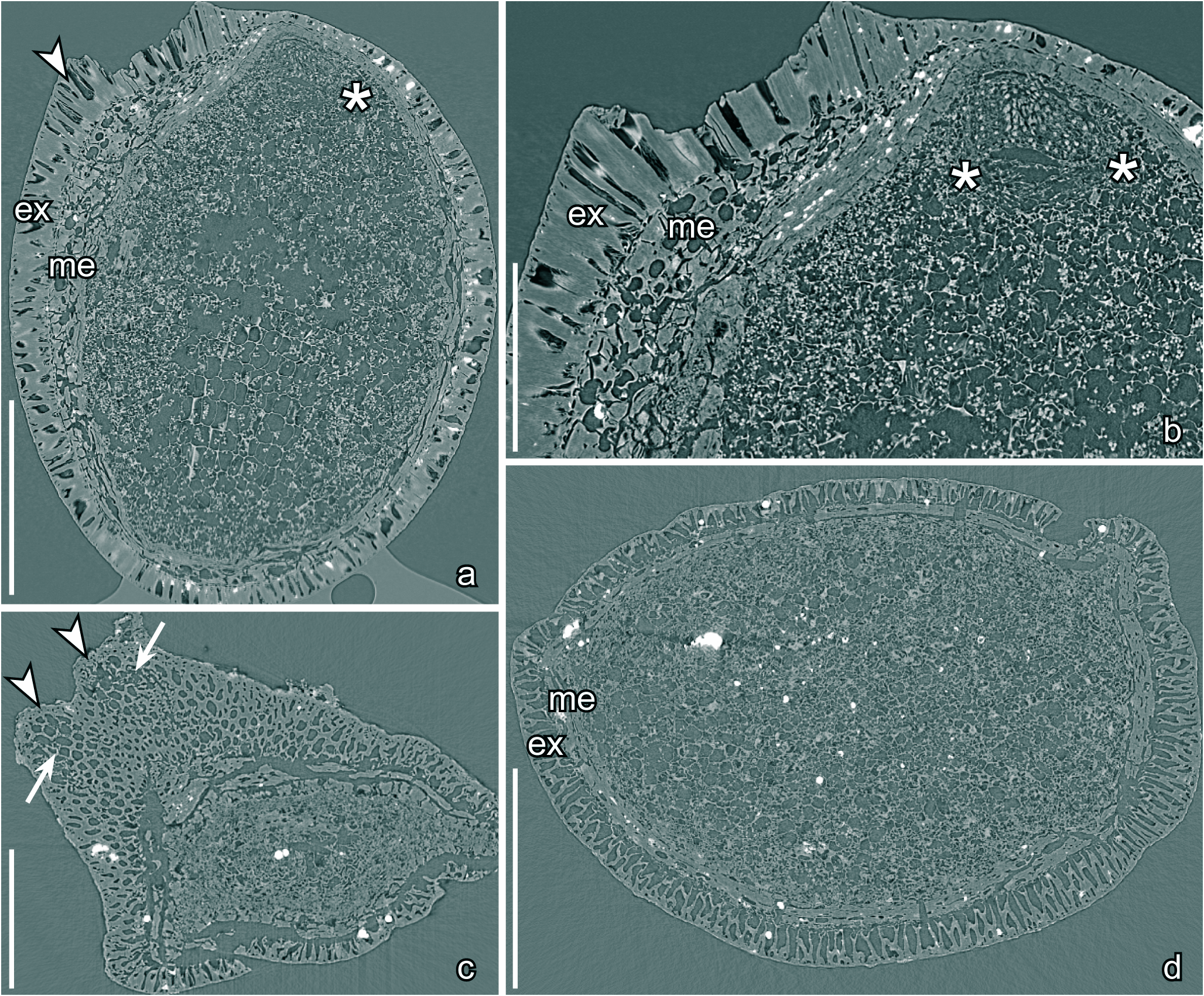
D e s c r i p t i o n a n d r e m a r k s. The species is based on about 120 isolated seeds of which 12 specimens were studied using SRXTM ( S105097 , S105098 , S170239 , S174035 , S174345 , S174353 , S174467 – S174470 , S174472 , S174474 ). There is no information on the fruits in which the seeds were borne. The seeds are small, anatropous, bitegmic and exotestal with bilateral symmetry. They are elliptic in lateral view ( Text-figs 7a, c View Text-fig , 8a–e View Text-fig ) and elliptical to circular in transverse section ( Text-figs 9d View Text-fig , 10e). The seed surface is smooth with a jigsaw puzzle-shaped pattern formed from the slightly raised undulate anticlinal walls of the exotesta cells ( Text-fig. 8g View Text-fig ). The seeds vary considerably in size and shape and they may belong to more than one natural species. However, there are transitional forms between the smaller more narrow type ( Text-fig. 8a–c View Text-fig ) and the larger more rounded type ( Text-figs 7a–c View Text-fig , 8d, e View Text-fig ) and all specimens are therefore included here in the same species .
The micropyle and hilum are very close together at the seed apex ( Text-figs 8b View Text-fig , 10a, d). The hilum is rounded triangular in outline without a hilar rim ( Text-fig. 8f View Text-fig ). The course of the raphe is not seen on the seed surface. The micropyle is formed from the inner integument and marked externally by a transverse slit in the testa ( Text-figs 8f View Text-fig , 10d).
The seed coat is composed of a thick exotesta, a thick mesotesta/endotesta, and a thin tegmen. The exotesta consists of a single layer of tall, columnar sclerenchyma cells. In the bulging areas close to the hilum the exotestal cells are up to about 180 µm tall ( Text-figs 9a–d View Text-fig , 10a, c, e), but are about 95 µm tall laterally and on the antiraphal sides of the seed. These exotesta cells become gradually shorter towards the micropylar slit and in the micropylar region ( Text-fig. 10a, b). They have slightly raised anticlinal walls and towards the outside (Textfig. 8g) and inside ( Text-fig. 7d View Text-fig ) the cells of the exotesta are strongly undulate forming stellate-undulate facets and a jigsaw puzzle-like pattern with rounded, undivided lobes.
In the bulging portion of the seed coat around the micropylar area the cells of exotesta have slightly thinner walls ( Text-fig. 9c View Text-fig ) and this part of the seed is sometimes abraded or collapsed. Also in this part of the seed the cells of the exotesta have straight anticlinal walls and polygonal facets ( Text-fig. 8f View Text-fig ). The mesotesta/endotesta consists of at least three layers of large parenchyma cells ( Text-figs 9a, b, d View Text-fig , 10e), most prominent below the bulging hilar zone ( Text-fig. 9b View Text-fig ). The tegmen is thin over most of the seed, but thicker towards the micropyle.
Nutritive tissue is well preserved in most specimens studied using SRXTM and in seven specimens ( S170239 , S174345 , S174468 , S174469 , S174470 , S174472 , S174474 ) a complete or almost complete embryo was observed ( Text-figs 9a, b View Text-fig , 10f). In all cases the embryo is tiny, about 110 µm long and 180 µm broad, with two rudimentary cotyledons ( Text-figs 9b View Text-fig , 10f), and an embryo to seed ratio of about 0.016. The cells of the embryo are much smaller than those of the surrounding nutritive tissue and each cell has a dense central structure ,
which we interpret as the remains of a nucleus ( Text-figs 9b View Text-fig , 10f). The nutritive tissue is cellular consisting of isodiametric and thin-walled cells, about 40 µm in diameter, with thin, straight or slightly undulate cell walls ( Text-figs 9a–d View Text-fig , 10b, c, e). In most specimens the cells of the nutritive tissue are filled with small granular bodies that we interpret as the remains of protein or lipid bodies ( Text-figs 9b, d View Text-fig , 10c, d, f). The cells are rarely completely empty ( Text-fig. 10a, b, e).
| R |
Departamento de Geologia, Universidad de Chile |
| T |
Tavera, Department of Geology and Geophysics |
No known copyright restrictions apply. See Agosti, D., Egloff, W., 2009. Taxonomic information exchange and copyright: the Plazi approach. BMC Research Notes 2009, 2:53 for further explanation.
|
Kingdom |
|
|
Phylum |
|
|
Class |
|
|
Order |
|
|
Family |
|
|
Genus |