
Sperchon tridentatus, Sokolow, 1940
|
publication ID |
https://doi.org/10.11646/zootaxa.4970.1.6 |
|
publication LSID |
lsid:zoobank.org:pub:7BDDAD83-B88D-4B51-B395-864970AB207C |
|
DOI |
https://doi.org/10.5281/zenodo.5918074 |
|
persistent identifier |
https://treatment.plazi.org/id/1A651378-FFFB-FFCC-FF69-FCEE3F87FE11 |
|
treatment provided by |
Plazi |
|
scientific name |
Sperchon tridentatus |
| status |
|
Key to species of the Sperchon tridentatus -group
1 Integument without muscle attachment sclerites............................................................. 2
- Integument with muscle attachment sclerites............................................................... 3
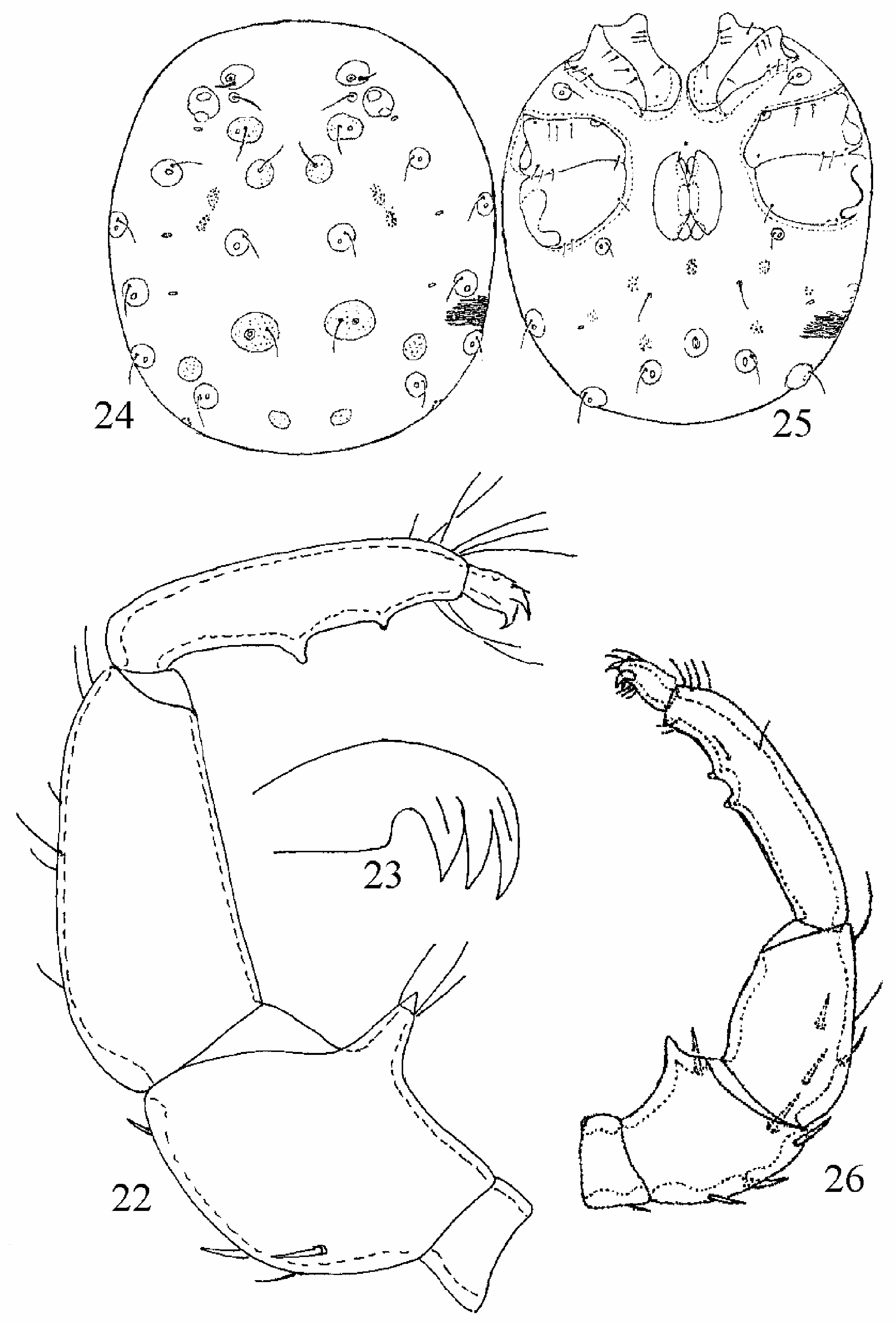
2 P-4 shorter than P-3, P-2 distoventral projection bearing three fine setae, P-3 ventral margin straight ( Fig. 22 View FIGURES 22–26 ), claw lamella with straight ventral margin ( Fig. 23 View FIGURES 22–26 ), excretory pore unsclerotized ( Sokolow 1940).......................... S. tridentatus
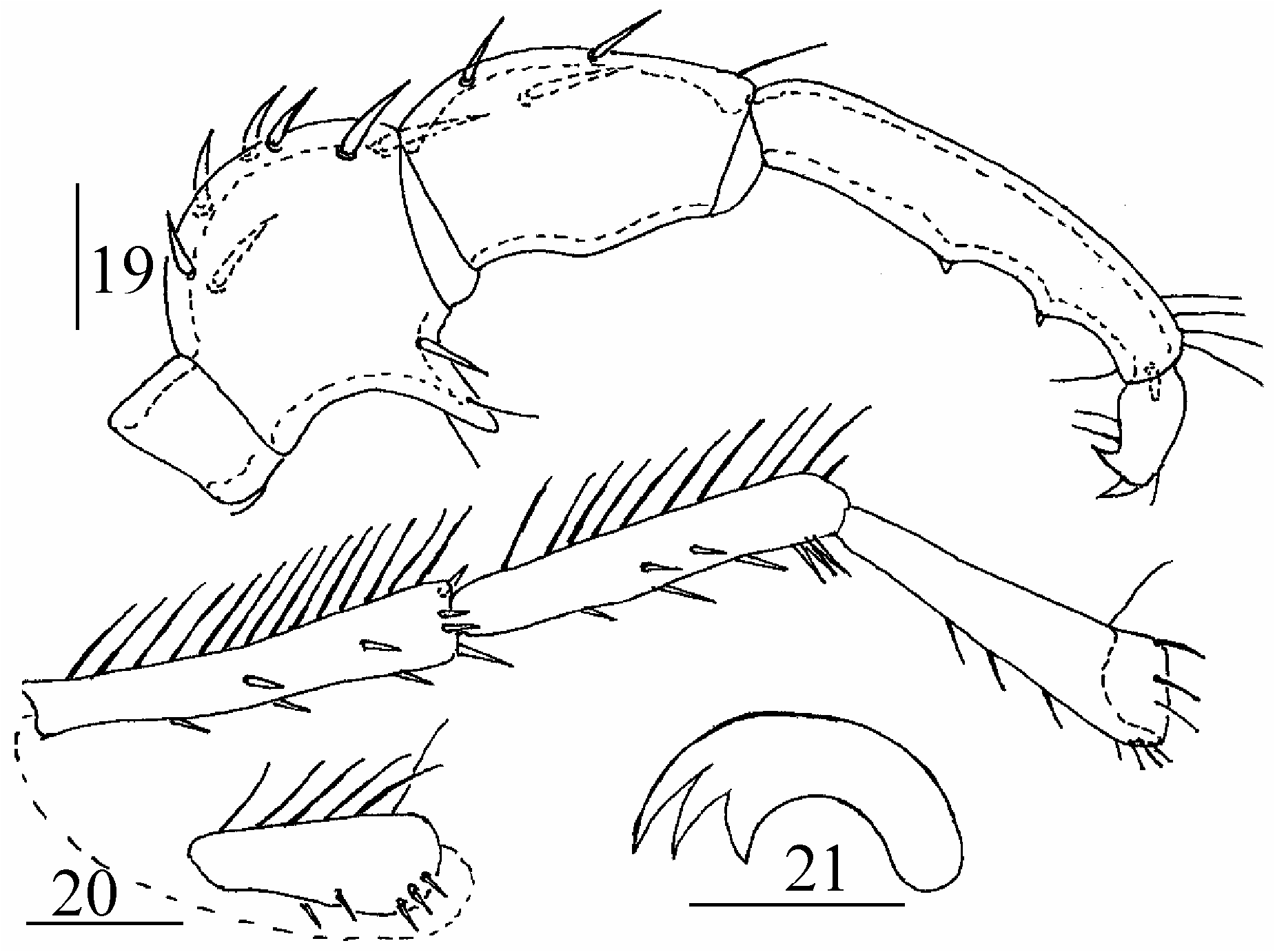
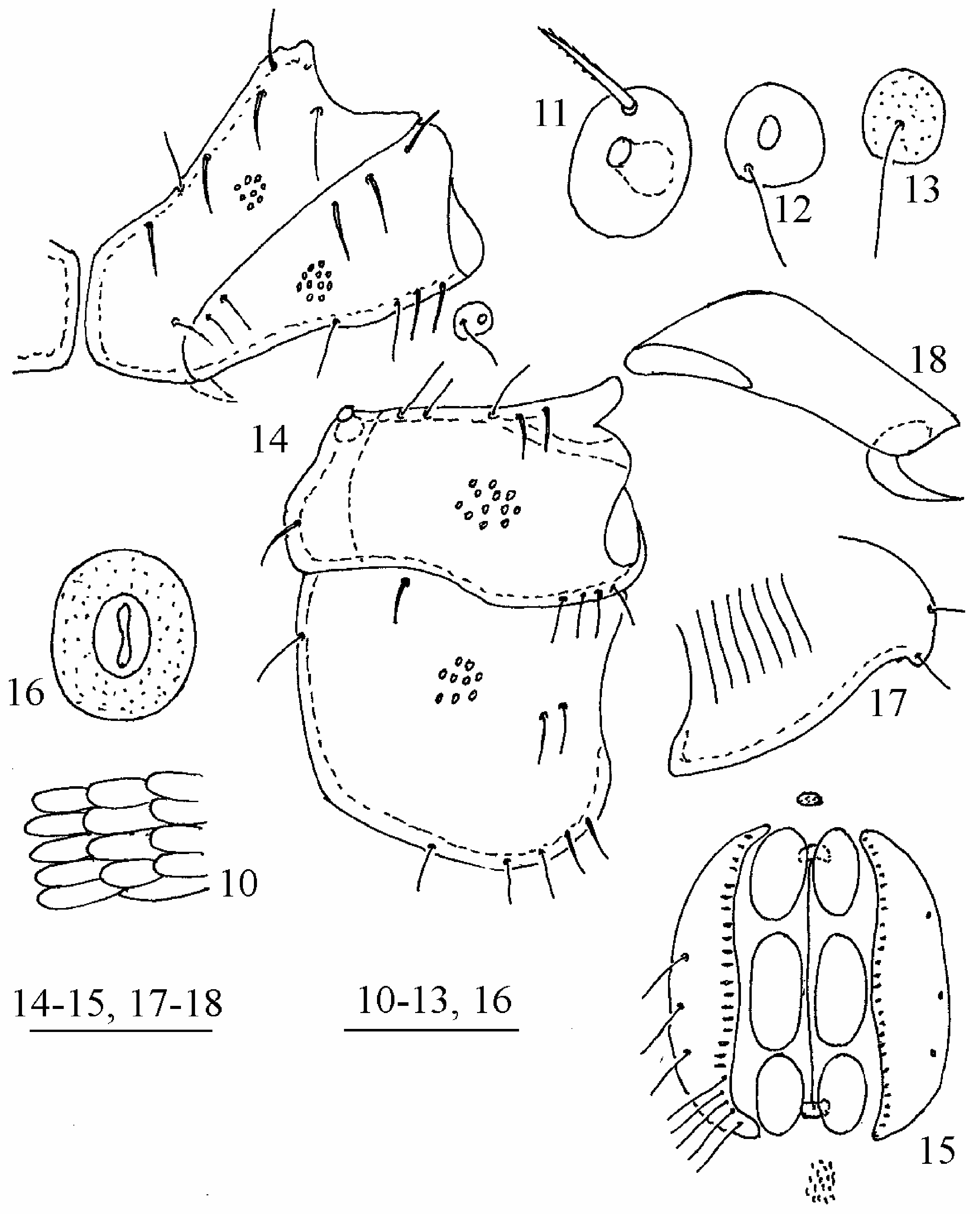
- P-4 longer than P-3, P-2 with thick seta near base of distoventral projection which bearing two fine setae ( Fig. 19 View FIGURES 19–21 ), P-3 ventral margin convex, claw lamella with concave ventral margin ( Fig. 21 View FIGURES 19–21 ), excretory pore sclerotized ( Fig. 16 View FIGURES 10–18 )...... S. smiti sp.n.
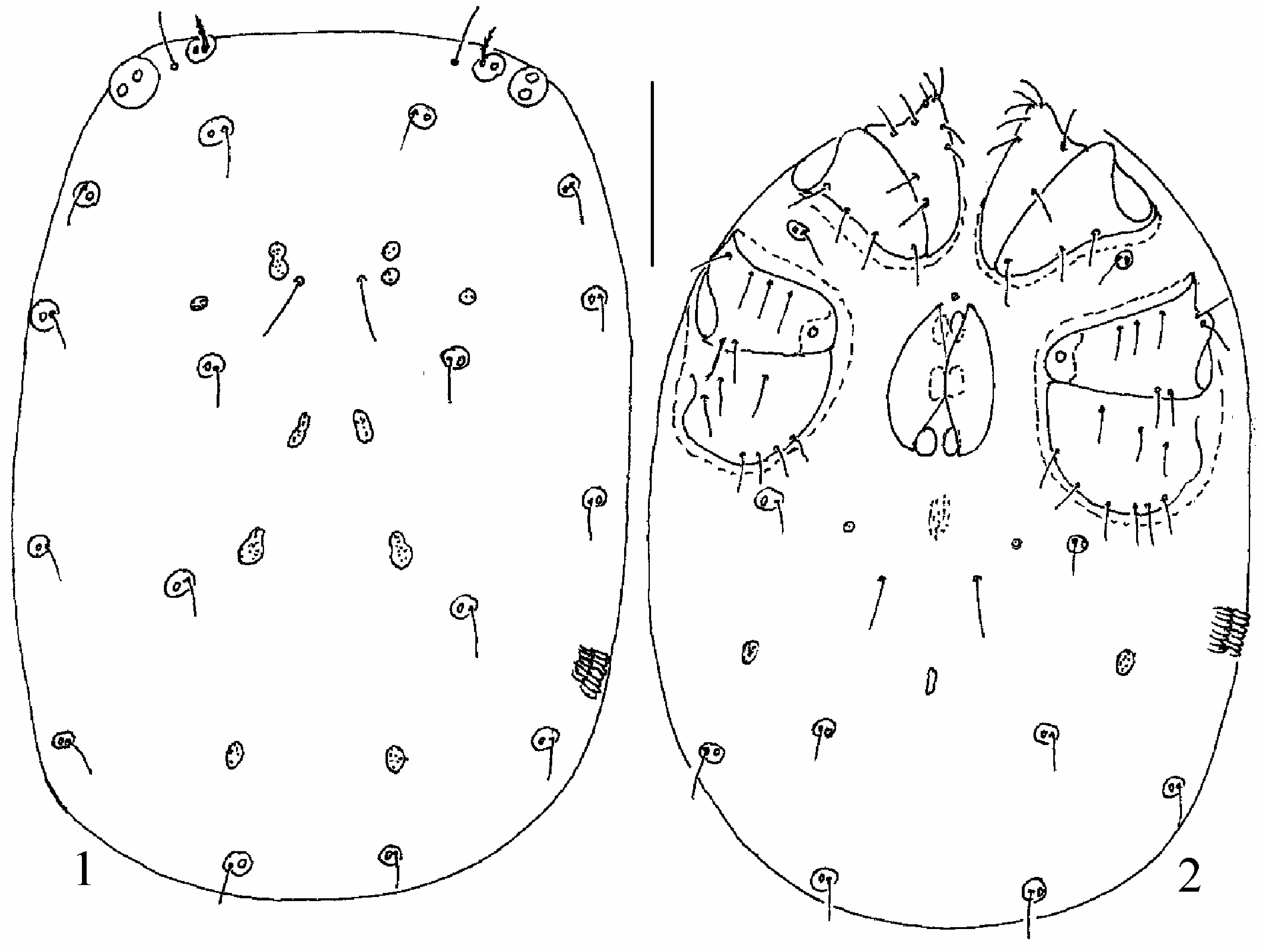
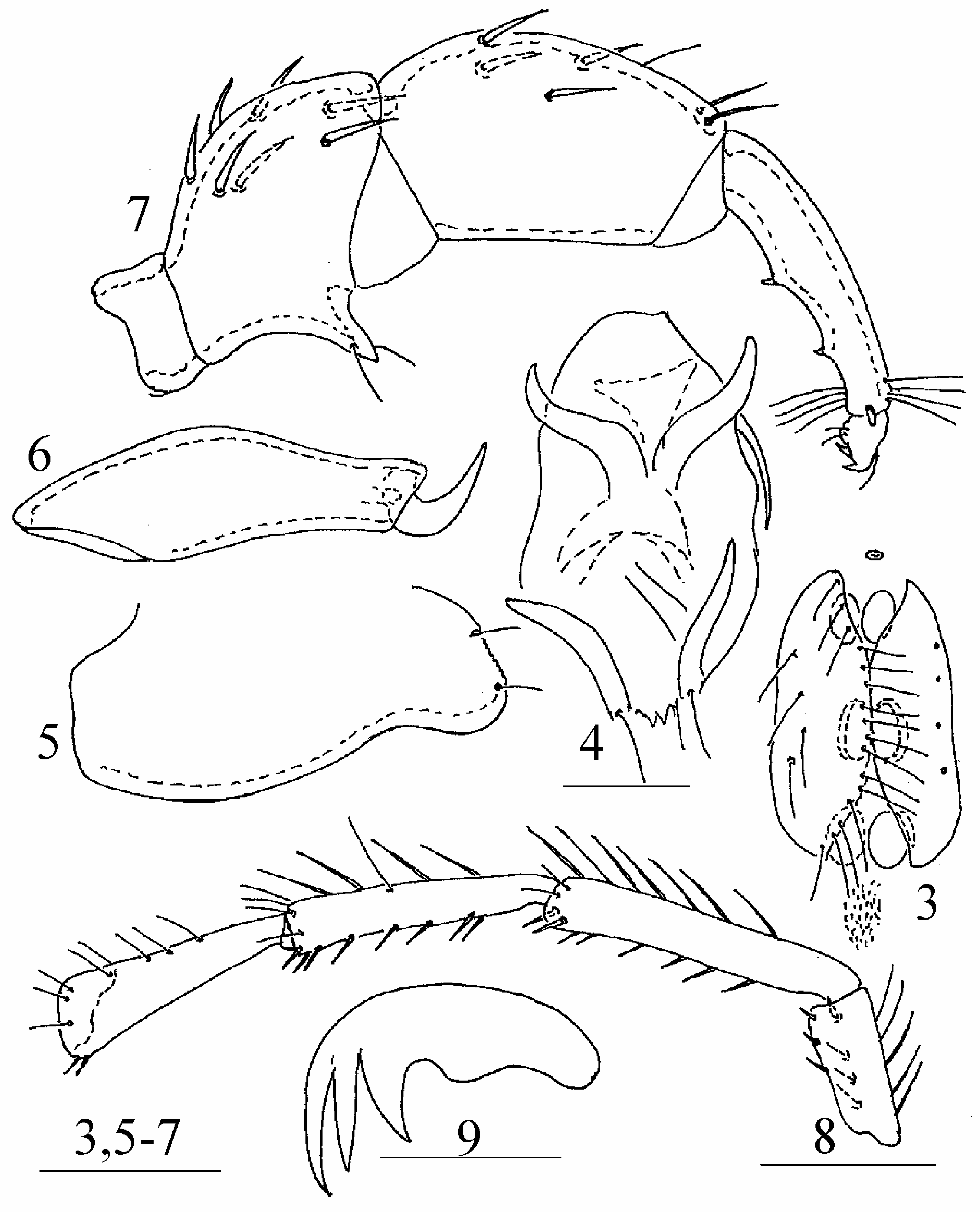
3 All dorsocentral sclerites free and sclerotized ( Fig. 1 View FIGURES 1–2 ), P-2 with rather long ventrodistal projection bearing two fine setae ( Fig. 7 View FIGURES 3–9 ), all genital acetabula subequal in size and distinctly separated ( Fig. 3 View FIGURES 3–9 ), excretory pore ( Fig. 2 View FIGURES 1–2 ) unsclerotized.................................................................................................... S. pesici sp. n.
- Dorsocentral sclerites dc.1 and dc.2 unsclerotized, dc.3 fused with glandulartia Sci ( Fig. 24 View FIGURES 22–26 ), P-2 distoventral projecting short and without fine setae, with single thick seta near base of projection ( Fig. 26 View FIGURES 22–26 ), ac.2 larger than ac.1 and ac.3, all acetabula slightly separated, excretory pore ( Fig. 25 View FIGURES 22–26 ) sclerotized ( Tuzovskij 2003)................................... S. minor
No known copyright restrictions apply. See Agosti, D., Egloff, W., 2009. Taxonomic information exchange and copyright: the Plazi approach. BMC Research Notes 2009, 2:53 for further explanation.
|
Kingdom |
|
|
Phylum |
|
|
Class |
|
|
Order |
|
|
Family |
|
|
Genus |