
Triconia derivata ( Heron & Bradford-Grieve, 1995 )
|
publication ID |
https://doi.org/ 10.11646/zootaxa.4286.3.3 |
|
publication LSID |
lsid:zoobank.org:pub:87BCC981-D033-43FF-A961-63EC3EE11F0B |
|
DOI |
https://doi.org/10.5281/zenodo.6015132 |
|
persistent identifier |
https://treatment.plazi.org/id/1B2C7A05-FFD3-FFCE-DAA0-69CBFA376C6E |
|
treatment provided by |
Plazi |
|
scientific name |
Triconia derivata ( Heron & Bradford-Grieve, 1995 ) |
| status |
|
Triconia derivata ( Heron & Bradford-Grieve, 1995)
( Figs. 1–4 View FIGURE 1 View FIGURE 2 View FIGURE 3 View FIGURE 4 , 9 View FIGURE 9 A, B, C)
Synonymy. Oncaea conifera Moulton, 1973 , ("bumped" only) p. 142, 145, 147, 148, 150–154, Figs. 4 View FIGURE 4 Ac, g, k, 4Bo, s, w (female).
Oncaea derivata Heron & Bradford-Grieve, 1995 , p. 25, 29 (female), Figs. 9 View FIGURE 9 h–j, 10a–l, 11a, 25c (female). Oncaea derivata Heron & Frost, 2000 , p. 1021, 1028–1032, Figs. 1 View FIGURE 1 C (P2–P4 endopod terminal spine set in female), 8F–I (male).
Type locality. Southwest Pacific , off New Zealand, 36°18.5'S, 165°05.5'E GoogleMaps
Material examined. Four females ( MABIK CR000235312 – CR000235315) dissected on ten slides. Two males ( MABIK CR000235316 – CR000235317) dissected on ten slides. All specimens collected from NE equatorial Pacific, station BN 09-02-01 (10°30'N, 131°20'W) in August 2009 by D.J. Ham.
Description of female. Body length (measured in dorsal view): 1160-1244 µm, based on 4 specimens (illustrated female: 1244 µm).
Exoskeleton moderately chitinized. Prosome about 1.9 times length of urosome excluding caudal rami, about 1.7 times urosome length including caudal rami. P2-bearing somite with dorsoposterior projection viewed in lateral aspect ( Fig. 1 View FIGURE 1 B). Numerous integumental pores and sensilla on prosome ( Fig. 1 View FIGURE 1 A, B). Pleural areas of P4-bearing somite with pointed posterolateral corners in dorsal view.
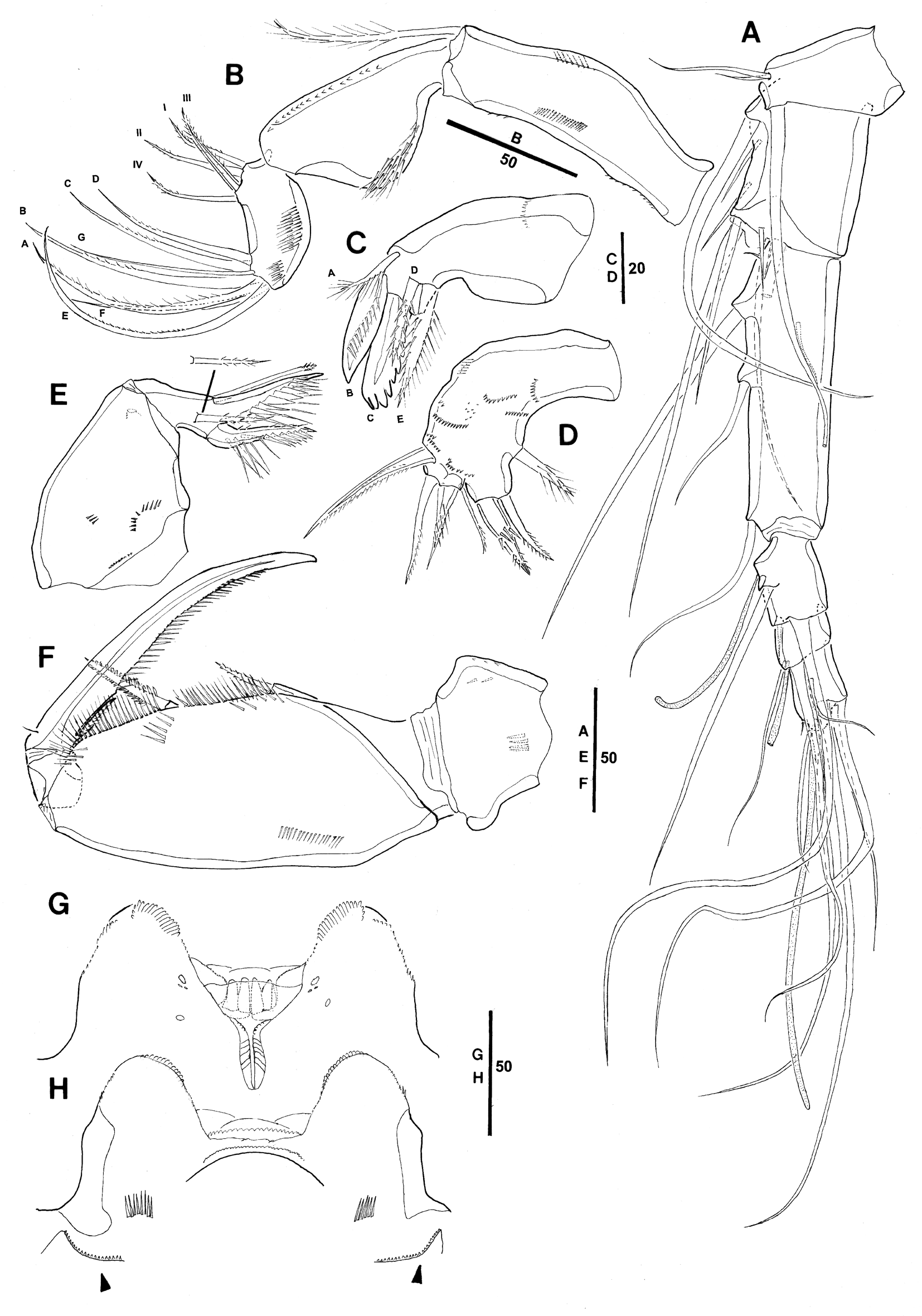
Genital double-somite ( Fig. 1 View FIGURE 1 C) elongate flask-shaped; about 1.95 times as long as maximum width (measured in dorsal aspect) and 2.1 times as long as postgenital somites combined; largest width measured at about 1/4 the distance between anterior and posterior, posterior part tapering gradually. Paired genital apertures located at about 1/3 the distance from anterior margin of genital double-somite. Pore pattern on dorsal surface as indicated in Fig. 1 View FIGURE 1 C.
Anal somite slightly wider (about 1.4) than long; about as long as caudal rami ( Fig. 1 View FIGURE 1 C). Caudal rami ( Fig. 1 View FIGURE 1 C) 2.3 times as long as wide; caudal seta VI about 3/4 length of seta IV, seta VII longer than seta III, plumose and biarticulate at base. Inner margin of CR with fringe of long, fine spinules.
Antennule ( Fig. 2 View FIGURE 2 A) six-segmented. Armature formula: 1-[3], 2-[8], 3-[5], 4-[3+ae], 5-[2+ae], 6-[6+(1+ae)].
Antenna ( Fig. 2 View FIGURE 2 B) three-segmented, distinctly reflexed. Coxobasis with row of long, fine spinules along outer and inner margins and with few additional denticles on proximal and distal part of outer margin. Endopod segments unequal in length; proximal endopod segment subtriangular forming outer lobate outgrowth bearing spinular patch, with row of denticles along posterior inner margin. Distal endopod segment distinctly shorter than proximal endopod segment, with narrow cylindrical base and two patches of short spinules along outer margin; lateral (proximal) armature with four elements: 1 bipinnate spiniform seta (III) and 3 curved setae, setae I and IV sparsely pinnate; distal armature consisting of 7 elements: 1 long curved unipinnate seta (E), curved setae A and B similar in length, length decreasing from seta C to D, all elements unipinnate, slender bare setae F and G nearly equal in length, slightly shorter than seta D.
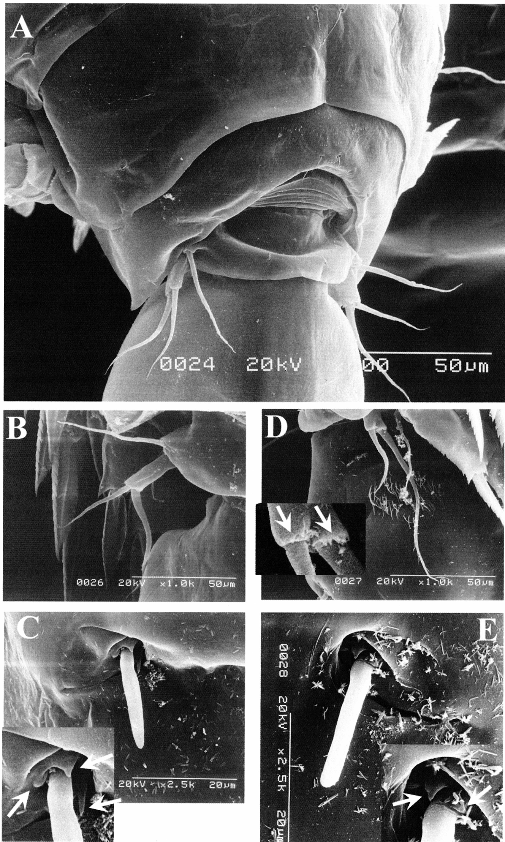
Labrum ( Fig. 2 View FIGURE 2 G, H) distinctly bilobed. Each lobe with row of minute denticles around outer ventral margin and dentiform processes converging and decreasing in size medially. Lobes separated by medial concavity covered anteriorly by several transverse hyaline lamellae, which are undulating. Posterior wall of medial concavity ornamented with four long, sclerotized, dentiform processes (“teeth”). Anterior surface ( Fig. 2 View FIGURE 2 H) with paired row of long setules either side of median swelling and paired integumental pockets latero-posteriorly (arrowed in Fig. 2 View FIGURE 2 H), free margin of pockets surrounded by minute denticles [the three-dimensional shape of these pockets is difficult to discern and illustrate under a light microscope, but has been shown in detail for other oncaeid species, such as Oncaea venusta , by scanning electron microscopy (cf. Böttger-Schnack 2001, fig. 9A, C, D]; large secretory pore posterior to median swelling (not figured). Posterior surface ( Fig. 2 View FIGURE 2 G) with group of three secretory pores located on proximal part of each lobe and an additional one on midregion.
Mandible ( Fig. 2 View FIGURE 2 C) with surface of coxa ornamented with row of spinules. Gnathobase with 5 elements, numbered using capital letters in Fig. 2 View FIGURE 2 C: element A subdistal ventral corner much shorter than ventral blade B, with long setules along dorsal margin; ventral blade B strong and broad, with row of setules on posterior surface; dorsal blade (C) strong and broad, with dentiform processes around distal margin and along distal two-thirds or half of dorsal margin; dorsal elements setiform, the shorter spinulose (D), the longer multipinnate (E).
Maxillule ( Fig. 2 View FIGURE 2 D) indistinctly bilobed, with numerous spinules on anterior and posterior surfaces. Inner lobe subcylindrical, with 3 elements; outermost one spiniform, swollen at base, fringed with coarse spinules, other two setiform and bipinnate; innermost one multipinnate and located along concave inner margin at some distance from other elements. Outer lobe with 4 elements; 2 outermost elements setiform and pectinate, longer than other 2 elements, which are pectinate or with sparse spinules; innermost element shortest.
Maxilla ( Fig. 2 View FIGURE 2 E) with surface of syncoxa ornamented with spinule rows and one large secretory pore. Allobasis produced distally into slightly curved claw bearing 2 rows of very strong spinules along medial margin; outer margin with strong seta extending almost to tip of allobasal claw, ornamented with few strong spinules distally, tip of seta with tubular extension; inner margin with slender pinnate seta (figured separately in Fig. 2 View FIGURE 2 E) and strong basally swollen spine, which is ornamented with single row of shorter spinules along outer margin in addition to double row of very strong spinules along the inner margin.
Maxilliped ( Fig. 2 View FIGURE 2 F) with surface of syncoxa ornamented with few spinules. Basis robust, inner margin with 2 spiniform spinulose setae, distal seta 1.5 times longer and stouter than proximal one; fringe of long pinnules between distal seta and articulation with endopod, row of long spinules between proximal and distal seta; few short transverse rows of setules on anterior surface and additional longitudinal row near outer margin. Proximal endopod segment unarmed. Distal endopod segment (claw) with row of pinnules on proximal 5/6 of concave margin; accessory armature consisting of minute, naked seta on outer proximal margin and unipectinate spine fused basally to inner proximal corner of claw.
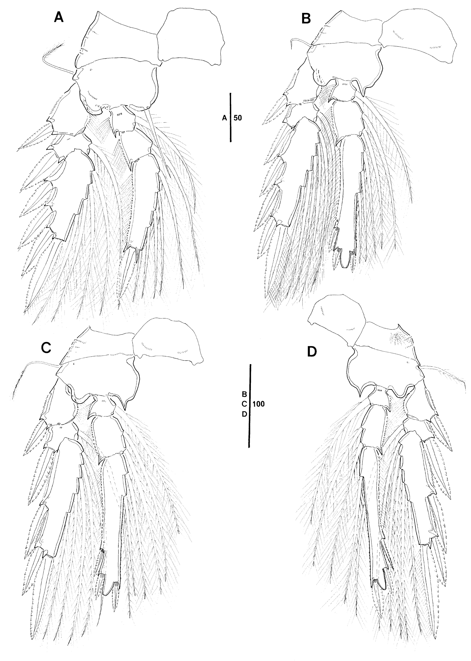
Swimming legs 1–4 biramous ( Fig. 3 View FIGURE 3 A–D), with 3-segmented rami. Intercoxal sclerites well developed, posterior face ornamented with paired row of denticles near distal corner in P2–P4. Coxae and bases of P1–P4 with surface ornamentation as in Fig. 3 View FIGURE 3 A–D. Coxae of P1–P4 with raised secretory pore on posterior face near outer distal corner. Bases with short naked (P2) or plumose (P1, P3, P4) outer seta; with anterior secretory pore near outer proximal corner; inner portion slightly produced medially in P2–P4. Coxa of P4 with tuft of long fine setules posteriorly at outer posterior surface. Inner basal seta on P1 spiniform and naked. Respective legs without distinct length differences between exopod and endopod (P1) or with endopod slightly longer than exopod (P2–P4). Bases of spines on exopod and endopod segments anteriorly surrounded by small spinules.
Leg armature formula (Roman numerals indicate spines, Arabic numerals indicate setae):
Exopods. Outer margin of exopod segments with well-developed serrated hyaline lamella; inner margin of proximal exopod segments with long setules. Secretory pore located on posterior surface of distal segments. Hyaline lamellae on outer spines well developed; outer and distal spines of P1 with subapical tubular extension; this extension lacking on proximalmost spine of P1 exp-3. Distal spine shorter than distal exopod segment in all legs.
Endopods. Outer margin of endopod segments with fringe of long setules. Distal endopod segments with single secretory pores on posterior surface; distal margin of P2–P4 produced into conical process, process with apical pore. Distal (inner) spine reaching only slightly beyond tip of conical process in P2, clearly reaching beyond the tip in P3 and P4. Length data of endopodal spines of four females as shown in Table 1; length ranges of outer subdistal spine (OSDS) and outer distal spine (ODS) relative to distal spine (DS) are given in Table 2.
P5 ( Figs. 1 View FIGURE 1 C, D, 9B) comprising long, free exopod segment and plumose outer basal seta, which is about same length as inner exopodal seta. Exopod about 2.3 times longer than wide, bearing 2 naked, spiniform setae, inner seta 1.7 times longer than outer seta.
P6 ( Figs. 1 View FIGURE 1 C, 9C) represented by operculum closing off each genital aperture; armed with long spine and three minute processes (arrowed in inset of Fig. 9 View FIGURE 9 C).
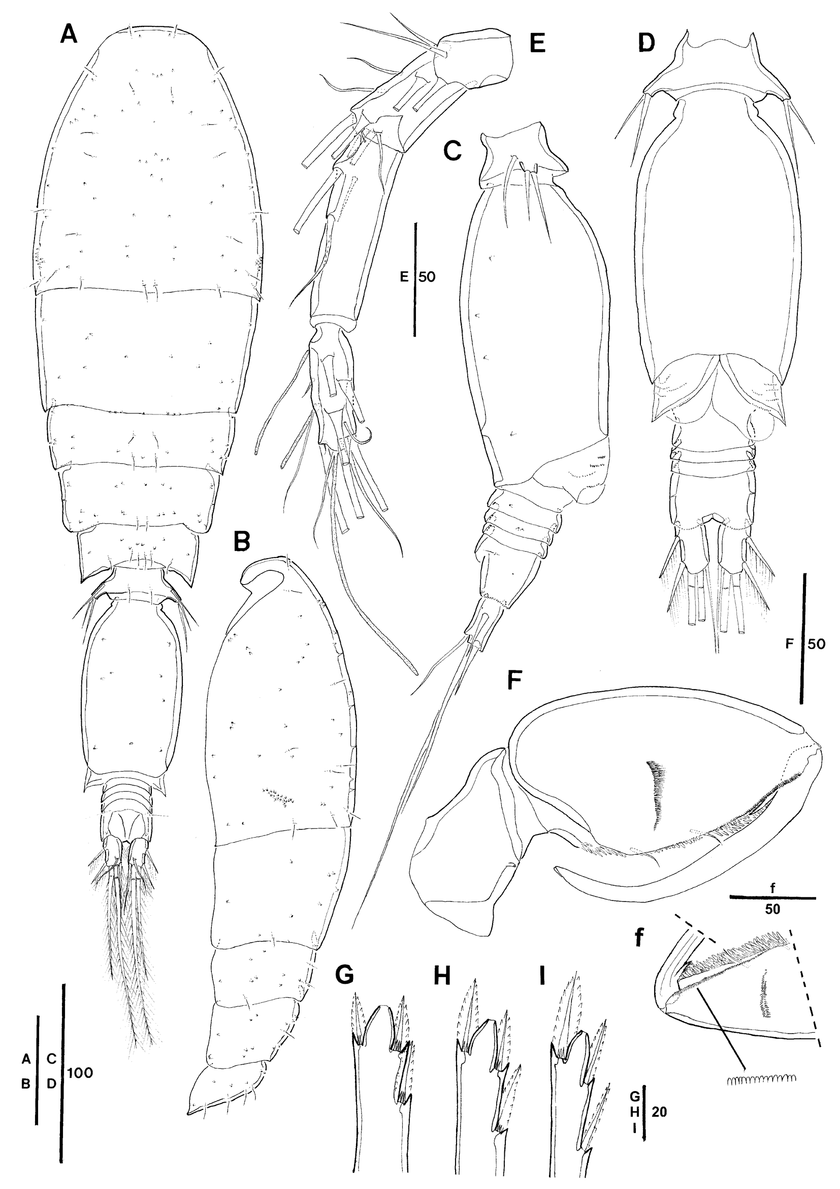
Description of male. Body length: 768-791 µm, based on 2 specimens (illustrated male: 768 µm). Sexual dimorphism in antennule, maxilliped, P5, P6, genital segmentation and CR; slight dimorphism in endopodal spine lengths on P2.
Prosome 1.9 times length of urosome, excluding caudal rami, 1.7 times urosome length, including caudal rami ( Fig. 4 View FIGURE 4 A). Integumental pores on prosome and urosome as figured ( Fig. 4 View FIGURE 4 A). Cephalosome with conspicuous posterolateral pattern of about 20 hooded pores ( Fig. 4 View FIGURE 4 B).
Female Male
spec 1 spec 2 spec 3 spec 4 spec 1 spec 2 spec 3 P2 OSDS 27.9 31.3 31.6 31.3 19.9 21.0 20.0 ODS 27.9 30.1 32.1 30.5 18.8 19.9 18.4 DS 14.0 17.6 17.3 16.2 11.8 11.8 12.5 P3 OSDS 33.8 33.1 33.5 32.7 21.3 22.4 20.2 ODS 31.6 32.0 33.1 33.8 21.3 22.1 18.8 DS 20.2 23.9 23.9 22.8 15.4 15.8 14.7 P4 OSDS 35.3 36.8 dmgd 39.3 23.5 26.8 24.8 ODS 33.8 33.8 39.7 23.0 24.6 21.3 DS 39.4 37.9 38.6 25.7 25.4 22.8
continued.
Female Male
spec 1 spec 2 spec 3 spec 4 spec 1 spec 2 P2 OSDS 34.9 32.4 34.3 31.3 22.1 20.6 ODS 29.4 28.7 32.7 28.7 19.9 18.4 DS 27.0 25.7 27.6 23.9 19.1 17.3 P3 OSDS 36.8 34.9 37.5 32.4 25.0 22.1 ODS 33.8 33.8 35.7 29.8 21.3 21.3 DS 37.1 35.3 36.8 34.9 26.1 22.1 P4 OSDS 36.8 39.3 47.4 38.6 25.7 22.1 ODS 35.7 35.7 39.0 34.2 24.1 21.3 DS 50.7 49.3 57.7 50.0 32.7 29.0 Length to width ratio of genital somite 1.7:1. Caudal rami ( Fig. 4 View FIGURE 4 C, D) relatively shorter than in female, about 1.4 times longer than wide. Surface of genital flaps ornamented with several rows of small spinules. Anal somite 1.7 times wider than long, relatively wider than that of female.
Antennule ( Fig. 4 View FIGURE 4 E) with distal segment corresponding to fused segments 4–6 of female, including minute element. Armature formula 1-[3], 2-[8], 3-[4], 4-[11+2ae+(1+ae)].
Maxilliped ( Fig. 4 View FIGURE 4 F) 3-segmented, comprising syncoxa, basis and 1-segmented endopod. Syncoxa without surface ornamentation, with single secretory pore at inner distal margin. Basis robust, particularly inflated in proximal half forming bulbous swelling; anterior surface with 1-2 transverse spinular rows and small flat spinules (with rounded tips) along distal part of inner margin (figured separately in Fig. 4 View FIGURE 4 f); posterior surface with rows of spatulate setules of graduated length along palmar margin; with 2 small naked setae of equal length inserted within longitudinal cleft. Endopod drawn out into long curved claw, concave margin unornamented; accessory armature consisting of short, unipectinate spine basally fused to inner proximal corner of claw; claw with minute hyaline apex ( Fig. 4 View FIGURE 4 F).
Swimming legs 1–4 ( Fig. 4 View FIGURE 4 G–I, only P2–P4 enp-3) with armature and ornamentation as in female, length data of endopodal spines of two males as shown in Table 1; length ranges of outer subdistal spine (OSDS) and outer distal spine (ODS) relative to distal spine (DS) are given in Table 2. Relative spine lengths of OSDS and ODS on P2 slightly shorter than that of female ( Table 2).
Triconia hirsuta Triconia derivata
NW Pacific1) Equatorial Pacific SW Pacific2) NE Pacific3) Equatorial Pacific NE Pacific3) Equatorial Pacific Female Female Male Female Female Female Male Male
size range (mm) 0.95-1.04 1.07-1.16 0.72 1.11-1.20 1.05–1.15 1.16–1.24 0.67–0.74 0.77–0.79
genital (double-)somite L:W ratio 1.73 1.63 1.61 1.83 1.95 1.62 1.72 (dorsal view)
L ratio spines OSDS:DS 1.58–1.78** 1.77–2.00* 1.60–1.78* 1.22 1.35 1.25–1.31* 1.15/1.19* P2 enp-3 ODS:DS 1.68–1.79** 1.71–2.00* 1.47–1.69* 1.22 1.15 1.09–1.20* 1.04/1.06* L ratio spines OSDS:DS 1.27–1.47** 1.38–1.67* 1.38–1.42* 0.93 1.05 0.93–1.02* 0.96/1.00* P3 enp-3 ODS:DS 1.32–1.38** 1.34–1.56* 1.28–1.40* 0.93 0.87 0.85–0.97* 0.82/0.97* L ratio spines OSDS:DS 0.91–1.04** 0.90–1.02* 0.91–1.09* 0.83 0.80 0.73–0.82* 0.76/0.79* P4 enp-3 ODS:DS 0.86–0.91** 0.86–1.03* 0.89–0.97* 0.67 0.65 0.68–0.72* 0.73/0.74* L ratio CR: AS 0.88 0.81 0.88 0.81 0.94 0.88 0.75 Wi et al. 2010; 2) Heron & Bradford-Grieve 1995; 3) Heron & Frost 2000
= for number of specimens measured see Table 1
= values from reexamination of type material (see under “Remarks” of T. hirsuta )
P5 ( Figs. 4 View FIGURE 4 A, C, 9A) exopod not delimited from somite, shorter than that of female, outer basal seta about as long as inner exopodal seta; length ratio between inner and outer exopodal setae smaller (1.5:1) than that of female.
P6 ( Fig. 4 View FIGURE 4 D) represented by posterolateral flap closing off genital aperture on either side; covered by pattern of spinules; posterolateral corners protruding laterally and discernible in dorsal aspect ( Fig. 4 View FIGURE 4 A).
Remarks. The morphology of female T. derivata from the tropical Pacific agrees in most parts with the original description of the species by Heron & Bradford-Grieve (1995). Yet the present specimens appear to differ in a few morphometric characters, which seem to be more similar to its sibling T. furcula (Farran, 1936) as redescribed in detail by Heron & Bradford-Grieve (1995). The aforementioned authors separated co-occurring females of T. derivata and T. furcula in New Zealand waters by a number of slightly different morphometric characters, such as (1) the size of the dorsal “hump” of the P2-bearing somite, (2) the length of the genital double-somite relative to the total length of three posterior segments of the prosome, (3) the length to width ratio of the anal somite, (4) the length of the anal somite relative to the length of the CR, (5) the length of the outer basal seta on P5, and also by (6) the presence or absence of a sclerotised ridge on the prosome near the apex in lateral view (Heron & Bradford- Grieve 1995, p. 24, key p. 14). Our female specimens from the tropical Pacific are similar to T. derivata from the New Zealand waters in the first two characters, but appear to be more similar to T. furcula in characters (3) and (4). Some characters of the females from our study were intermediate between the two species (character 5) or were not clearly discernible (character 6). Moreover, the form of the genital double-somite in dorsal view had a more tapering posterior part than that figured by Heron & Bradford-Grieve for T. derivata (their figs. 9i, 10b), suggesting some similarity to T. furcula (their fig. 8c & d). Slight morphometric differences were also found in the proportional lengths of the innermost element on the outer lobe of the maxillule, which is slightly shorter in our specimens than that figured by Heron & Bradford-Grieve for either T. derivata (their fig. 10g) or T. furcula (their fig. 8i).
Support for a positive species identification of T. derivata in our study was given by the observed differences in endopodal spine lengths between the two sibling species, which were not mentioned in Heron & Bradford- Grieve´s study. The proportional lengths of endopodal spines on P4 reported for T. derivata from the New Zealand waters (calculated from Heron & Bradford-Grieve 1995, fig. 11a) and the northeastern Pacific (calculated from Heron & Frost 2000, fig. 1C) both fall into the range of variation examined in our specimens from the equatorial Pacific ( Table 2). The spine lengths on P4 enp-3 of T. furcula are different, with the outer subdistal spine (OSDS) and outer distal spine (ODS) being similar in length to the distal spine (DS) (cf. Heron & Bradford-Grieve 1995, fig. 9c), while in T. derivata both spines are shorter than the distal spine ( Table 2). The proportional spine lengths on the endopods of P2 and P3 are not informative in this respect, because they are similar for both species in question and fall in the range of variation indicated for our specimens from the equatorial Pacific. More individuals from different locations need to be studied for intraspecific variation in morphometric characters in order to define the interspecific differences between T. derivata and related species more clearly.
The present study includes additional morphological characters not described, mentioned or figured in earlier descriptions of T. derivata . Most notably, a complete description of the labrum is provided, including the number of blunt “teeth” on the posterior wall of the medial concavity in T. derivata ( Fig. 2 View FIGURE 2 G), as well as the paired integumental concavities (“pockets”) on the anterior face (arrowed in Fig. 2 View FIGURE 2 H). Both characters are of significance for the classification of the genus Triconia within the family Oncaeidae , and support the morphological definition of the conifera -subgroup ( Böttger-Schnack & Schnack 2013, table 3). Other additional micro-structural characters described for T. derivata in the present account include ornamentation details of the female antenna, as well as those on the intercoxal sclerite(s) of P2–P4 (posterior view), which were not provided by Heron & Bradford-Grieve (1995), but might be of importance for species identification in the future.
The male of T. derivata was first described by Heron & Frost (2000) based on specimens collected in coastal waters at high latitudes in the NE Pacific (Juan de Fuca Strait, Washington DC). They were seemingly identified by the proportional lengths of endopodal spines on P2–P4, which were described as “being similar to the female”, but not figured for the male. In the present account, we also used this character to assist in preliminary identification of the males of T. derivata , although the proportional spine lengths of OSDS and ODS on the endopod of P2 were relatively shorter than that of the female, which was not noted by Heron & Frost (2000). Heron & Frost´s figure of the male (their fig. 8F-I) also showed minor morphometric differences as compared to specimens from our study: (1) the width to length ratio of the anal somite is smaller in the equatorial Pacific (1.7 times wider than long), compared to the northeastern Pacific (about 2.3 times wider than long), and (2) the length of the inner exopodal seta on P5 of our specimens was relatively shorter (only 1.5 times longer than outer seta) compared to the northeastern Pacific, where the inner seta was about 2 times longer than the outer one. However, the length ratios of the outer basal seta to the inner exopodal seta on P5 of male specimens are similar for both areas (about 1.1 times, calculated from Table 2).
All females of T. derivata , originally described from the southwestern Pacific Ocean had various sizes of a tumorous growth on the mid-dorsal surface of the cephalosome ( Heron & Bradford-Grieve 1995, their fig. 9h), an abnormality which had also been recorded by Moulton (1973, “bumped form”) for about half of the specimens collected in the Indian Ocean, while it was absent in the other half of the individuals (cf. Moulton 1973, his table 5). Heron & Bradford-Grieve (1995) noted that the “bump” was absent on specimens from the Panama Basin (their fig. 9j) and was found only in 2 out of 3 specimens examined in the western Atlantic (Florida Strait); the same authors recorded T. derivata also from the western equatorial Atlantic (off Liberia, sample 10- 0-20 in their table 4) and from the eastern equatorial Pacific (sample 1 and sample 2 in their table 4), which is close to the sampling area of the present study, but gave no information about the presence or absence of a “bump” in these specimens. In the present study from the northeastern equatorial Pacific, none of the females of T. derivata examined had a tumorous growth on the cephalosome, and it was also absent in females collected in the northeastern Pacific (inland waters of Washington DC; Heron & Frost 2000). Thus it may be concluded that the “bump” only irregularly occurs in the species and cannot be used for separating it from its congeners during routine counts.
The records of T. derivata to date are summarized in Fig. 11 View FIGURE 11 , indicating a wide zoogeographical distribution.

No known copyright restrictions apply. See Agosti, D., Egloff, W., 2009. Taxonomic information exchange and copyright: the Plazi approach. BMC Research Notes 2009, 2:53 for further explanation.
|
Kingdom |
|
|
Phylum |
|
|
Class |
|
|
Order |
|
|
Family |
|
|
Genus |
Triconia derivata ( Heron & Bradford-Grieve, 1995 )
| Cho, Kyuhee, Böttger-Schnack, Ruth, Kim, Woong-Seo & Lee, Wonchoel 2017 |
Oncaea derivata
| Heron & Frost 2000 |
Oncaea derivata
| Heron & Bradford-Grieve 1995 |