
Metarhabditis giennensis, Abolafia & Peña-Santiago, 2019
|
publication ID |
https://doi.org/ 10.11646/zootaxa.4652.1.8 |
|
publication LSID |
lsid:zoobank.org:pub:D90C7864-D755-4E42-8370-6EFBF4DECB6A |
|
DOI |
https://doi.org/10.5281/zenodo.5673940 |
|
persistent identifier |
https://treatment.plazi.org/id/7EBAC9DB-A148-4A1A-97E5-3B796D0839BF |
|
taxon LSID |
lsid:zoobank.org:act:7EBAC9DB-A148-4A1A-97E5-3B796D0839BF |
|
treatment provided by |
Plazi |
|
scientific name |
Metarhabditis giennensis |
| status |
sp. nov. |
Metarhabditis giennensis View in CoL sp. n.
( Figs 1–3 View FIGURE 1 View FIGURE 2 View FIGURE 3 )
Material examined. Two females and eleven males, generally in acceptable condition.
Measurements. See Table 1 View TABLE 1 .
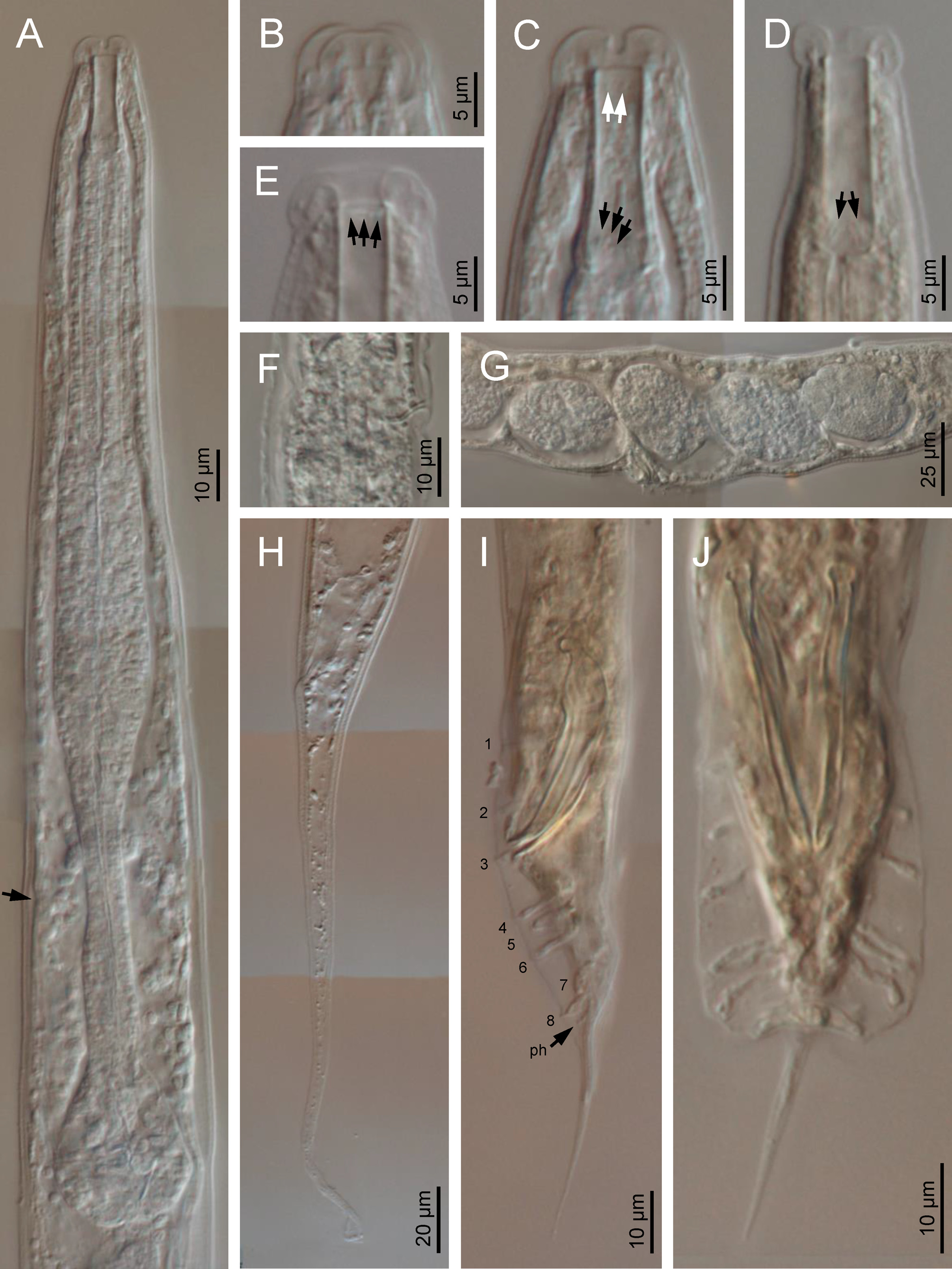
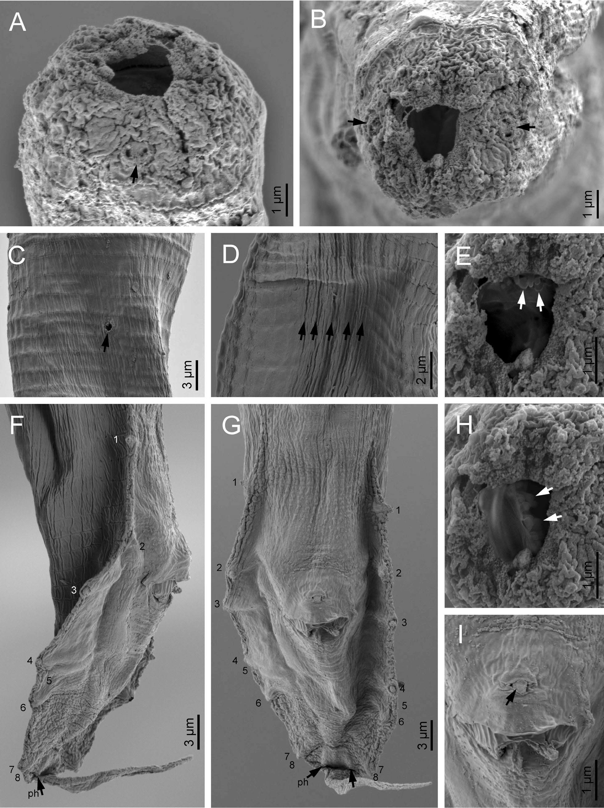
Description. Adult: Moderately slender to slender (a = 22–37) nematodes of small to medium size, body 0.77– 1.16 mm long. Upon fixation, habitus nearly straight or somewhat curved ventrad. Cuticle 2–3 µm thick, bearing fine but perceptible transverse striation. Lateral fields barely discernible under LM, occupying ca one-tenth (11%) of mid-body diameter. Lip region offset from the adjacent body by an appreciable depression or weak constriction: lips swollen and rounded, grouped in pairs, with acute labial and cephalic sensilla; primary axils deep, U-shaped; secondary axils shallow. Amphids very small, oval, located at the base of lateral lips. Stoma rhabditoid, 1.6–2.3 times the lip region diameter long and 5.0–7.0 times longer than wide: cheilostom with barely refringent rhabdia, slightly shorter than lip height; gymno-promesostegostom (buccal tube) tubular, straight, occasionally somewhat collapsed at mid-length after fixation, with gymnostomatal rhabdia slightly more refringent at its anterior end; gymnostom bearing minute denticles anteriorly; metastegostom with glottoid apparatus bearing minute denticles, three per valve, two of them longer; telostegostom with minute rounded rhabdia. Pharynx also rhabditoid: pharyngeal corpus subcylindrical, 1.6–2.2 times the isthmus length, with metacorpus slightly swollen, tubular or somewhat fusiform; isthmus comparatively thick, weakly narrowing until its junction with the bulb; basal bulb pyriform, with both valvular apparatus and posterior haustrulum well developed. Cardia very small, surrounded by intestinal tissue. Nerve ring situated at 66–79% of neck length, at level of isthmus. Excretory pore at 66–77% of neck length, also at level of isthmus, adjacent to the hemizonid. Deirids poorly visible, posterior to excretory pore, at 71–76% of neck length, at level of isthmus. Intestine without distinct specializations but with slightly thinner walls at cardiac part.
Female: Reproductive system didelphic-amphidelphic, the anterior branch in sinistral position to intestine and the posterior branch in dextral position. Ovaries 95–150 µm long, with a flexure at their distal portion. Oviducts 56–108 µm, tubular and distally differentiated in a spheroid spermatheca having coarse sperm cells inside. Uteri 94–108 µm long, 2.1–2.8 times as long as the corresponding body diameter, tubular, with abundant uterine eggs. Vagina short, 9–14 µm long, extending inwards 23–30% of body diameter. Vulva slightly protruding. Rectum short, 1.2–1.7 times the anal body width (ABW); three large gland-like cells are distinguishable around the intestinerectum junction. Tail conical-elongate with acute terminus, 8.2–11.1 times the rectum length. Phasmids located at 12–13% of tail length.
Male: Reproductive system monorchic, with testis reflexed ventrad anteriorly. Spicules paired and symmetrical: manubrium rounded, well developed and ventrad bent, short conoid calamus, and slightly ventrally curved lamina with ventrally bent acute tip in lateral view. Gubernaculum well developed, slightly curved, about one-half of the spicule length, with thin corpus. Three small gland-like cells are distinguishable around the anterior end of the cloaca. Bursa leptoderan, on each side bearing eight genital papillae plus a fine phasmid located posterior to the GP 8, at 73% of tail length (1+1+1/3+2+ph). One middle papillae located anterior to the cloacal opening. Tail conical, slightly curved ventrad, ending in an acute, filiform, 15–25 µm long tip extending out of the bursa.
Diagnosis. The new species is characterized by its 1.01–1.16 mm long body in females and 0.77–0.98 in males, cuticle with very fine transverse striation, lip region 9–14 µm broad and consisting of six swollen rounded lips fused in pairs, stoma 14–25 µm long with gymno-promesostegostom tubular, pharynx with metacorpus slightly swollen and isthmus slender, nerve ring, excretory pore and deirids at isthmus level, female reproductive system didelphicamphidelphic, vulva equatorial ( V = 49–50), female rectum 1.2–1.7 times the anal body diameter, female tail conical-elongate with acute tip (123–199 µm, c = 5.8–8.2, c’ = 8.2–11.1), male tail conical (34–56 µm, c = 15.5–25.7, c’ = 2.4–3.5) with a filiform tip, leptoderan bursa, 32–41 µm long spicules with rounded and ventrad bent manubrium, and 17–20 µm long gubernaculum.
Type locality and habitat. Spain, Jaén province , Jaén town, Puente de la Sierra (GPS coordinates: 37°42’36.5”N and 3°45’33.2”W, elevation 439 m), from decaying wood from dead trees ( Populus alba L.) in a riverbank forest between an orchard and the Quiebrajano river GoogleMaps .
Type material. Two females (holotype and paratype) and ten males (paratypes) deposited in the nematode collection of the Departamento de Biología Animal, Biología Vegetal y Ecología, Universidad de Jaén, Spain; one male paratype deposited in the nematode collection of the Swedish Museum of Natural History , Stockholm ( Sweden) .
Relationships. In the morphology of spicules (dorsally humped and rounded and ventrad bent manubrium), the new species resembles M. adenobia ( Poinar, 1971) Sudhaus, 2011 and M. andrassyana Tahseen, Hussain, Tomar, Shah & Jairajpuri, 2004 . Nevertheless, it can be distinguished from M. adenobia by its broader (5–6 vs 2–4 µm) lip region, offset by depression (vs continuous), longer female tail (123–199 vs 93–115 µm), shorter spicules (23–41 vs 40–53 µm) and gubernaculum (17–20 µm vs 20–26 µm), and from M. andrassyana by its more slender female tail (c’ = 8.2–11.1 vs c’ = 5.0–7.9), and male tail with longer filiform part (more than one-half vs less than one-half of the conoid part).
Etymology. The specific name refers to the locality where the species was found.
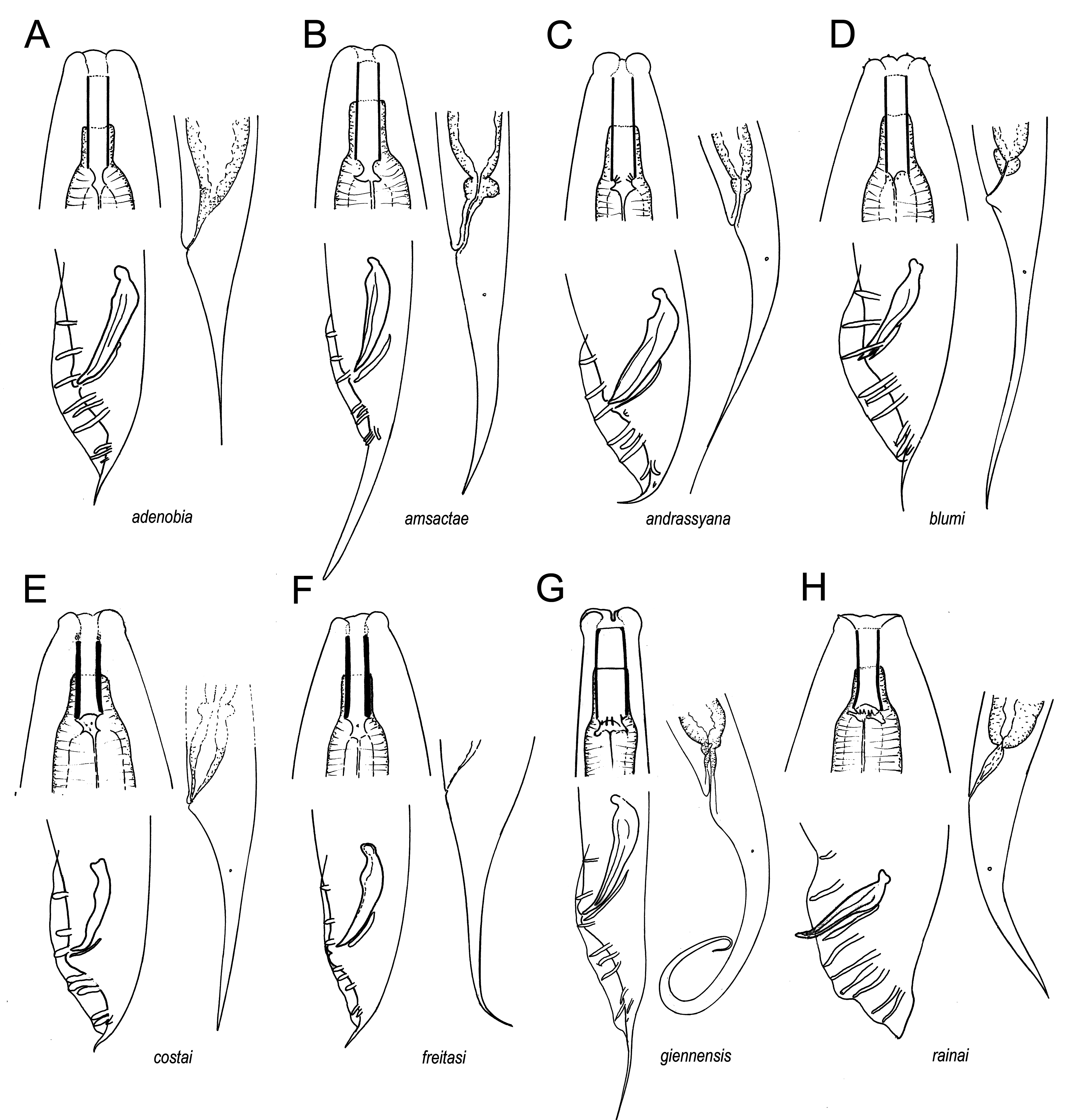
Comments on the genus Metarhabditis . Morphologically, Metarhabditis species form a homogeneous taxon, easily distinguishable from its relatives, and characterized by a combination of diagnostic features, among others those referring lip region morphology, stoma structure, female tail length, male tail shape, and, in particular, the morphology of spicules ( Fig. 4 View FIGURE 4 ). Species separation is often based on small differences (see below the key to their identification), but some appreciable interspecific variation occurs in several characters. Thus, and regarding spicule morphology, four species ( M. blumi , M. costai , M. freitasi and M. rainai ) show kidney-like manubrium, one species ( M. amsactae ) presents small rounded manubrium slightly bent ventrad, and three species ( M. adenobia , M. andrassyana and M. giennensis sp. n.) bear spicules with rounded and bent ventrad manubrium.
The biology of the group also displays relevant and remarkable diversity. Four species ( M. adenobia , M. amsactae , M. blumi and M. rainai ) are associated with insects ( Poinar 1971, Carta & Osbrink 2005, Ali et al. 2011, Shaheen et al. 2011, Park et al. 2012, Pervez et al. 2012, Asif et al. 2013, Tomazini et al. 2013, Khalaf 2018), three ( M. andrassyana , M. blumi and M. costai ) have been found in manure ( Sudhaus 1974, Asif et al. 2 013), and M. giennensis sp. n. is herein recorded in decaying wood. Significantly, three species ( M. blumi , M. costai and M. freitasi ) have been reported in association with external otitis in cattle ( Martins 1985, Bossi et al. 2015, Barbosa et al. 2016, Silva et al. 2016).

| GP |
Instituto de Geociencias, Universidade de Sao Paulo |
| V |
Royal British Columbia Museum - Herbarium |
No known copyright restrictions apply. See Agosti, D., Egloff, W., 2009. Taxonomic information exchange and copyright: the Plazi approach. BMC Research Notes 2009, 2:53 for further explanation.
|
Kingdom |
|
|
Phylum |
|
|
Class |
|
|
Order |
|
|
Family |
|
|
Genus |