
Ribautia combinata, Pereira, Luis A., Uliana, Marco & Minelli, Alessandro, 2006
|
publication ID |
https://doi.org/ 10.5281/zenodo.171440 |
|
DOI |
https://doi.org/10.5281/zenodo.6255015 |
|
persistent identifier |
https://treatment.plazi.org/id/1D5687B5-666E-FFD8-3A5C-BD6FFDE9FE82 |
|
treatment provided by |
Plazi |
|
scientific name |
Ribautia combinata |
| status |
sp. nov. |
Ribautia combinata n. sp.
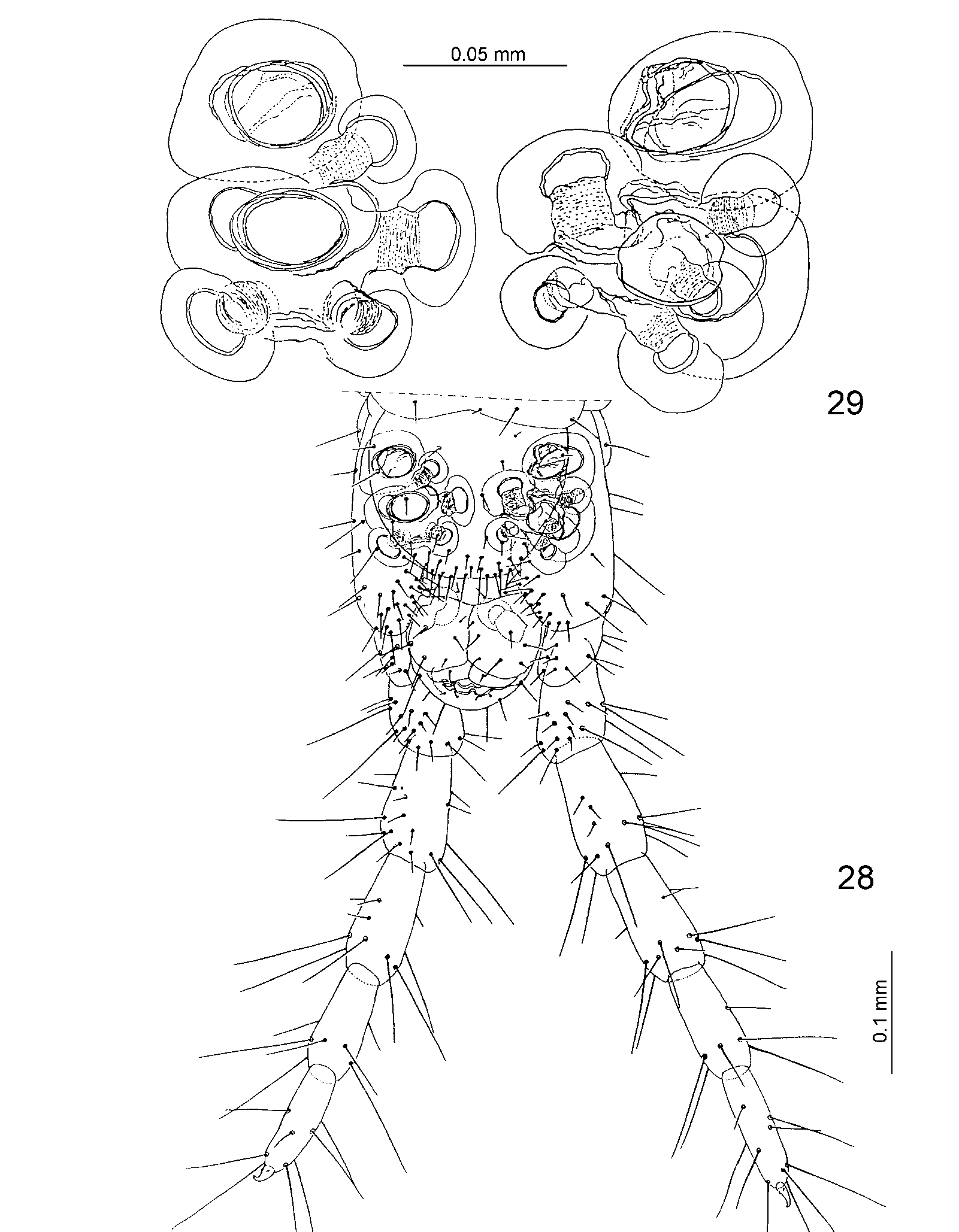
( Figs. 1–29 View FIGURES 1 – 6 View FIGURES 7 – 12 View FIGURES 13 – 17 View FIGURES 18 – 26 View FIGURES 28 – 29 )
Diagnosis: This is the only species of Ribautia having in each coxopleuron an anterior coxal organ with independent opening and 4–5 additional coxal organs grouped in one posterior cluster. Of the other Neotropical species in this genus, those most similar to Ribautia combinata n. sp. are R. limaensis Kraus, 1957 and R. silvana Kraus, 1954 . Differential characters are shown in table 1.
Type material examined: Holotype Ψ, 55 pairs of legs, body length 9 mm, from Peru: Loreto: Allpahuayo ca. 30 Km S Iquitos, Terra firme, primary tropical rainforest, 26.III.1998, S. I. Golovatch leg. ( MNHL).
Etymology: The specific epithet refers to the coxal pores of this species, which combine characteristics hitherto accepted as discriminating between Schizoribautia and Ribautia s.str. (see Introduction).
Description of Ψ holotype. 55 pairs of legs, body length 9 mm, maximum body width 0.5 mm. Colour (of preserved specimen in alcohol) yellowish.
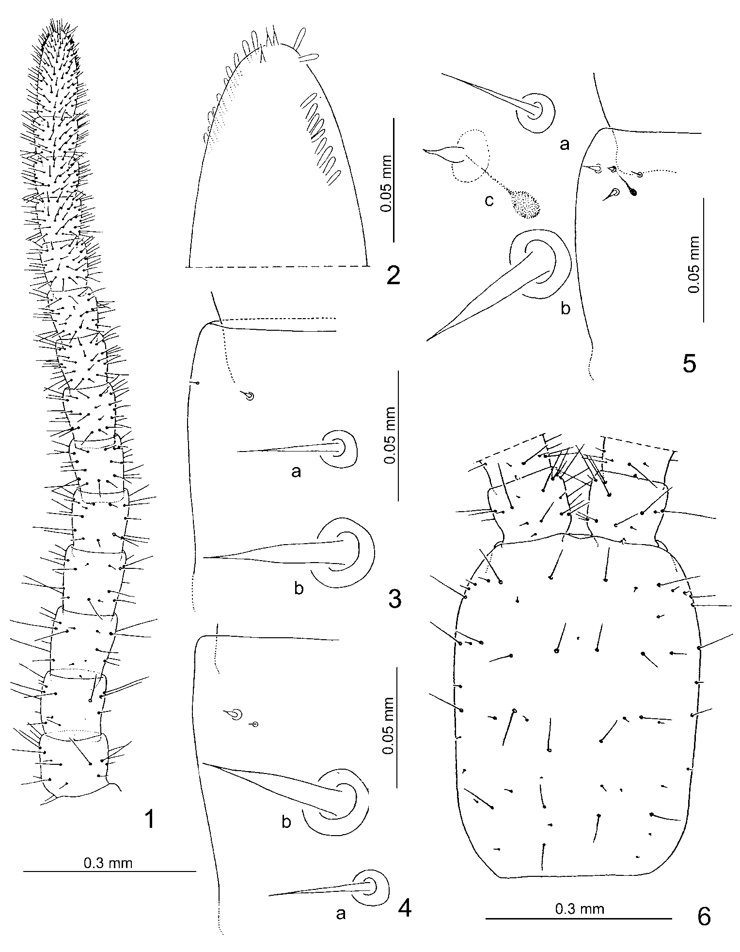
Antennae ca. 2.4 times as long as the cephalic plate, distally slightly attenuate; all articles (except the first) longer than wide. Setae on a.a. I to V–VI few and of different length, those of remaining articles progressively shorter and more numerous towards the tip of the appendage ( Fig. 1 View FIGURES 1 – 6 ). Terminal a.a. with ca. 11 claviform sensilla on the external border and ca. 10 on the internal border ( Fig. 2 View FIGURES 1 – 6 ). Distal end of this a.a. with ca. three very small specialised sensilla apparently not split apically ( Fig. 2 View FIGURES 1 – 6 ). Dorsal and ventral surface of a.a. II, V, IX and XIII with very small specialised sensilla. On the ventral side these sensilla are restricted to an internal lateroapical area and occur in two different types: a and b. Type a sensilla are very thin and not split apically, type b sensilla are thicker and very similar to those on the distal end of the terminal a.a. (a, b, Fig. 3 View FIGURES 1 – 6 ). Specialised sensilla on dorsal side are restricted to an external lateroapical area and of three types: a and b, respectively similar to a and b of ventral side, and type c sensilla, similar to type b but smaller and with a small dark 'root' (a, b, c, Fig. 5 View FIGURES 1 – 6 ). Distribution of sensilla as in table 2.
Cephalic plate approximately rectangular but sides curved, distinctly longer than wide (ratio 1.5:1), shape and chaetotaxy as in Fig. 6 View FIGURES 1 – 6 .
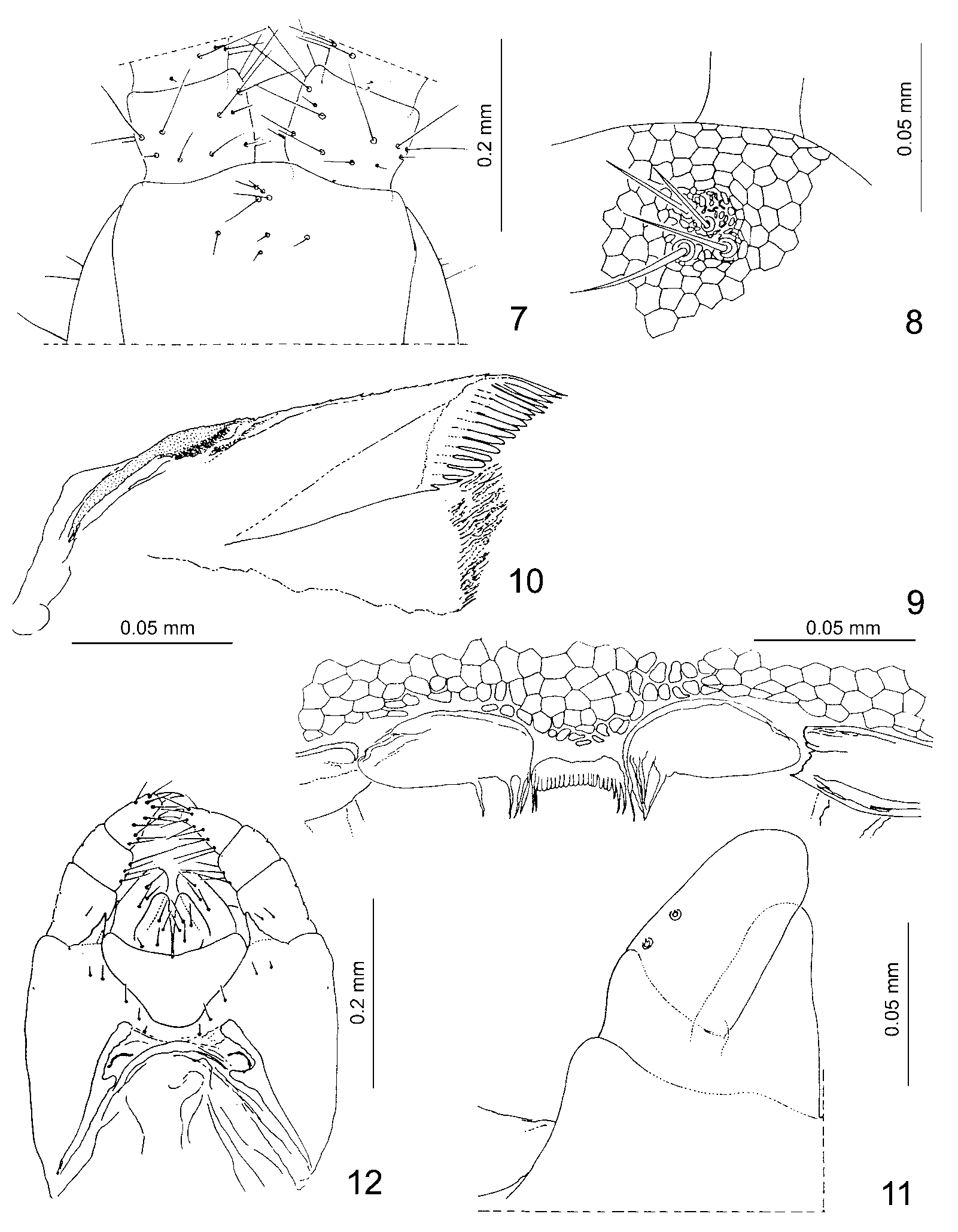
Clypeus with four setae on the clypeal area; middle part with four setae of similar size ( Fig. 7 View FIGURES 7 – 12 ). Surface of clypeal area very densely reticulated ( Fig. 8 View FIGURES 7 – 12 ).
Labrum: midpiece welldeveloped, sclerotised, with ca. 17 short, roundpointed teeth on the middle and 2+3 long hyaline filaments on the sides. Sidepieces with 4+5 long hyaline filaments ( Fig. 9 View FIGURES 7 – 12 ).
Mandible: pectinate lamella with ca. 17 hyaline teeth ( Fig. 10 View FIGURES 7 – 12 ).
First maxillae without lappets on coxosternum; telopodites with rudimentary lappets ( Fig. 11 View FIGURES 7 – 12 ). Coxosternum without setae; median projections of coxosternum subtriangular, well developed, with 4+5 setae of different length. Article II of telopodite with 3+3 ventral setae and 2+2 dorsal setae ( Figs. 11–12 View FIGURES 7 – 12 ).
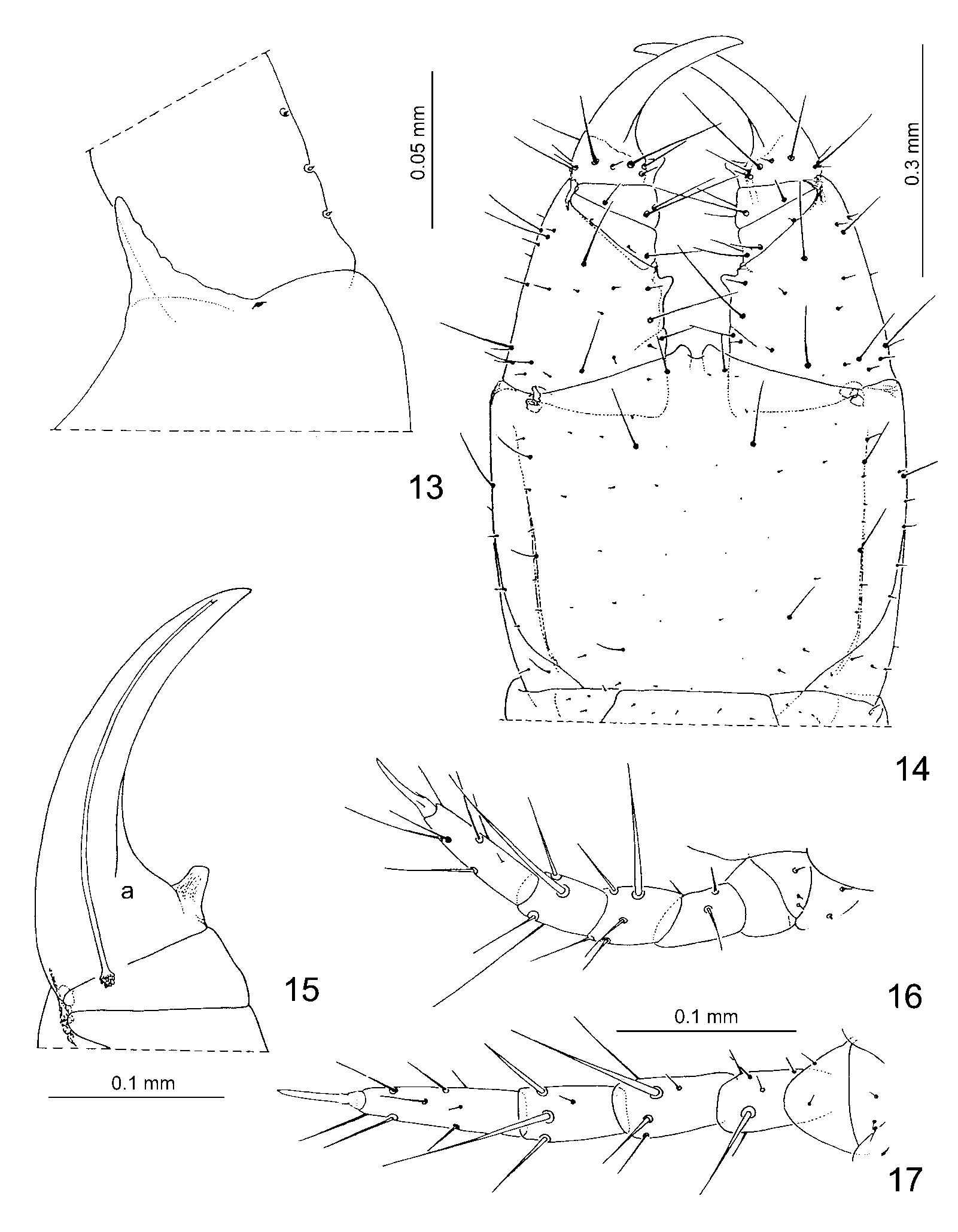
Second maxillae: coxites with 5+5 setae, joined medially only by a nonareolate membranous isthmus ( Fig. 12 View FIGURES 7 – 12 ). Telopodites with setae of uniform thickness, relative size of apical claw as in Fig. 12 View FIGURES 7 – 12 . Process of anterointernal corners of coxosternum well developed ( Fig.13 View FIGURES 13 – 17 )
Forcipular segment: when closed, telopodites project slightly beyond the level of the anterior margin of the head. Basal plate with an irregular transverse row of six large setae near the posterior margin, a few smaller setae dispersed on the remaining surface. Coxosternum with incomplete chitinous lines. Telopodites: trochanteropraefemur with a conspicuous, subtriangular and slightly pigmented apical tooth on the medial edge; femur and tibia without teeth; tarsungulum with a welldeveloped, slightly pigmented basal tooth; dorsal and ventral edges of the ungular blade not serrulate ( Figs. 14–15 View FIGURES 13 – 17 ). Calyx of poison gland as in figure 15; chaetotaxy of coxosternum and telopodites as in figure 14.
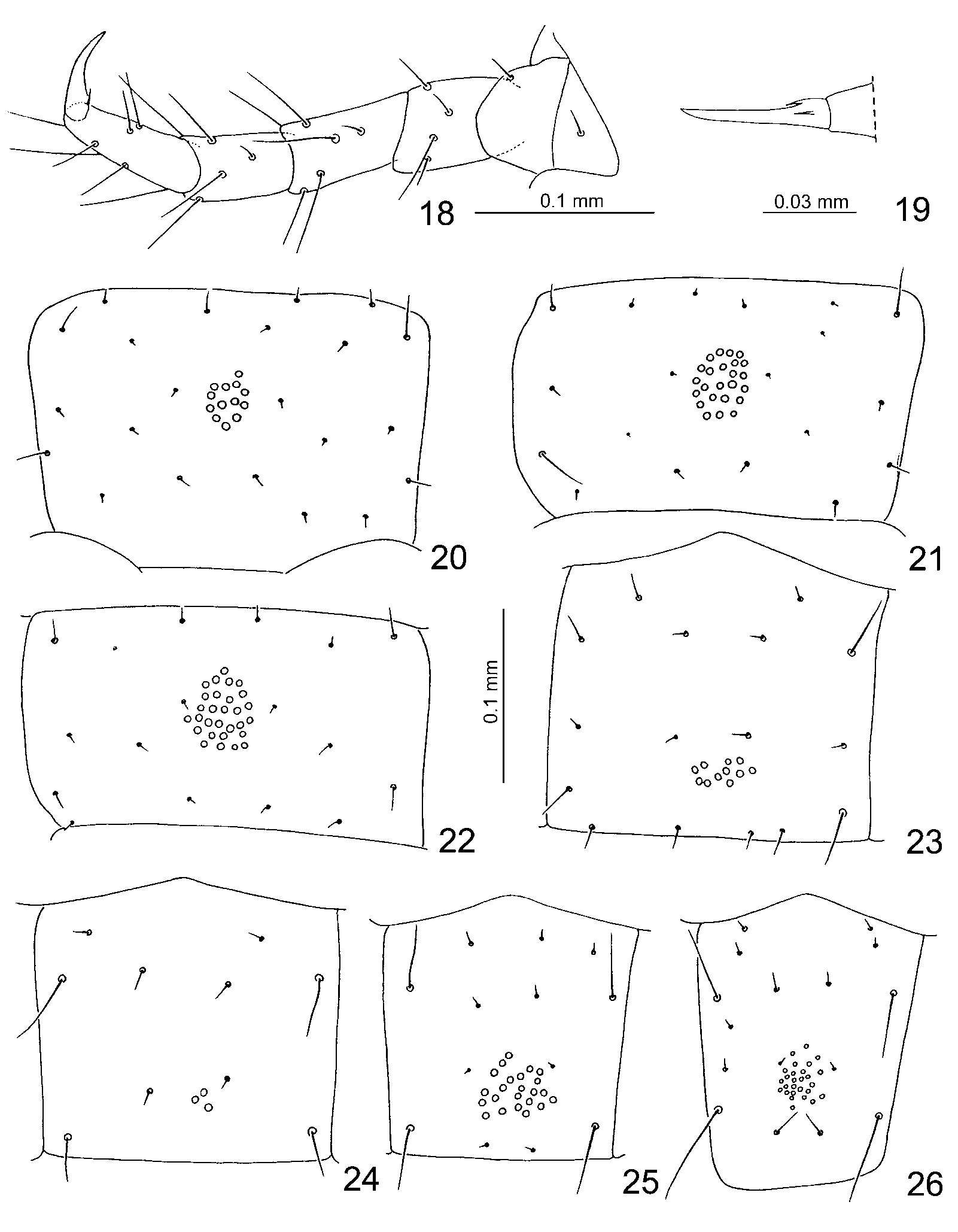
Walking legs: pairs I to IX–X with setae of different thickness ( Figs. 16–17 View FIGURES 13 – 17 ), remaining legs with setae of uniform thickness ( Fig. 18 View FIGURES 18 – 26 ). Claws ventrobasally with an anterior and a posterior parunguis ( Fig. 19 View FIGURES 18 – 26 ).
Sterna: pore fields present from the second to the antepenultimate sternum. All fields undivided. Form of fields changing along the trunk as in Figs. 20–26 View FIGURES 18 – 26 . Number of pores on selected sterna: on sternum II, 13 pores; on III, 24; on VII, 29; on XLIII, 11; on XLVII, 3; on LII, 23; on LIII, 26.
Last legbearing segment without pleurites at the sides of praetergum. Praesternum not divided along the sagittal plane; form and chaetotaxy of tergum and sternum as in Figs. 27–28. Coxopleura slightly protruding at the ventral distal end; setae small and numerous on internal distal edge, remaining surface with few larger setae. Each coxopleuron with an anterior independent coxal organ and a posterior cluster of ca. 5–6 coxal organs, both opening on the membrane between coxopleuron and sternum and covered by the latter ( Figs. 28–29 View FIGURES 28 – 29 ). Last legs with seven podomeres, shape and chaetotaxy as in Figs. 27–28. Praetarsus unguiform, smaller than those of the preceding legs.
Terminal segments: intermediate tergum with posterior margin convex, intermediate sternum seemingly covered by the sternum of the last legbearing segment, first genital sternum as in Fig. 28 View FIGURES 28 – 29 . Anal organs present.
ɗ. Unknown.
Barbieri, 1995, R. onycophaena Pereira, Foddai & Minelli, 2000 , R. rossi Chamberlin, 1957 and R.
No known copyright restrictions apply. See Agosti, D., Egloff, W., 2009. Taxonomic information exchange and copyright: the Plazi approach. BMC Research Notes 2009, 2:53 for further explanation.