
Culicoides marksi Lee & Reye
|
publication ID |
https://doi.org/10.5281/zenodo.204428 |
|
DOI |
https://doi.org/10.5281/zenodo.5673948 |
|
persistent identifier |
https://treatment.plazi.org/id/1D6FB737-FF94-FFA3-FF2F-FE205502F869 |
|
treatment provided by |
Plazi |
|
scientific name |
Culicoides marksi Lee & Reye |
| status |
|
Culicoides marksi Lee & Reye View in CoL
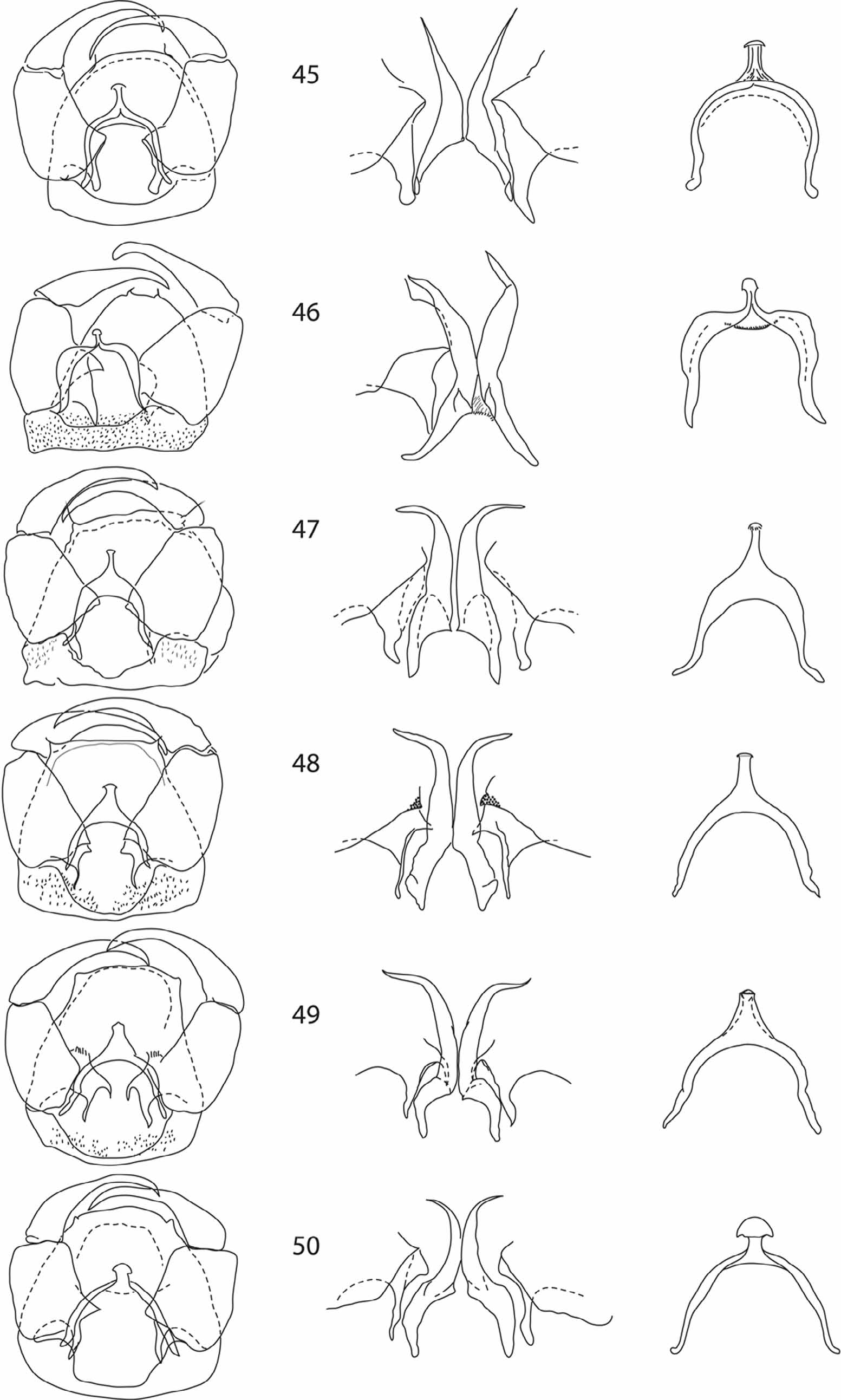
(Figs, 1, 7, 13, 19, 25, 26, 33, 39, 45, 51)
Culicoides marksi Lee & Reye 1953: 392 View in CoL
Type material examined. Holotype female: Australia, NSW, Yagobie 3.Dec.1951, Light trap, AL Dyce ( ANIC). Allotype: same data as holotype ( ANIC). Paratypes: same data as holotype ( 2 males, 9 females, all ANIC).
Non-type material examined. Australia, NSW, Yetman, 22.ii.63, A.L. Dyce & M.D. Murray ( 10 females, 10 males ANIC).
Diagnosis. Wing with 2 pale spots in cell M4, anterior spot boomerang shaped. Female with SCo distribution 3, 11–15, six SCh on 4–10, two on 11–15; spermathecae subspherical with proximal half non-sclerotised. Male with SCo distribution 3, 13–15, STl distribution 3–7,(8–9), STc distribution 4–10,(11–12); five SCh on 14; apical half of parameres straight, ventral membrane of ninth sternite bare. Pupal abdomen with paired lateral spines only on lpm and lasm, thorax with single lateral spine on di and dii; prothoracic horn covered with scales.
Female. Head. Eyes bare, separated by a distance of one facet or less ( Fig 1 View FIGURES 1 – 6 ), proboscis short. Palpus (fig 7) brown with five segments, segment 3 expanding apically then abruptly narrowed beyond a round, shallow sensory pit with several protruding capitate sensilla. Antennomeres ( Fig 13 View FIGURES 13 – 18 ) 4–10 barrel shaped, 11–15 cylindrical, short.
Thorax. Legs (fig 19) dark brown with dark knees, all femora pale from base to about midlength and with conspicuous pale subapical band, all tibiae with pale subbasal band and broadly pale subapically, tarsi pale. Wing (fig 25) strongly patterned with three pale spots in cell R5, the two proximal spots rarely reduced or absent (fig 26), two pale spots present in cell M4, proximal one boomerang shaped.
Abdomen. Three developed ovoid, similar-sized spermathecae, proximal half not sclerotised, ducts short (fig 33); sclerotised ring wider than long.
Male. Head. Eyes bare. Palp similar to female with shallow pit on segment 3. Antenna ( Fig 39 View FIGURES 39 – 44 ) with 2 rows of plume verticils on segment 3, single row on 4–12; antennomeres 13–15 elongate, subcylindrical and narrow.
Genitalia. ( Fig 45 View FIGURES 45 – 50 ) Ninth tergite with caudal margin convex. Ninth sternite with shallow caudomedial excavation, ventral membrane bare. Gonocoxite rectangular, longer than wide, dorsal root long and simple; ventral root reduced to a short point or rounded. Gonostylus slender, distally curving gently to a pointed apex. Aedeagus with distal process short with a rounded mushroom-shaped expansion apically. Parameres separate, with simple basal arms angled sharply to weakly swollen stem narrowing gradually to simple, sharp-tipped distal portion directed slightly lateroventrad.
Immatures. Fourth instar larvae and pupae of this species were adequately described by Kettle & Elson (1976).
Distribution. (fig 63) all states of Australia and Western Province of PNG.
Biology. Debenham (1978) summarised the known biology of this species. Immature stages live in the margins of water bodies, pupae float on the surface, unable to submerge. Adults feed on mammals and birds and have been implicated in the transmission of nine viruses. Adults are active mostly at dawn and dusk ( Bellis et al. 2004).
Remarks. Male and female specimens of this species can be distinguished from C. dycei and C. pseudostigmaticus by the presence of two pale spots in cell M4 and from all other members of the group by the boomerangshaped proximal spot in cell M4 of the wing. C. marksi may additionally be separated from species with two pale spots in cell M4 by the shape of the spermathecae in the female and by the bare membrane on the ninth sternite of the male. Pupae can be distinguished from C. parvimaculatus by the lateral spurs on all lpm tubercles from C. zentae by the lack of paired spines on dasm, dpmiv and dpmv and thoracic tubercules di and dii and from C. dycei by the presence of scales on the prothoracic horn. Immature stages of the remaining two species of Marksomyia are not known.


No known copyright restrictions apply. See Agosti, D., Egloff, W., 2009. Taxonomic information exchange and copyright: the Plazi approach. BMC Research Notes 2009, 2:53 for further explanation.
|
Kingdom |
|
|
Phylum |
|
|
Class |
|
|
Order |
|
|
Family |
|
|
Genus |
Culicoides marksi Lee & Reye
| Bellis, Glenn & Dyce, Alan 2011 |
Culicoides marksi
| Lee 1953: 392 |