
Culicoides zentae Bellis & Dyce
|
publication ID |
https://doi.org/ 10.5281/zenodo.204428 |
|
DOI |
https://doi.org/10.5281/zenodo.5673953 |
|
persistent identifier |
https://treatment.plazi.org/id/1D6FB737-FF9E-FFAB-FF2F-FEC850E3F957 |
|
treatment provided by |
Plazi |
|
scientific name |
Culicoides zentae Bellis & Dyce |
| status |
sp. nov. |
Culicoides zentae Bellis & Dyce View in CoL sp. nov.
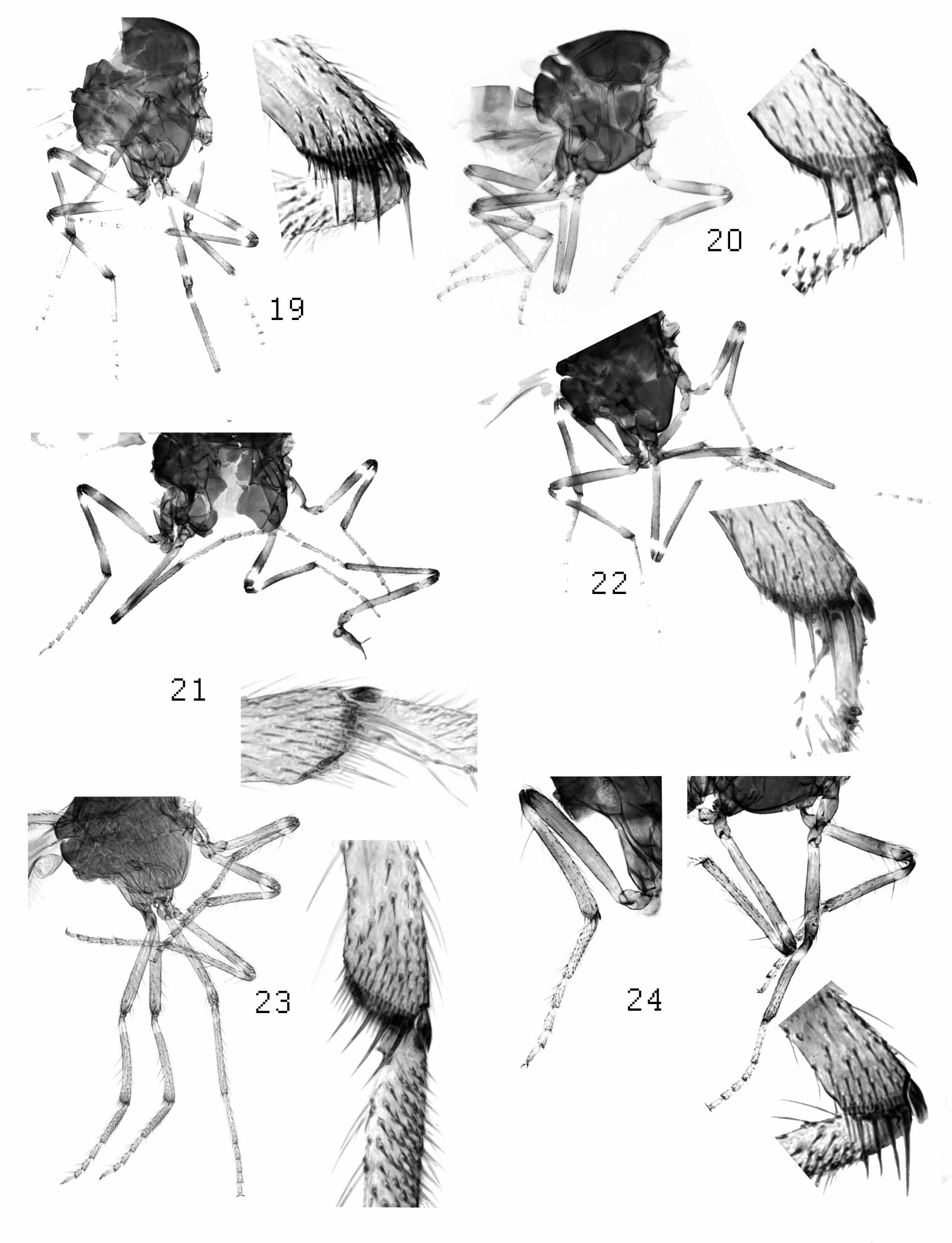
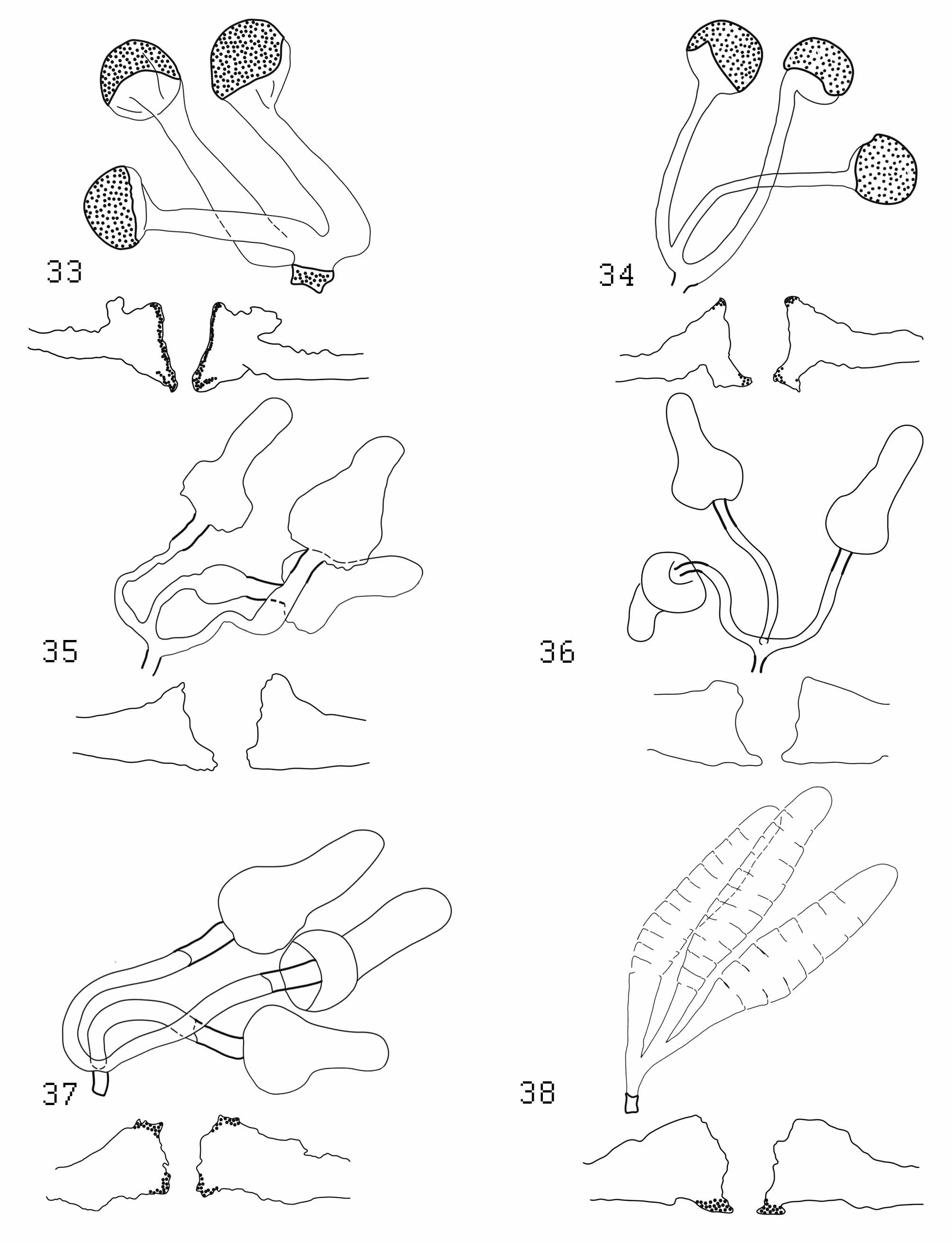
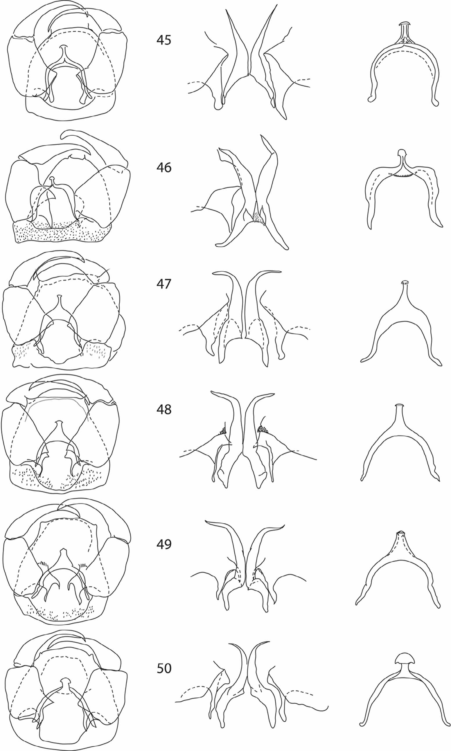
( Figs 3 View FIGURES 1 – 6 , 9 View FIGURES 7 – 12 , 15 View FIGURES 13 – 18 , 21 View FIGURES 19 – 24 , 28 View FIGURES 25 – 32 , 35 View FIGURES 33 – 38 , 41 View FIGURES 39 – 44 , 47 View FIGURES 45 – 50 , 53 View FIGURES 51 – 56 )
Type material. Holotype female, Australia Qld, Gilruth, Bore drain, sweep net, sunset, 26.vii.1963, A.L. Dyce (female, ANIC). Paratypes: Australia, Qld, Gilruth, Light trap, Bore drain, 26.vii.1963, A.L. Dyce & M.D. Murray (9 females 1 male ANIC); Charleville, 20.ii.1969, Truck trap run 18.30–19.00 hrs, I. Fanning & B. Kay (8 males ANIC); Charleville, 18.ii.1969, Wallal Bore, A.L. Dyce, (5 females, 1 male and associated pupae, ANIC); NT, Vic River Research Station, 16°24’S, 131°01’E, 28.x.2003, D. Cherry L.T. 03-1615 (1 female AQISNT).
Diagnosis. Wing with 2 pale spots in cell M4, anterior spot rounded or triangular. Female with SCo distribution 3, (4–6),7,(8),9,11–15, six SCh on 4–10, two to three on 11–15; spermathecae with unsclerotised, pear-shaped body and short, sclerotised necks. Male with SCo distribution 3, (4),13–15; STl distribution 3–6; STc distribution 4–6; six or seven SCh on 14; apical half of parameres strongly curved laterad and with triangular excavation medially, ventral membrane of ninth sternite with spicules present laterally, medially bare. Pupal abdomen with paired lateral spines on lpm, lasm and dasm, thorax with paired lateral spines on di. dii and div; prothoracic horn with scales covering basal third.
Female. Head. Eyes separated by a distance of about one facet ( Fig 3 View FIGURES 1 – 6 ); proboscis short. Palpus (fig 9) brown with five segments, segment 3 expanding apically then abruptly narrowed beyond a round, shallow sensory pit with several protruding capitate sensilla. Antennomeres (fig 15) 4–10 barrel shaped, 11–15 cylindrical, short.
Thorax. Legs (fig 21) dark brown with dark knees, all femora pale from base to about midlength and with pale subapical band, all tibiae with pale sub basal band and broadly pale subapically (less obvious on mid tibiae), tarsi pale. Wing (fig 28) strongly patterned with two ovoid to triangular pale spots in cell M4.
Abdomen. Three developed, pear-shaped spermathecae, unsclerotised and difficult to see in most specimens but necks strongly sclerotised and conspicuous (fig 35), ducts short, about as long as spermathecae; sclerotised ring tapering posteriorly.
Male. Head. Eyes bare. Palpus similar to female with shallow pit on segment 3. Antenna ( Fig 41 View FIGURES 39 – 44 ) with single row of plume verticils on antennomeres 3–12; antennomeres 13–15 elongate subcylindrical and narrow. Genitalia. (fig 47) Ninth tergite with caudal margin slightly convex. Ninth sternite with deep, U shaped caudomedial excavation, spicules confined to two posterior lateral patches. Gonocoxite short and broad, longer than wide, dorsal root long and simple; ventral root reduced to rounded point. Gonostylus slender, distally curving gently to a pointed apex. Aedeagus with distal process short with a slight apical rounded expansion. Parameres separate, with triangular excavation near base, basal arms roughly parallel, stem weakly swollen narrowing gradually to simple sharp-tipped distal portion curved lateroventrad to almost 90°.
Immatures. Larvae: unknown
Pupa. Head. (fig 60): Setae vm and vl with each group consisting of a long and a short seta; am (fig 61) large tubercule bearing a blunt spur, a stout seta and a sensillum; ad (fig 61) a large tubercule with a pair of unequal, stout setae.
Operculum. (fig 61): A single row of bluntly pointed thorns along lateral margins posterior to lateral angles, extending to about ¾ of operculum length; two groups of thorns either side of midline posterior to each am, narrowly connected anteriorly; oval structures between lateral rows of thorns extending between am ’s to anterior margin.
Thorax. (fig 57): Tubercules di, dii, and div with two pointed lateral spines and a stout seta; diii ridged with a small seta; dm a long, thin seta; dl long thin seta and short stout seta on protuberant base.
Prothoracic horn. (fig 62): Pigmented throughout but darker near base, on lateral tubercules and in distal third; folds and sharply pointed scales on basal third. Four to six (mean 5.18) terminal and one to three (mean 2) lateral spiracular openings.
Abdomen. (fig 58): Setae dpm i, iv and v with paired spines and stout setae, seta on i much shorter than on iv or v; ii and iii ridged tubercules with one rounded spine and no setae. lpm all with paired sharply pointed spurs, i and iii with short stout seta, ii with long thin seta; V ridged tubercules, i with short seta, ii and iii with long thin seta; dasm with paired rounded spines and long (i) or short (ii) seta; lasm similar to lpm. Anterior band of spicules present dorsally and ventrally but absent laterally.
Caudal segment. (fig 59): Encircling band of spicules anteriorly; median group of v-shaped spicules dorsally; apicolateral processes pigmented distally with spicules scattered over surface.
Etymology. This species is named for the late Ms. Zenta Liepa, formerly of CSIRO. Division of Entomology, in recognition for her assistance with the curation of Diptera at ANIC.
Distribution. (fig 65) Northern and eastern Australia, more common in drier, inland areas.
Biology. Pupae examined in this study were collected from the muddy margins of a 5x 3m pond constantly fed by warm, subterranean water. Adults are often collected in light traps suggesting a crepuscular or nocturnal activity. Muller et al. (1981) reported this species (as C. McMaster sp121) feeding on marsupials.
Remarks. . The adult morphology of this species is very similar to that of C. parvimaculatus but pupal morphology is markedly different indicating the usefulness of pupal morphology in defining species.
Males of this species can be distinguished from those of other members of Marksomyia by the triangular excavation on the medial part of the parameres. Females can be distinguished by the distribution of antennal SCo. Pupae can be distinguished from those of other species of Marksomyia by the paired, sharp lateral spines on abdominal tubercules dasm, dpmiv and dpmv and thoracic tubercules di and dii.
The wing of this species was illustrated by Dyce et al (2007) under the name Marksi gp sp no 1.


| ANIC |
Australian National Insect Collection |
No known copyright restrictions apply. See Agosti, D., Egloff, W., 2009. Taxonomic information exchange and copyright: the Plazi approach. BMC Research Notes 2009, 2:53 for further explanation.