
Pachyprotasis maculoscutellata Zhong & Wei
|
publication ID |
https://doi.org/10.11646/zootaxa.3914.1.1 |
|
publication LSID |
lsid:zoobank.org:pub:AAD0A1E5-4DFE-4853-A071-BA62D1F91D25 |
|
DOI |
https://doi.org/10.5281/zenodo.6096610 |
|
persistent identifier |
https://treatment.plazi.org/id/1E1BEF31-CD50-FFF7-FE9F-F90105D9F9E5 |
|
treatment provided by |
Plazi |
|
scientific name |
Pachyprotasis maculoscutellata Zhong & Wei |
| status |
sp. nov. |
Pachyprotasis maculoscutellata Zhong & Wei , sp. nov.
( Figs. 16, 17 View FIGURES 14 – 17 , 47 View FIGURES 44 – 49 , 63 View FIGURES 60 – 65 , 81 View FIGURES 80 – 83 , 96, 110, 126)
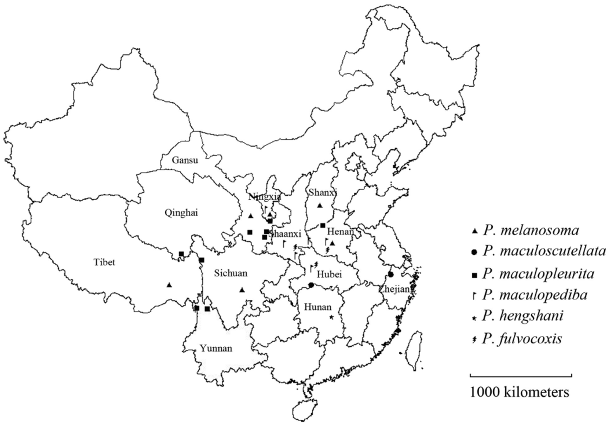
Distribution. China (Hunan and Zhejiang) ( Fig. 126 View FIGURE 126 ).
Material examined. Holotype, Hunan: ♀, Shimen ( 29°55′N, 110°46′E), 0 7.1994, Zhiwei Liu leg. ( CSCS); paratypes: 1 ♀, Mt. Huping, Shimen ( 29°55′N, 110°46′E), 0 1.05.2000, Yihai Zhong leg.; 7 ♀, Shimen ( 29°55′N, 110°46′E), 0 7.1994, Zhiwei Liu leg.; Zhejiang: 1 ♀, Mt. Tianmu ( 30°23′N, 119°71′E, alt. 350–1000 m), 0 6.05.1963, Gentao Jin leg.
Etymology. The specific epithet ( maculoscutellata ), refers to the distinct white spot on the medial mesoscutellum. It is an adjective.
Female ( Figs. 16, 17 View FIGURES 14 – 17 ): Body length 9.0 mm. Body black, with white pattern as follows: labrum, clypeus, supraclypeal area, inner orbit connected to an oblique spot on temple, anterior margin of tegula, a quite small spot on middle of lateral mesoscutal lobe, a medial stripe on mesoscutellum, middle of mesoscutellar appendage, medial metascutellum, upper metepisternum, a medial stripe on each posterior margin of terga 3–6, posterolateral margin of each tergum, posterior margin of each sternum; legs black, with white pattern as follows: underside of fore leg from apical half of femur to claw, an irregular spot on middle trochanter, underside of middle tibia and tarsus, a large basal spot on outer of hind coxa, hind trochanter connected to base of hind femur. Wings hyaline, stigma and veins dark brown.
Labrum and clypeus with sparse and shallow punctures; punctures on vertex and frons minute, not dense, space between punctures smooth, shiny; mesoscutum with dense punctures and sculpture, shiny; punctures on upper mesepisternum deep, large, but not dense, on lower part dense and minute, shiny; mesepimeron with distinct sculpture, slightly shiny; metapleruron with dense, shallow and minute punctures, slightly shiny ( Fig. 81 View FIGURES 80 – 83 ); medial mesoscutellum smooth, punctures neartly absent, laterally with sparse, deep and large punctures; apex of mesoscutellar appendage with sparse, deep and large punctures, sculpture faint, space between punctures distinctly shiny; terga with distinct sculpture and luster, laterally with sparse and shallow punctures; outer hind coxa with dense and distinct punctures.
Anterior margin of labrum with a medial shallow notch; clypeus roundly incised to about 2/5 of its medial length, lateral lobe acute; malar space wider than diameter of median ocellus; inner margins of eyes slightly convergent downwards; supra-antennal tubercle indistinct; frontal area distinctly elevated, as high as top of eye in lateral view, frontal ridge round; median and lateral fovea pit-shaped, shallow ( Fig. 47 View FIGURES 44 – 49 ); interocellar and postocellar furrow broad and shallow; postocellar area slightly elevated, 1.8 times as broad as long, lateral postocellar furrows shallow; head slightly narrowed behind eyes in dorsal view ( Fig. 63 View FIGURES 60 – 65 ). Antenna slightly shorter than combined length of thorax and abdomen, flagellomere 1 slightly shorter than flagellomere 2, some apical flagellomeres compressed. Mesoscutellum slightly elevated, lateral carina blunt, middle carina of mesoscutellar appendage acute. Hind tarsomere 1 nearly as long as combined length of following 4 tarsomeres. Fore wing with middle petiole of anal cell 1/2 as long as basal part of anal cell, as long as vein R+M; petiole of hind wing anal cell shorter than 1/2 length of vein cu-a.
Ovipositor sheath shorter than hind tarsomere 1 in lateral view, valvula 3 round at apex and slightly longer than valvifer 2. Lancet with 18 serrulae (Fig. 96), base of each serrula distinctly elevated, middle serrula each with 2 basal denticles and 5–6 distal denticles (Fig. 110).
Male. Unknown.
Discussion. This species is similar to P. micromaculata and may be distinguished as follows ( P. micromaculata in parentheses): terga 3–6 posterior with distinct yellow-white triangular spot (all terga completely black); basal 1/ 7 of hind femur yellow-white (hind femur completely black); labrum and clypeus completely yellow-white (labrum and clypeus with distinct black spots). It is also similar to P. nitididorsata and may be distinguished as follows ( P. nitididorsata in parentheses): supraclypeal area black (supraclypeal area white); median mesoscutal lobe completely black (apex of median mesoscutal lobe black with a white arrow-shaped-spot); hind coxa black, outer part with a white spot (hind coxa white, outer part with a black stripe); base of hind femur white (hind femur completely black). Except for the serrulae, reliable morphological differences between these three species were not found.


No known copyright restrictions apply. See Agosti, D., Egloff, W., 2009. Taxonomic information exchange and copyright: the Plazi approach. BMC Research Notes 2009, 2:53 for further explanation.
|
Kingdom |
|
|
Phylum |
|
|
Class |
|
|
Order |
|
|
Family |
|
|
Genus |