
Lepidodermella acantholepida, Suzuki, Takahito G., Maeda, Masako & Furuya, Hidetaka, 2013
|
publication ID |
https://doi.org/ 10.11646/zootaxa.3691.2.3 |
|
publication LSID |
lsid:zoobank.org:pub:E24A5702-6A02-4AFD-B80D-CCE646F9C8E2 |
|
DOI |
https://doi.org/10.5281/zenodo.5674199 |
|
persistent identifier |
https://treatment.plazi.org/id/1F30878C-4A1A-BF49-69F9-11534B2AC56B |
|
treatment provided by |
Plazi |
|
scientific name |
Lepidodermella acantholepida |
| status |
sp. nov. |
Lepidodermella acantholepida View in CoL n. sp. Suzuki and Furuya
[New Japanese name: kagiurokoitachimushi] (Figs. 1,2)
Type locality. Japan, Honshu, Shiga Prefecture, Otsu (35º 08' 5.40ʺ N, 135º 54' 51.18ʺ E). 24, June 2011, collected by Suzuki.
Type specimens. Holotype, mounted in glycerol, is deposited in Lake Biwa Museum (LBM1360000013). All specimens were collected by T.G. Suzuki. Eight paratype specimens are in the first author’s collection: two specimens site 3, 21 June, 2011; 4 specimens site 3, 1 July, 2011; Five additional specimens were collected from the Karasuma Peninsula: Japan, Honshu, Shiga Prefecture, Kusatsu (35º 4' 29.70ʺ N, 135º 56' 2.28ʺ E: 3 specimens collected 18 June 2012; 2 specimens, 22 June, 2012.
Etymology. Latin adjective, describing the characteristic spined scale of the dorsal end plate.
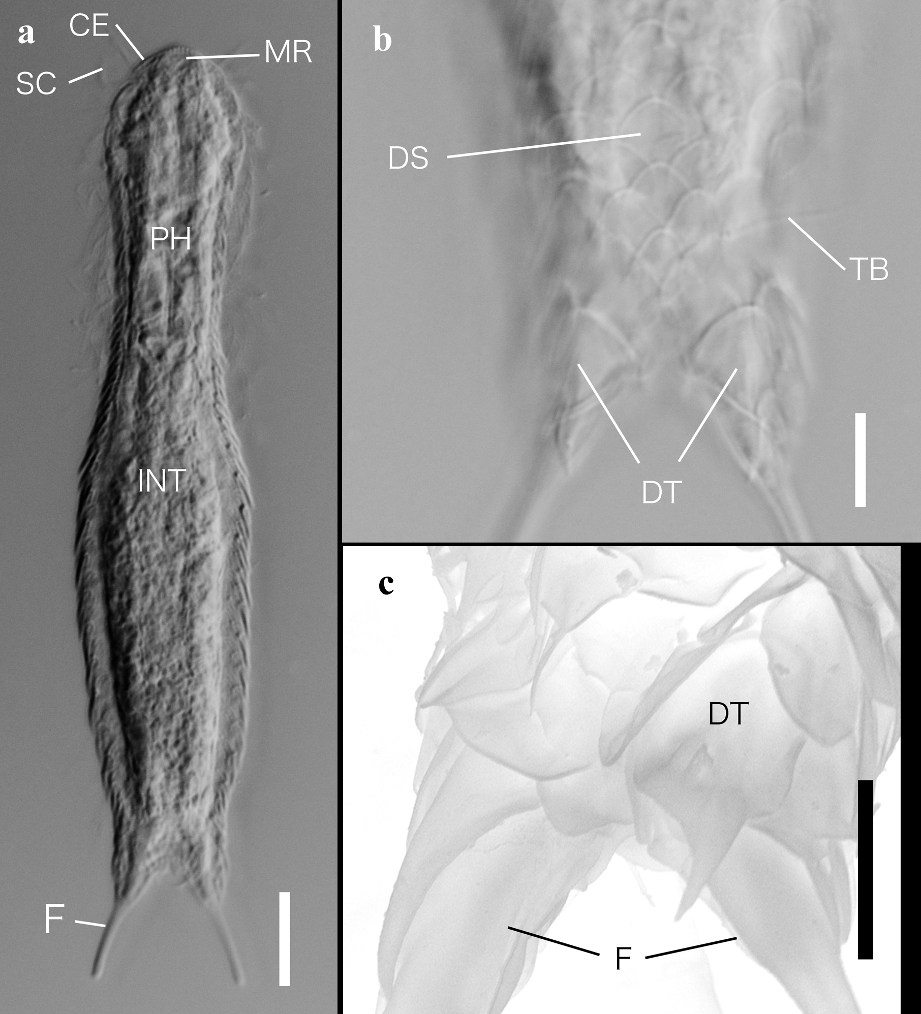
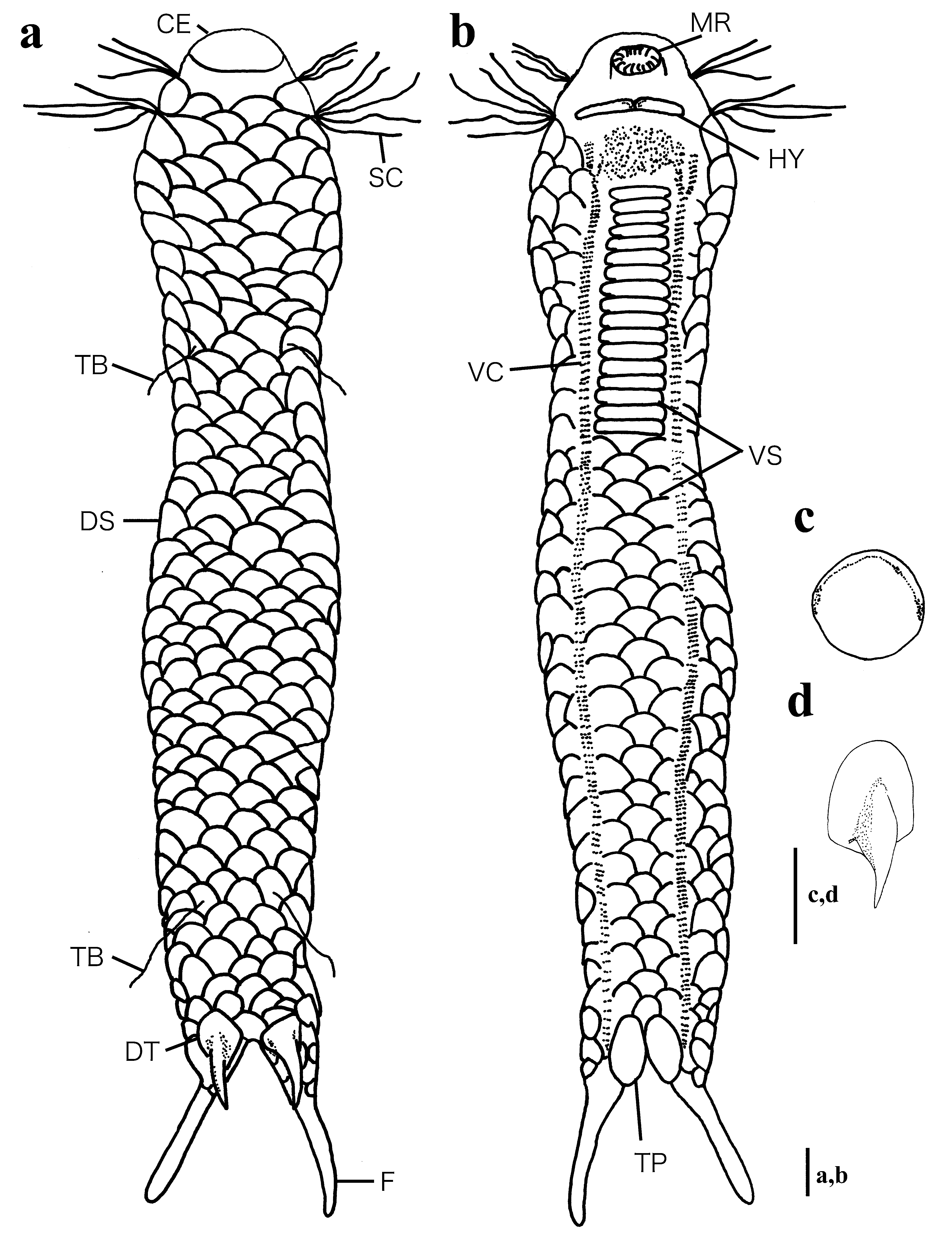
Diagnosis. Medium-sized Lepidodermella of total length 140.0–147.4 μm including furca of 15.2–19.9 μm beginning at U86.0; pharyngeo-intestinal junction (PhIJ) at U29.7; head slightly five- lobed, with cephalion, hypostomion, and two pairs of sensory ciliary bundles. Eye-spots absent. Body enveloped by flat, rounded, scales. Two dorsal terminal plates with a claw-like spine. Two types of ventral scale between two columns of ventral cilia: rectangular anteriorly around the PhIJ at U31 and oval posteriorly. Rectangular scales in 1 longitudinal row. Oval scales in 3 longitudinal alternating rows. A pair of flat, oval terminal plates cover posterior end of ventral scales.
Description. Based on an adultspecimen (holotype), 144.6 μm in total length ( Fig. 1 View FIGURE 1 ). Body medium-sized, tenpin-shaped with distinct head, neck, and trunk region. Widths of head / neck / trunk / caudal base are 22.9 / 16.0 / 32.7 / 17.4 μm at U10 / U29 / U58 / U84, respectively. Length of distal furca 17.2 μm. Pharynx 36.9 μm in length, from posterior edge of mouth to junction with intestine; PhIJ at U 29.7. Head with 5 shallow lobes and well developed cephalion. Ventral hypostomion located at posterior of mouth. Hypostomion divided into right and left sides, each of length 5.6 μm ( Fig. 2 View FIGURE 2 ).
Sensory organs: Two pairs of cephalic ciliary tufts isolated on either side of head, each consisting of seven or more sensory cilia: anterior pair 11 μm in length, posterior 20 μm. Eye-spots absent. Tactile bristles 10 μm in length at U25.0 on neck and U84.5 caudally.
Cuticular armature: Body covered dorsally with 28–32 columns of flat scales in 5–7 rows, except for regions of cephalion and furca. Dorsal scales flat, round, without spine, mean diameter 5.8 μm. Scales overlap, dorsally shape and size identical except for a pair of dorsal terminal plates which are rhombic, 6.7 μm in length, 5.1 μm in width, with a thick, simple spine of length 5.8 μm. Two types of ventral scale covering region between two columns of ventral cilia: one column of 17 rectangular scales of 7.1 μm in width, 2.3μm in length, located from 13.4U to 33.7U; 3 columns of 18 flat, oval scales, each 3.8 μm in length, 5.4 μm in width. A pair of oval terminal plates covering posterior end of ventral scales, their width 3.0 μm, length 9.4 μm; larger than other ventral scales.
Ventral ciliation: Densely packed field of cilia at posterior edge of hypostomion (07 U) extending posteriorly as two longitudinal bands, ending at U 88.
Digestive tract: Mouth opening with maximum and minimum diameters 8.2 μm and 5.2 μm. Pharynx cylindrical, shape width 11μm. Intestine straight, slightly wider anteriorly (9.7 μm), narrowing gradually over its length to 7.0 μm. Anus ventral, at U 83.4.
Remarks. Lepidodermella acantholepida n. sp. is similar to L. squamata (Dujardin) in the shape of body and scales. Lepidodermella squamata has a wide geographical distribution in Europe, Russia, Asia, America, and Africa (Schwank & Bartsch 1990). Amato and Weiss (1982) reported many variations in the shape and arrangement of scales in L. squamata , and Kånneby, Todaro and Jondelius (2012) suggest that the currently recognized L. squamata is probably composed of several cryptic species complexes. However, L. acantholepida n. sp. is easily distinguished from L. squamata by its hypostomion (linear pair vs. curved pair) and its pair of spined scales, which are absent in L. squamata (Schwank & Bartsch 1990) .
Lepidodermella acantholepida n. sp. is similar to six species in dorsal scale shape and the presence of spines: cf. L. amazonica Kisielewski, L. broa Kisielewski, L. intermedia Kånneby, L. minor chaetifer Kisielewski, and L. spinifera Tretyakova. Lepidodermella amazonica , L. broa , and L. minor chaetifer are recorded from Brazil (Kisielewski 1991). Lepidodermella acantholepida n. sp. differs from L. amazonica in body length (140.0–147.4 μm vs. 199–200 μm), scale shape (oval vs. with antero-lateral horns), tactile bristles on the neck (a pair vs. absent), and spine at dorsal terminal plate (claw-like spine vs. no spine; Kisielewski 1991). Lepidodermella acantholepida n. sp. differs from L. broa in the ventral scales between the ciliary bands (rectangular and oval vs. absent) and spine at dorsal terminal plate (claw like spine vs. no spine; Kisielewski 1991). Lepidodermella acantholepida n. sp. differs from L. minor chaetifer in the number of spined scale (2 vs. 6), ventral terminal plate (flat vs. keeled spined), and ventral scales between ciliary bands (rectangular and oval vs. absent; Kisielewski 1991).
Lepidodermella intermedia was described from Mount Njulla, Swedish Lapland. Lepidodermella acantholepida n. sp. differs from L. intermedia in body length (140.0–147.4 μm vs. 114 μm), the shape of the dorsal spined scale (large rhombic vs. small rounded) and the keeled scale (absent vs. dorsal plate near the cilia and terminal plate; Kånneby et. al. 2011).
Lepidodermella spinifera is recorded from Europe and Northern Asia excluding China (Tretyakova 1991). Lepidodermella acantholepida n. sp. differs from L. spinifera in scale shape (round vs. in the shape of a gourd or distended rectangle), terminal plate (oval vs. square) and spined scale (dorsal terminal plate vs. ventral scale; Tretyakova 1991).
No known copyright restrictions apply. See Agosti, D., Egloff, W., 2009. Taxonomic information exchange and copyright: the Plazi approach. BMC Research Notes 2009, 2:53 for further explanation.