
Stictolecanium cranstoni Kondo & Gullan
|
publication ID |
https://doi.org/10.5281/zenodo.293984 |
|
DOI |
https://doi.org/10.5281/zenodo.5323175 |
|
persistent identifier |
https://treatment.plazi.org/id/1F5487B7-FFDC-FFA7-29F7-C9B0BD358F21 |
|
treatment provided by |
Plazi |
|
scientific name |
Stictolecanium cranstoni Kondo & Gullan |
| status |
sp. nov. |
Stictolecanium cranstoni Kondo & Gullan sp. nov.
( Figs 1 View FIGURE 1 D & E, 4).
Proposed common names. Spanish: Escama blanda de Cranston; English: Cranston’s scale.
Type material examined. Holotype, adult female. Stictolecanium cranstoni Kondo & Gullan , Chile, Peulla, 41˚05'13.7" S, 72˚00'54.7" W, 241 m, 17.ii.2006, voucher No. TK0242, T. Kondo, ex Gevuina avellana Mol. , 1(1) ( USNM). Paratypes, same data as holotype, adult female 1(1) ( USNM), and second-instar male nymphs 2(2) ( USNM).
Description. Adult female (measurements based on n=2).
Unmounted material ( Fig. 1 View FIGURE 1 D & E). Adult female in life about 4–5 mm long, 3–4 mm wide, oval, moderately convex. Insect covered in a thin layer of clear wax.
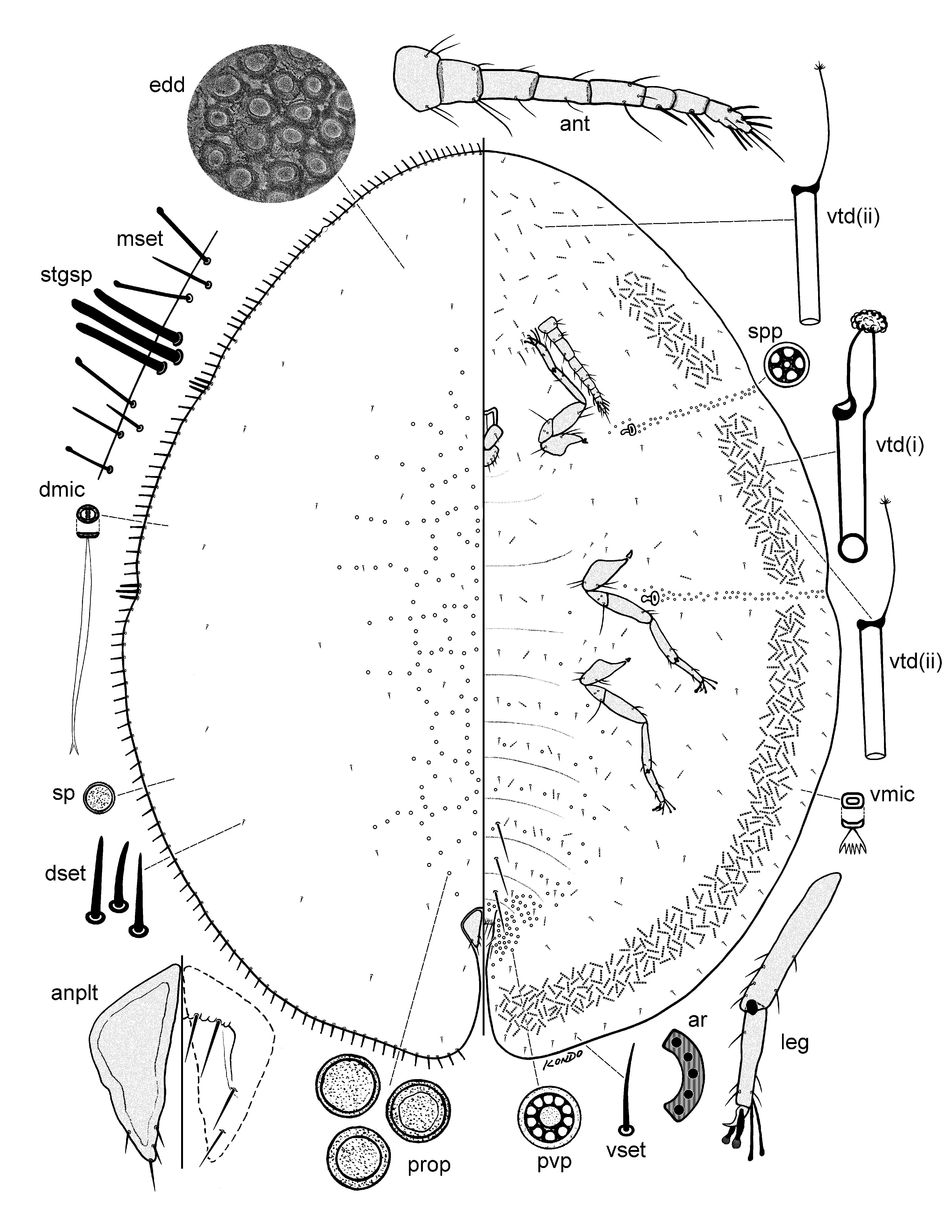
Slide-mounted material ( Fig. 4 View FIGURE 4 ). Body 4.0– 4.7 mm long, 3.1–3.5 mm wide, rather convex, elongate oval in shape.
Dorsum. Derm areolated. Dorsal setae sharply spinose, straight or slightly bent, each 7.5–17.5 mm long. Dorsal microducts oval, with a clear septum seen under high magnification, each about 2.5 μm wide, scattered over dorsum. Simple pores each 2.0–3.0 μm wide, scarce, scattered over dorsum. Cribriform plates absent. Dorsal tubular ducts, dorsal tubercles and pocket-like sclerotizations absent. Preopercular pores circular to irregularly circular in shape, with a granulated surface, each 7.5–8.8 μm wide, present on mid-dorsal area in a reticulated pattern. Anal plates together quadrate, with smooth rounded outer angles, plates located at about 1/ 5 of body length from posterior margin, each plate 175–178 μm long, 73–78 μm wide, anterolateral margin 100–105 μm long, posterolateral margin 123–125 μm long, with 4 slender setae on dorsal surface, i.e., 1 subapical seta on outer margin, 2 subapical setae on inner margin and 1 apical seta; with 2 pairs of fringe setae on ventral side and with 2 subapical setae on each side; hypopygial setae not detected. Anal ring with 10 setae. Sclerotic area around anal plates absent.
Margin. Marginal setae of 2 types; (i) slender, straight, capitate setae, each 36–45 μm long, arranged in a single row, and (ii) sharply spinose setae with a pointed apex, each seta 20–33 μm long, present in an irregular single row, intermixed with type (i) setae; total number of marginal setae ( type i + ii) totalling 25–41 on each side between anterior and posterior stigmatic areas. Stigmatic clefts shallow, with 3 stigmatic spines per stigmatic area, each bluntly spinose, and 25–55 μm long, generally with median setae longest. Eyes 26–28 µm wide, located on dorsal margin.
Venter. Derm entirely membranous. Perivulvar pores each 8.0–9.0 μm wide, with 10 loculi, abundant on perivulvar area; in sparse transverse rows on abdominal segments, and with a pore mesad to each mesothoracic and metathoracic coxa. Spiracular pores each 5.0–6.0 μm wide, with 5 loculi, present in a narrow band as wide as peritreme (about 1 or 2 pores wide), with line of pores extending laterally from each spiracle to body margin, with several pores in a linear group anterior to each spiracle extending from each spiracle towards mid area between spiracle and nearest coxa. Ventral microducts scattered evenly throughout, each about 2.5 μm wide. Ventral tubular ducts of 2 types present: (i) larger duct with a short and broad inner ductule with flower-shaped terminal gland, present around body in an almost complete submarginal band, except absent from spiracular furrows and from head region between eyespots; (ii) a smaller duct with a thin inner ductule with a small and branched terminal gland, rather sparse throughout on thorax and abdomen, with a few around mouthparts, more abundant and fairly evenly distributed on head region, and with a few intermixed with larger ducts in submarginal band; with neither duct type present around margins near ventral submarginal setae. With about 7 pairs of setae on area between antennae; ventral submarginal setae slender, straight or slightly bent, each 10–25 μm long; long pairs of ventral median setae present on last 3 abdominal segments, each 107–163 μm long; other setae slender, similar in shape to submarginal setae, each 5–15 μm long, present in single transverse rows across abdominal segments and sparse on thorax and elsewhere. Spiracles rather small, anterior spiracular peritremes each 47–55 μm wide, posterior peritremes each 60–70 μm wide, without a sclerotization around each spiracle. Legs well developed, each coxa 190–265 μm long, trochanter + femur 300–355 μm long; tibia + tarsus 340–405 μm long, with tibio-tarsal scleroses; claw 37–40 μm long, without a denticle. Tarsal digitules similar, knobbed, one slightly thicker than other; claw digitules similar, broad, with apical dilations, each longer than claw. Antennae each 500–550 μm long, 8 segmented, with fleshy setae present on last 3 antennal segments. Mouthparts normally positioned between coxae; clypeolabral shield 175–210 μm wide; labium 1 segmented, with 4 pairs of labial setae.
Diagnosis. The adult female of S. cranstoni can be diagnosed by the combination of the following features: (1) insect in life covered in a thin clear waxy layer; (2) dorsal derm areolated; (3) cribriform plates absent; (4) preopercular pores present on mid-dorsum in a reticulated pattern; (5) dorsal tubercles and dorsal tubular ducts absent; (6) marginal setae of two types, i.e., capitate and sharply spinose, more or less intermixed, present in an irregular row; (7) stigmatic spines bluntly spinose, three per stigmatic cleft, median seta generally slightly longer than lateral setae; (8) antennae well developed, 8 segmented; (9) legs well developed, small, with tibio-tarsal scleroses; (10) ventral tubular ducts of two types, the larger forming an almost complete submarginal band; and (11) perivulvar pores with 10 loculi, present on vulvar area, and on mid areas of venter and with a few around mesothoracic and metathoracic coxa. Stictolecanium cranstoni can be easily separated from all other species currently included in the genus by the lack of cribriform plates and the presence of differentiated stigmatic spines. The only other species of Stictolecanium with differentiated stigmatic spines is S. entrerrianum Granara de Willink (Granara de Willink, 2006), but the two can be easily separated by the character states given in the key to species of Stictolecanium provided above.
Etymology. The species is named after the entomologist Dr Peter Cranston, who kindly invited the first author on a collecting trip to Chile, during which the species was collected.
Biology. Insects found on the leaves of the host, Gevuina avellana (Proteaceae) .

| USNM |
Smithsonian Institution, National Museum of Natural History |
No known copyright restrictions apply. See Agosti, D., Egloff, W., 2009. Taxonomic information exchange and copyright: the Plazi approach. BMC Research Notes 2009, 2:53 for further explanation.
|
Kingdom |
|
|
Phylum |
|
|
Class |
|
|
Order |
|
|
SuperFamily |
Coccoidea |
|
Family |
|
|
Genus |