
Anomala Samouelle
|
publication ID |
https://doi.org/ 10.11646/zootaxa.3872.5.9 |
|
publication LSID |
lsid:zoobank.org:pub:41BDCF15-0657-4CC8-82DC-37D04C5DDE38 |
|
DOI |
https://doi.org/10.5281/zenodo.6141590 |
|
persistent identifier |
https://treatment.plazi.org/id/214087F6-FF8C-0832-FF26-2E48CB1007EE |
|
treatment provided by |
Plazi |
|
scientific name |
Anomala Samouelle |
| status |
|
Anomala Samouelle, 1819: 191 . Type species Melolontha frischii Fabricius, 1775 . Full synonymy see Zorn 2006a: 256.
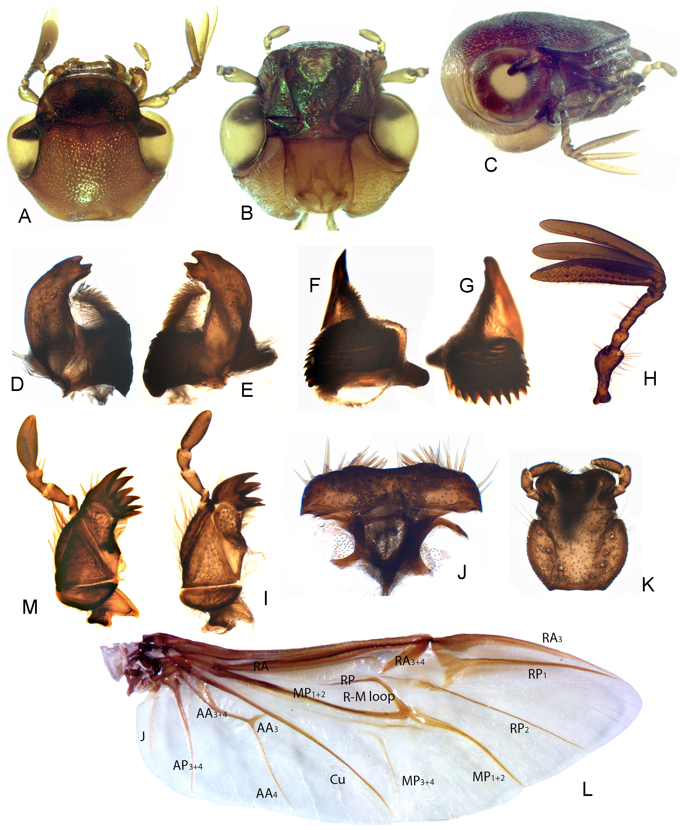
Diagnosis. Anomala can easily be separated from remaining genera of Australian Rutelinae by having the tergite and sternite of the 7th abdominal segment completely fused into a ring without visible sutures ( Fig. 2J View FIGURES 2 A – J ), and by a combination of 9 antennomeres ( Fig. 1H View FIGURES 1 A – M ) and the base of the labrum horizontal and separated by membrane from the clypeus.
Diagnostic combination. Length 6.0–18.0 mm. Body elongate oval, convex; dorsum glabrous, green or brown often with iridescent shine. Head with completely visible frontoclypeal suture ( Fig. 1A View FIGURES 1 A – M ). Labrum ( Fig. 1J View FIGURES 1 A – M ) separated by exposed membrane from clypeus, horizontal at base and not extending far beyond clypeus ( Fig. 1C View FIGURES 1 A – M ); anterior edge emarginate medially with dense bristles laterally. Antenna with 9 antennomeres ( Fig. 1H View FIGURES 1 A – M ). Mandibles multidentate apically, asymmetrical ( Figs 1A–G View FIGURES 1 A – M ) with right mandible having larger and more complex mola ( Fig. 1F View FIGURES 1 A – M ); prostheca membranous with dense fringe of marginal setae. Maxilla ( Figs 1A, 1M, 1I View FIGURES 1 A – M ) with lacinia absent; galea well developed bearing several heavily sclerotised teeth apically; terminal maxillary palpomere without sensory palp organ. Labium ( Figs 1B, 1K View FIGURES 1 A – M ) heavily sclerotised, ligula weakly emarginate medially, without median projection or process.
Prothorax transverse; prosternum ( Fig. 2B View FIGURES 2 A – J ) short in front of coxae with narrow prosternal process; procoxal cavities open externally and internally. Scutellar shield well developed ( Fig. 2E View FIGURES 2 A – J ). Mesoventrite flat, broad anteriorly, separated from small mesanepisternum by complete sutures ( Fig. 2D View FIGURES 2 A – J ). Mesoventral process narrow, extending beyond middle of mesocoxal cavity. Mesocoxal cavity obliquely transverse; laterally closed by broad mesepimeron. Mesotrochantin narrow, exposed. Metaventrite with complete discrimen. Elytra 1.3 times as long as wide and 2.5 times as long as pronotum, with fine strial punctures; epipleura glabrous, obsolete near apices; apical half with membranous border ( Fig. 4E View FIGURES 4 A – K ).
Hind wing ( Fig. 1L View FIGURES 1 A – M ) about 2.6 times as long as wide; radial bending zone relatively short, setose; R-M loop quite strong; apical field about 0.4 times total wing length, with two RP veins, MP1+2 very long and detached to base; medial field with 4 free veins, MP3+4 extremely narrow and detached, AA vein forms a basal cell with Cu; anal lobe well-developed, anal notch absent, jugal veins reduced to one.
Abdomen with 7 visible segments (II–VIII, ventrites 1–7) ( Figs 2I, J View FIGURES 2 A – J ); segments I and II connate and mostly hidden under very large and flat metacoxae ( Fig. 2H View FIGURES 2 A – J ); intercoxal process indistinct. Abdomen with 7 pairs of spiracles, terminal spiracle exposed and placed on evenly sclerotised and fused sternite and tergite VII. Pygidium convex in profile, apex narrowly rounded.
Male genitalia trilobate, symmetrical; parameres short, simple, weakly bilobed; phallobase with laterobasal apodemes; penis simple, with slender struts; endophallus with exposed setal patch. Spiculum gastrale short and “Y” shaped ( Fig. 4K View FIGURES 4 A – K ), associated plates large, not overlapping at centre. Ovipositor very short ( Fig. 4J View FIGURES 4 A – K ); gonocoxites sclerotised, kidney shaped, without gonostyli; small spermatheca and a pair of large sclerotised vaginal glands open to short vagina and bursa copulatrix.
Comments. The diagnosis and the diagnostic combination provided above are based on the Australian representatives of Anomala only.


No known copyright restrictions apply. See Agosti, D., Egloff, W., 2009. Taxonomic information exchange and copyright: the Plazi approach. BMC Research Notes 2009, 2:53 for further explanation.
|
Kingdom |
|
|
Phylum |
|
|
Class |
|
|
Order |
|
|
Family |
Anomala Samouelle
| Jin, Mengjie, Weir, Tom, Ślipiński, Adam & Pang, Hong 2014 |
Anomala
| Zorn 2006: 256 |
| Samouelle 1819: 191 |