
Chaetonotus dadayi Schwank, 1990
|
publication ID |
https://doi.org/ 10.11646/zootaxa.5213.2.1 |
|
publication LSID |
lsid:zoobank.org:pub:D6E00223-3F93-4173-B03E-FA24F1E87072 |
|
DOI |
https://doi.org/10.5281/zenodo.7361888 |
|
persistent identifier |
https://treatment.plazi.org/id/226087BF-FFBE-FFF6-3FE1-661DFCA3FD6F |
|
treatment provided by |
Plazi |
|
scientific name |
Chaetonotus dadayi Schwank, 1990 |
| status |
|
Chaetonotus dadayi Schwank, 1990 View in CoL
Synonym: Chaetonotus similis: Daday (1905) View in CoL , not Zelinka (1889)
C. dadayi View in CoL - d‘Hondt et al. (2006)
( Figs. 2–6 View FIG View FIG View FIG View FIG View FIG , Table 2 View TABLE 2 )
Examined Material
Photomicrographs available at the Museum of Biological Diversity - MDBio - IB/UNICAMP, Brazil under access numbers ZUEC GCH 73 to 104. i) Photomicrographs of 4 specimens (adults), collected in gravel sediment from the Água Limpa stream, Diamantina, Minas Gerais, Brazil (ZUEC GCH 101 to 104), ii) 14 specimens (adults) collected in a dam with floating vegetation at the Serra do Japi Ecological Station, Jundiaí, São Paulo, Brazil (ZUEC GCH 87 to 100), iii) 13 specimens (subadults and adults), collected in an urban lagoon with floating vegetation ( Eichhornia sp. ) in Paulínia, São Paulo, Brazil (ZUEC GCH 73 to 85), v) one specimen (adult) collected at the Billings Dam (lentic) in Santo André, São Paulo, Brazil (ZUEC GCH 86).
Emended diagnosis of the species
Chaetonotus with a 203 to 240 μm body length; adhesive tubes 27 to 32 μm long. Pentalobed head with two pairs of sensory ciliary tufts. Cephalion, epi- and hypopleurae present; hypostomium not observed. Strong and short rod-like reinforcements lining the walls of the mouth ring, pharynx with posterior dilation, and a narrow pharyngealintestinal junction. Scales distributed in 12 to 14 columns, with 20 to 26 scales. Pentalobed dorsal scales, increasing in size from neck to trunk and decreasing at the base of the furca. Rounded scales with small spines in the interciliary ventral area, two rows of unilobed oval scales in the ventral region of the furca. Dorsal spines with denticles positioned in the final third of the body; spines increasing in size from neck to trunk, decreasing at the base of the furca; two pairs of long spines without dorsal and ventral denticles at the base of the furca. Spines without denticles in the furcal base greater than the length of the adhesive tubes.
Description
The description is based on 18 individuals studied alive and documented digitally. Morphometric variability is shown in Table 2 View TABLE 2 .
Habitus
Tenpin-shaped with body length of 203–240 μm (figs. 2A, 4A, 5A, 6A). Pentalobed head 21–26 μm long, with two pairs of sensory tufts located latero-ventrally (with 5 to 6 cilia measuring 18–21 μm in U6 and U7). Subterminal mouth (8–11 μm in diameter) with mouth ring with rod-like cuticular reinforcements (fig. 2C). Head width 33–41 μm (U10), neck width 29–37 μm (U28) with slight constriction. Trunk width in half of the body 38–47 μm (U50) and at the base of the furca 27–33 μm (U82).
Well developed cephalion (20–25 μm long and 19–23 μm wide) covering the entire dorso-anterior part of the body (U1 to U11, figs. 2B,C, 4B, 5A,C,E). Paired epi- and hypopleurae, 18–20 μm and 23–25 μm in length (figs. 2C, 4B, 5E). Hypostomium not visible. V-shaped furca, adhesive tubes 28–34 μm in length and distally dilated (1–2 μm wide) (figs. 3A,B, 5D,F).
Scales
Body covered by 12 to 14 columns, with 20 to 26 scales per row (fig. 2A, 6A). Dorsal scales pentalobed scales posteriorly concave (figs. 4C,D, 5A,C, 6A–D), maintaining the shape along the body. Dorsal scales increase in size from the anterior to the middle body region: 7 μm (U22) to 11 μm (U58) in width, and then subsequently decrease to 6 μm (U59) (fig. 5A,B). Ventrolateral scales increase in size along the body, anterior with 4 μm in length (U10), middle with 8 μm (U43), and posterior with 9 μm (U67). Four rounded scales at the ventral base of the furca (U80) (8 μm x 4 μm and 4 μm x 3 μm), and a pair of single-lobed oval scales located at the base of the adhesive tubes (U95) (3 μm wide) (figs. 3B,C, 5F, 6G).
Spines
All spines arise from pentalobed scales, insert in a single central point on the scales, and possess a denticle (2 μm in length) in the final third (at 19–24 μm from the base) of the length (30–32 μm long) (figs. 2D, 4E, 5A,B, 6A-E). The distal part of the spine, from denticle to the apex, continues the shape of the basal part with a smooth concavity, progressively increasing in length along 4/5 of body in the dorsal region (7 μm at U25, 16 μm at U42, 32 μm at U61), decrease to 5 μm from U84 to U97 ( Fig. 6F View FIG ), and increase again in a last pair of spined (31-35 μm) at the base of the furca (figs. 3A,C, 2D, 5B). The length of this last pair of spines slightly longer than the adhesive tube. Lateral spines also increase in length along the body (18 μm at U18, 26 μm at U65, 32 μm at U68), but the penultimate pair of lateral spine longer than the last one (and also slightly longer than the adhesive tube) (figs. 2A, 5A).
Two rows of spines in the final part of the dorsal region of the body (U76), 5–9 μm long (figs. 3A, 5D, 6F), followed by two pairs of 25 μm–27 μm long and thick spines, and two small 3–4 μm long spines between them at U81 (fig. 3A). A row of four spines (5–7 μm) in the terminal ventral part of the body (U93), followed by 2 pairs of 7–9 μm long, thin spines, without denticule at U95 ( Figs. 3B,D View FIG , 5F View FIG , 6G View FIG ).
Ventral Interciliary area
Ventral interciliary area covered with rounded and unilobed scales from U6 to U95 ( Figs. 2E View FIG , 3D View FIG ). The scales and spines increase in length along the ventral body, respectively, from 2 μm and 6 μm (U19) to 4 μm and 8 μm (U59).A pair of longitudinal locomotor ciliary bands are present between U5 and U60 (fig. 2E). The ciliary bands converge medially at the base of the furca (U93) (fig. 2E). The length of the locomotor cilia increases from 6 μm around U7, to 7 μm at about U28 and finally to 9 μm at U66 (fig. 2E).
Internal Morphology
Pharynx 42–43 μm in length (U5–U22), width of the anterior region 19–21 μm, middle region 8–9 μm, posterior region 30–33 μm, presenting clear dilation at UXY (fig. 2C). Narrow small pharyngeal intestinal junction with a diameter of 12–15 μm (U20); intestine 80 μm (U20 to U80).
Probably parthenogenetic, presence of paired very large eggs (13 μm in length, 9μm in width - from U46 to U56) simultaneously present per intestine side.
Taxonomic comments
Daday (1905) identified some chaetonotids in Estância Postillon, close to the border of Paraguay and Brazil, as Chaetonotus similis Zelinka (1889) . Later, Schwank (1990) pointed out that these specimens identified by Daday (1905) could not be assigned to the same species described by Zelinka (1889), because they have two caudal spines projecting beyond the adhesive tubes and decided to create a new species - Chaetonotus dadayi Schwank (1990) . Several years later, d'Hondt et al. (2006) presented the first photographic record of a C. dadayi specimen from French Guiana.
Individuals from the different populations studied here share a series of characteristics with those described by Daday (1905), Schwank (1990) and d'Hondt et al. (2006). These are: i) the whole dorsal and dorso lateral regions covered by spined scales in the form of shields, arched anteriorly and rounded posteriorly; ii) spines on the back of the head and neck are smaller than those on the trunk, which gradually become longer and abruptly decrease in size in the final region of the body; and iii) lateral spines of the base of the furca are longer than the adhesive tubes. On the other hand, our specimens also have slight morphological variations, mainly related to morphometric features such as body length, head, neck and pharynx length and width ( Table 2 View TABLE 2 ). It is important to highlight that some comparisons were compromised, since the original and subsequent descriptions do not describe or schematize some structures present in the specimens. Finally we consider the specimens studied by us as belonging to the species C. dadayi .
Distribution
French Guiana ( d'Hondt et al., 2006), Paraguay ( Daday, 1905), Brazil: Paulínia, Santo André, Diamantina (present study) and Jundiaí (Guidetti, et al. 2021) (fig.14).

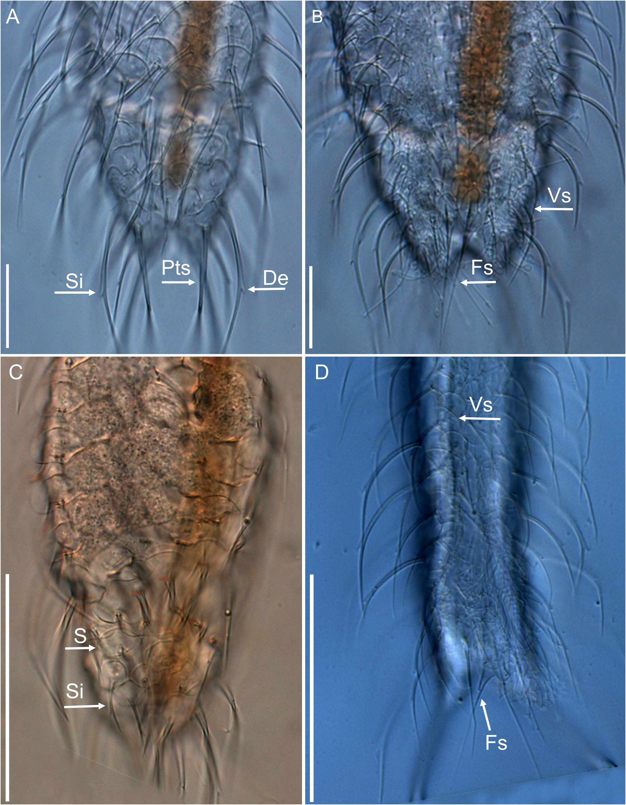
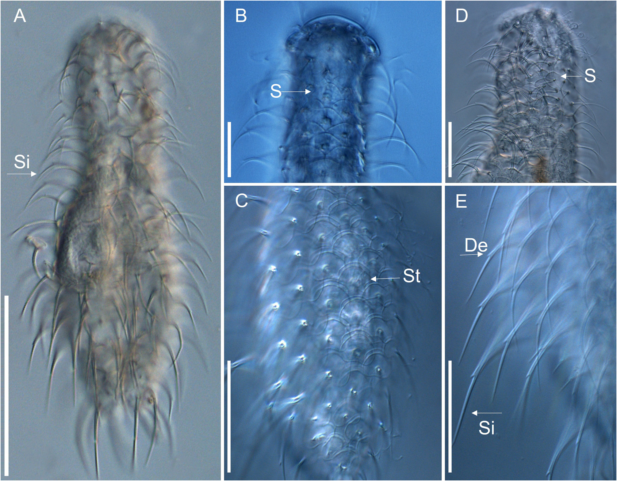
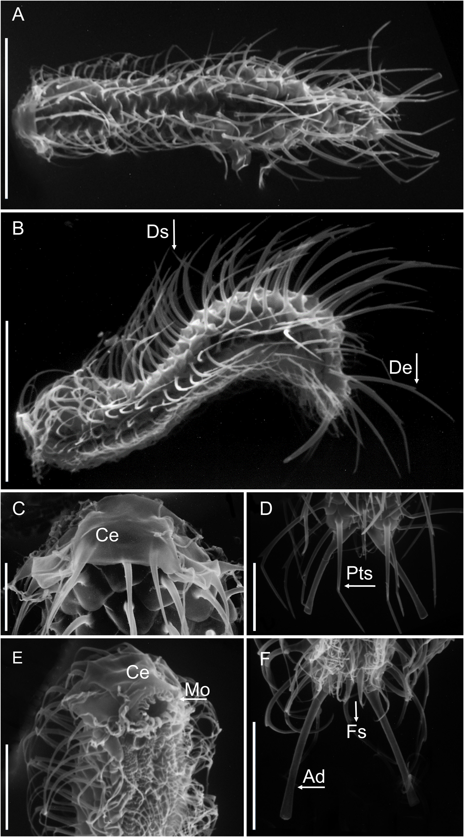
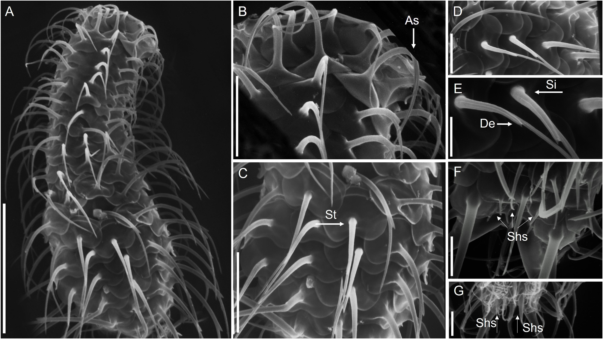
TABLE 2. Morphometric characteristics of Chaetonotus dadayi Schwank (1990) from Paraguay (Daday, 1905), French Guiana (d'Hondt et al., 2006) and Brazil (present study). "–": lack of information; X: mean measurements of Brazilian specimens; N: number of Brazilian specimens measured. Measurements in μm.
| Characteristics | Daday (1905) | d'Hondt et al., (2006) | Present study | X | N |
|---|---|---|---|---|---|
| Body length (without adhesive tubes) Total body length Head length Neck length Pharynx length Anterior dorsal spine length Trunk dorsal spine length Posterior dorsal spine length Length of the small dorsal spines at the base of the furca | 140 – 7 13 48 11 19 37 – | 187 227 – 5 52 – – 51 11 | 156–206 203–240 21–26 4 –7 42–43 5–8 14–26 30–34 5–8 | 169 216 21 4 43 7 16 32 5 | 18 18 18 18 18 18 18 11 18 |
| Anterior lateral spine length Trunk lateral spine length Posterior lateral spine length | 9 26 35 | 29 34 41 | 16–19 22–26 27–31 | 18 26 32 | 18 18 18 |
| keeled spine length Adhesive tube length Head width Mouth width Neck width Trunk width Trunk width at the furca base Anterior scale width Trunk scale width Width of small dorsal terminal scales | 25 23 – – – 50 – – – – | – 34 50 11 52 57 36 – – – | 32–27 27–32 33–41 8–11 29–37 38–47 27–33 5–7 9–12 5–7 | 28 27 32 8 29 40 28 7 11 6 | 18 18 18 18 18 18 18 18 18 18 |
No known copyright restrictions apply. See Agosti, D., Egloff, W., 2009. Taxonomic information exchange and copyright: the Plazi approach. BMC Research Notes 2009, 2:53 for further explanation.
|
Kingdom |
|
|
Phylum |
|
|
Order |
|
|
Family |
|
|
Genus |
Chaetonotus dadayi Schwank, 1990
| Salgado, Kelly Fernanda Acosta, Minowa, Axell Kou & Garraffoni, André Rinaldo Senna 2022 |
C. dadayi
| Schwank 1990 |
Chaetonotus similis:
| Daday 1905 |