
Pantepuisaurus rodriguesi
|
publication ID |
https://doi.org/ 10.5281/zenodo.185553 |
|
DOI |
https://doi.org/10.5281/zenodo.5673891 |
|
persistent identifier |
https://treatment.plazi.org/id/235D9D0C-FFF0-FFC1-FF73-F959FE6EFE36 |
|
treatment provided by |
Plazi |
|
scientific name |
Pantepuisaurus rodriguesi |
| status |
|
Pantepuisaurus rodriguesi species novum
Figs. 5–9 View FIGURE 5 View FIGURE 6 View FIGURE 7 View FIGURE 8 View FIGURE 9
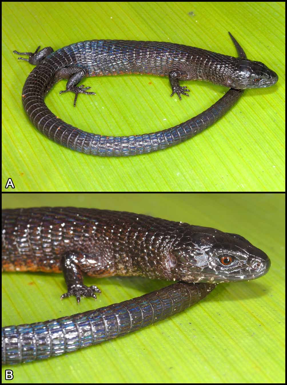
Holotype. IRSNB 2650 (field number PK 2044), an adult male collected by Paul Benjamin, Philippe J. R. Kok and Claudius Perry, 28 November 2007 at 14h55, summit plateau of Mount Maringma, Cuyuni-Mazaruni District, Guyana (05° 12’ 57”N, 060° 35’ 07”W, 2080 m).
Etymology. The specific epithet is a noun in the genitive case, honoring Brazilian herpetologist Miguel Trefaut Rodrigues (Universidade de São Paulo, São Paulo, Brazil) for his huge contribution to the knowledge of the family Gymnophthalmidae using both morphological and molecular approaches.
Diagnosis. In addition to the generic characteristics, the new species is also characterized by the following features: four supraoculars; 15 smooth temporal scales; scales around midbody 32; dorsal scales sharply keeled, in 33 transverse rows; ventral scales in 20 transverse rows; trunk length 2.6x length of forelimb; dorsum and flanks completely black, iris red.
Description of the Holotype. An adult male 58.3 mm SVL. Head length 19 % SVL, 1.38x head width, head width 1.45x head height. Head slightly wider than neck, temporal region not distinctly swollen, neck approximately as wide as anterior body. Snout blunt. Body oval in transverse section, slightly depressed, slightly wider than high. Tail complete, 93.3 mm, 1.60x SVL, circular in cross-section, tapering toward the tip. Limbs well-developed, forelimbs 19% SVL, hindlimbs 29% SVL. Limbs pentadactyl, all fingers and toes with terminal claw. Limbs short, not overlapping when adpressed along the body. Distance between forelimbs and hindlimbs (trunk length) = 29.2 mm, 50 % SVL, 2.6x forelimbs ( Fig. 5 View FIGURE 5 ).
Tongue lanceolate, anterodorsally covered by oblique (chevron-shaped), anteriorly converging plicae, posterodorsally covered by large scalelike papillae; tip bifid, smooth; one pair of infralingual plicae after fork. Anterior teeth conical, most of posterior ones bicuspid, a few of them tricuspid.
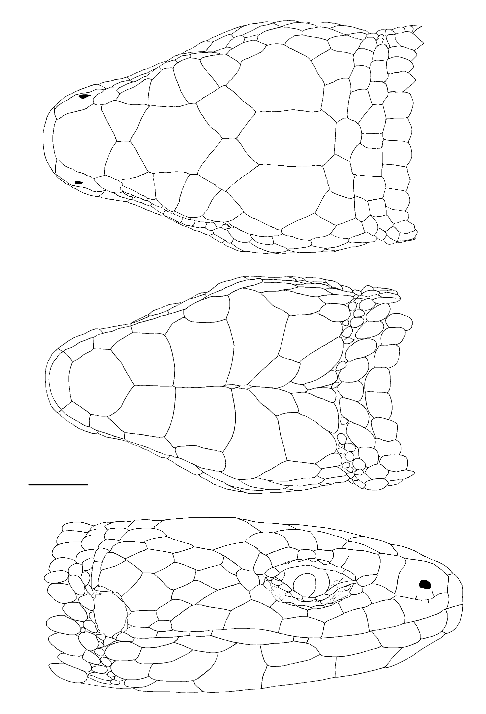
Rostral roughly hexagonal, twice as wide as high, visible from above, in broad contact with frontonasal. Frontonasal single, heptagonal, laterally in contact with nasal and loreal, not in contact with first supraocular. Prefrontals roughly pentagonal, as broad as long, with a moderately long medial suture, laterally in broad contact with first supraocular and the loreal, touching the anterior point of the second supraocular on the right, separated from it by first supraocular-frontal contact on the left. Frontal hexagonal, 1.4x as long as wide, anteriorly as wide as posteriorly, laterally markedly concave; frontal laterally in contact over the entire length of second supraocular, touching the first supraocular on the left, separated from it by prefrontal-second supraocular contact on the right, in contact with third supraocular. Frontoparietals pentagonal, 1.3x as long as wide with moderately long medial suture; each frontoparietal in contact with frontal, distinctly separated from second supraocular by frontal-third supraocular contact, in broad contact with third supraocular, parietal and interparietal, and narrow contact with the fourth supraocular. Interparietal heptagonal, posteriorly projecting, 1.7x as long as wide, with parallel sides, distinctly longer than parietals. Interparietal and parietals form a jagged, irregular, posterior margin. A row of five smooth occipitals of various shapes, shorter and wider than dorsal neck scales, the central smallest. Four supraoculars, first smallest, about 3/4 of the fourth; second and third larger, subequal. Five superciliaries, first much larger than the others, with a short incomplete suture running dorsad from ciliaries on the right side and a similar very short suture on the left side; second and third smallest, subequal, fourth distinctly larger, about twice the size of the fifth ( Fig. 6 View FIGURE 6 ).
Nasal entire, with two short divisions running ventrad from naris to supralabial on the left side, these two short divisions are incomplete, running from the first supralabial, on the right side; naris just below centre, directed laterally. Loreal quadrangular, slightly anteriorly inclined, in contact with nasal, frenocular, one preocular, first superciliary, first supraocular, prefrontal and frontonasal, separated from supralabials by frenocular-nasal contact. Two small preoculars between first superciliary and first subocular. Frenocular roughly quadrangular, longer than wide, about half-size the loreal, in contact with first supralabial. Four suboculars. Two postoculars, the lower smallest. Seven supralabials on both sides, fifth highest, fourth below middle of the eye; a very short, inconspicuous division on the first supralabial on the right side (running ventrad from nasal); suture between second and third supralabials in contact with first subocular, suture between first and second supralabials in contact with frenocular ( Fig. 6 View FIGURE 6 ).
Upper eyelid with three ciliaries on both sides, second very large, apparently resulting from the fusion of several smaller scales. Ocular recess with median row of three scales separating median ciliary from superciliaries. Lower eyelid with a semi-transparent, slightly pigmented, palpebral disc of two long vertical palpebral scales ( Fig. 6 View FIGURE 6 ).
Temporal region with 15 irregularly shaped, juxtaposed, smooth scales on both sides. Temporals arranged in four oblique rows. Ear opening moderately large, vertically semi-circular with a straight posterior margin, bordered by a few small, juxtaposed, smooth scales. Auditory meatus moderately deep, tympanum mostly transparent, slightly pigmented ( Fig. 6 View FIGURE 6 ).
Mental trapezoidal, its posterior edge almost straight, slightly concave. Postmental large, heptagonal, laterally in contact with first and second infralabials. Three pairs of large genials, first two pairs in contact with each other medially. First pair in lateral contact with second and third infralabials; second pair in lateral contact with third infralabial only, separated from fourth and fifth infralabials by a large sublabial scale, medial suture of second pair slightly longer than that of first pair; third pair in lateral contact with fifth infralabial, separated medially by two small scales. A pair of enlarged postgenials separated medially by scales of varying sizes and widely separated from infralabials. Seven infralabials on the right side, six on the left side, fourth scale highly reduced on both sides. One row of imbricate, smooth pregulars with rounded posterior margins. Five rows of gulars, four of them with two transversely enlarged scales. Collar row with seven scales, central one distinctly larger, others decreasing in size laterally, forming a slightly scalloped margin. Gular fold distinct, concealing one row of very small scales ( Figs. 6 View FIGURE 6 , 7 View FIGURE 7 B).
Head scales with numerous small pits, especially concentrated on rostral, frontonasal, supraoculars, and lateral head scales, also present on ventral head scales, and body and limbs scales. Scales on nape posterior to occipitals longer than wide, with a broad, flat keel, mucronate, in transverse rows, grading posteriorly into dorsal body scales. Sides of neck with medium-sized, roughly subequal, smooth, juxtaposed scales in about eight oblique rows, not distinctly decreasing in size posteriorly but interrupted by a row of smaller scales 2/3 the way before arm insertion, which weakly form annuli with nape and gular scales.
Dorsals imbricate, pentagonal, some appearing hexagonal because of imbrication, elongate, sharply keeled, mucronate, except very few isolated scales. Dorsal scales in transverse rows only, 33 rows between the interparietal and the posterior edge of hindlimbs (including row of occipitals). Middorsal scales not distinctly narrower than adjacent dorsals. A zone of small juxtaposed scales before arm insertion. Flank scales similar to, but slightly narrower than, dorsal scales, most of flank scales are not mucronate but have a straight or slightly rounded posterior margin; no abrupt demarcation between dorsals and laterals. Lateral scales becoming slightly smaller just before leg insertion. No row of ventrolateral scales (see Myers & Donnelly 2008) can be detected; some very small, longitudinally oval or rounded scales intercalated in the interstitial skin between the transverse rows of lateral; these small scales are more conspicuous along the first anterior third of flank and before leg insertion. All transverse rows of dorsals correspond to one row of laterals, except at midbody on the right side where one transverse row of dorsals corresponds to two rows of laterals, and before leg insertion and after arm insertion.
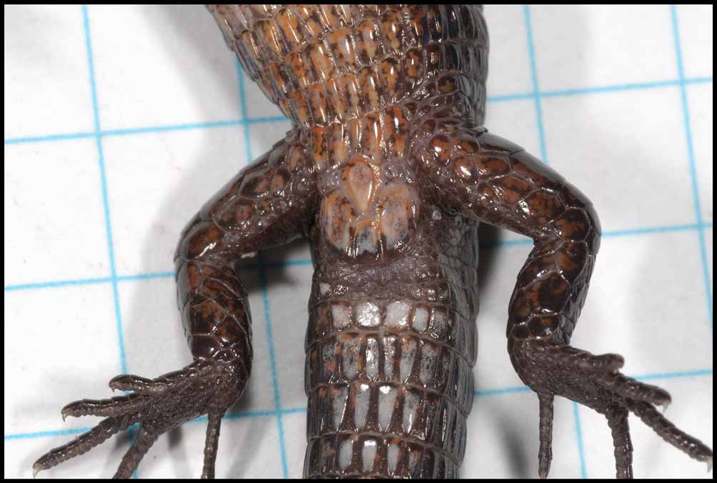
Ventrals imbricate, broadly keeled, longer than wide, mucronate except very few isolated scales, in transverse rows only ( Fig. 7 View FIGURE 7 A). Most transverse rows of ventrals correspond to a transverse row of laterals and of dorsals (exceptions occur at midbody). Twenty transverse rows between collar and preanal plate, 32 scales around midbody. Preanal plate consisting of six scales, one anterior, five posterior scales, most lateral posterior scales narrow, distinctly smaller than adjacent scales ( Fig. 8 View FIGURE 8 ). Four preanal pores. Four femoral pores on the right side, three on the left side, each femoral pore between three scales.
Scales on tail keeled, not mucronate (except proximally), slightly imbricate, roughly rectangular, as long as dorsal scales, in transverse rows only, in uninterrupted annuli. Pigmented subcaudal scales keeled, slightly imbricate, slightly mucronate, roughly rectangular, in transverse rows only.
Dorsal surfaces of upper and lower arms with smooth, variably polygonal scales, ventral surface of upper arms with small, juxtaposed scales, ventral surface of lower arms with scales similar to those on dorsal surface, except at the base of hands where scales are juxtaposed and distinctly smaller. Thighs with a row of enlarged, smooth, roughly hexagonal scales on anterior surface, bordered ventrally by two rows of smaller polygonal scales diminishing in size ventrad (scales of first row smooth, scales of second row broadly keeled); bordered dorsally by 2–3 rows of keeled scales diminishing in size dorsad, grading into small, smooth (but with many very small asperities and pits that give a rugose aspect), slightly granular, juxtaposed scales, which cover posterodorsal and posterior surfaces of thighs; these scales increase in size ventrally. Shanks ventrally and posteriorly with large, smooth, polygonal, imbricate scales. Scales on upper and anterior surfaces of shanks slightly smaller, imbricate, rugose (see above), and strongly keeled.
Dorsal surface of hands and digits with imbricate, smooth scales, dorsal surface of feet with imbricate, broadly keeled scales, except a few anterodorsally; palm and soles covered with small, smooth, slightly protuberant, juxtaposed, polygonal scales. Two enlarged, subequal, thenar scales on inner margin of palm below pollex, each with produced inner edge, similar scales occur at the base of Toes I and III–IV; two slightly enlarged hypothenar scales on outer margin of palm. Subdigital lamellae divided basally and single on distal halves on most digits.
Subdigital lamellae as follows (Roman numbers indicate digits, Arabic numbers indicate paired or single lamellae on left/right; lower ungual sheath scale is omitted from the counts): hands I 4 /4, II 7 /7, III 9 /9, IV 10/11, V 6/7; feet I 5 /5, II 8 /8, III 11/10, IV 16/16, V 9/8.
Color of Holotype in life. Dorsal and lateral surfaces completely black; closer examination shows that black scales on body and neck are peppered with brown or reddish brown. Upper surface of head black. Tympanum light pink. Arms, legs and tail black. Iris red ( Fig. 5 View FIGURE 5 ). Tongue dark gray in its anterior two-thirds, whitish with gray flecks on its posterior third. Palms and soles black. Underside of head and throat black with irregular light gray markings; venter orangish brown, ventral scales peppered with black; underside of upper legs black; underside of lower legs black with irregular orangish brown markings; underside of tail black, except proximally where some scales are white ( Figs. 7–8 View FIGURE 7 View FIGURE 8 ).
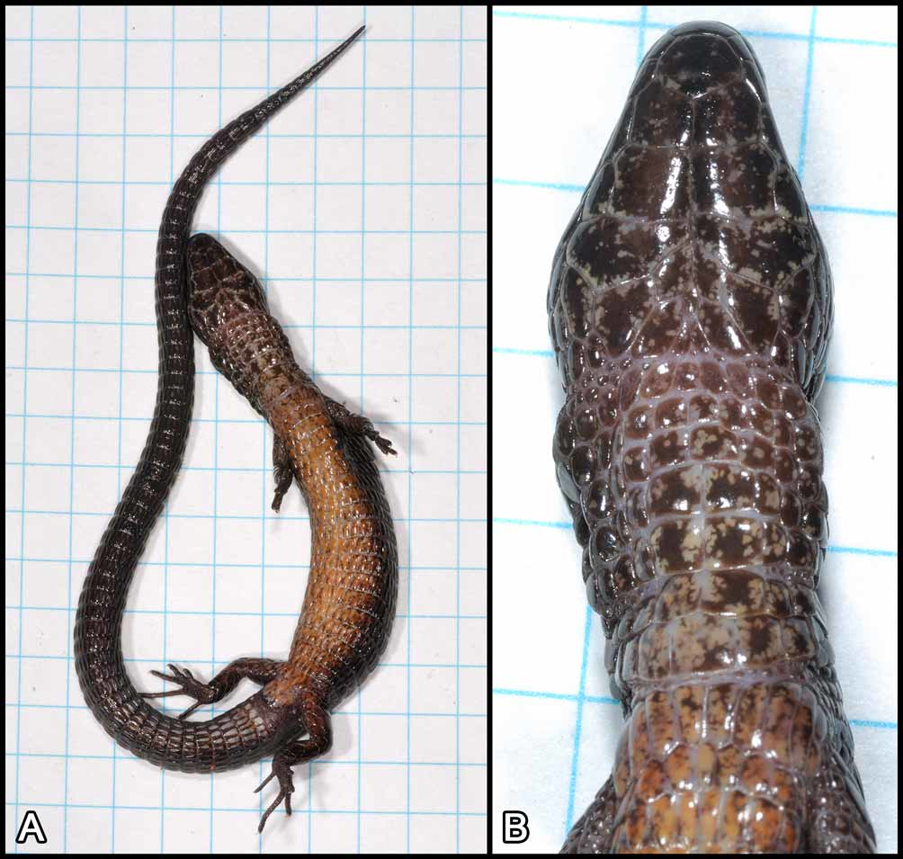
Color of Holotype in preservative. After four months in 70 % ethanol, the holotype became very dark brown, the light gray markings on the underside of head and chest are white; venter is whitish peppered with dark gray; tympanum is transparent, no other noticeable change.
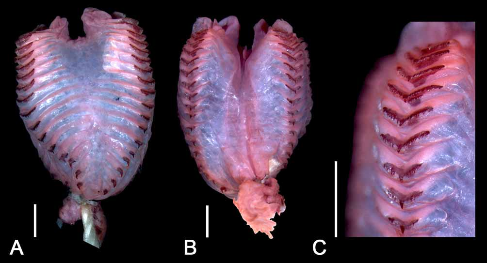
Hemipenis. The hemipenes of the holotype were everted in the field. The left organ was subsequently removed, sent to and prepared by Pedro Nunes (Universidade de São Paulo, São Paulo, Brazil), currently working on a PhD thesis on the hemipenial morphology in the family Gymnophthalmidae . The hemipenis was inflated with blue petroleum jelly and the spicules were stained with Alizarin Red ( Fig. 9 View FIGURE 9 ). The left hemipenis is 6.9 mm long, with a greatest width of 4.5 mm. The organ is weakly bilobed, with each of the short divisions terminating in 2–3 protuberances. Sulcus spermaticus medial, broad, poorly defined. Asulcate surface with a conspicuous series of 15–18 curved transverse plicae (tissue ridges) encircling the side of the organ and terminating laterally on the sulcate surface, most plicae being laterally shortly interrupted by a narrow nude space. A wide nude space between the ca. 8–9 distal plicae on the asulcate surface. All plicae bear mineralized spicules; plicae located on the proximal part of the hemipenis bear a single spicule, the number of spicules is increasing from the proximal part towards the tip resulting in plicae bearing a series of several uniform spicules interrupted by a larger central one ( Fig. 9 View FIGURE 9 ).
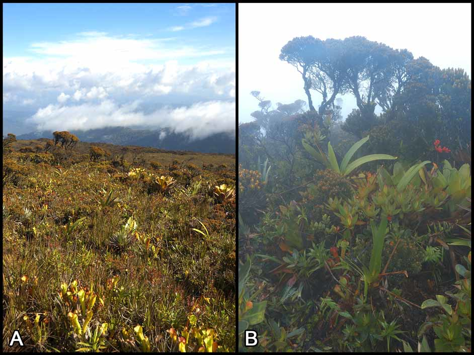
Distribution and ecology. The species is currently known only from the type locality, the summit of Maringma tepui in Guyana ( Fig. 1 View FIGURE 1 ). The only specimen available was collected in the afternoon (14h55), during a sunny spell, in a sunny spot, on the ground, among dense vegetation ( Fig. 3 View FIGURE 3 ). No other specimen was observed. The only other lizard species recorded from the summit of Mount Maringma is Arthrosaura hoogmoedi Kok, 2008 .

| IRSNB |
Institut Royal des Sciences Naturelles de Belgique |
No known copyright restrictions apply. See Agosti, D., Egloff, W., 2009. Taxonomic information exchange and copyright: the Plazi approach. BMC Research Notes 2009, 2:53 for further explanation.