
Physocyclus paredesi Valdez-Mondragón, 2010
|
publication ID |
https://doi.org/ 10.11646/zootaxa.3866.2.2 |
|
publication LSID |
lsid:zoobank.org:pub:C8987D65-67A6-4F1C-97EA-3C0D5FB3FF0D |
|
DOI |
https://doi.org/10.5281/zenodo.5672991 |
|
persistent identifier |
https://treatment.plazi.org/id/241D950E-0568-FFE1-FF41-FC39FE22FE16 |
|
treatment provided by |
Plazi |
|
scientific name |
Physocyclus paredesi Valdez-Mondragón, 2010 |
| status |
|
Physocyclus paredesi Valdez-Mondragón, 2010 View in CoL
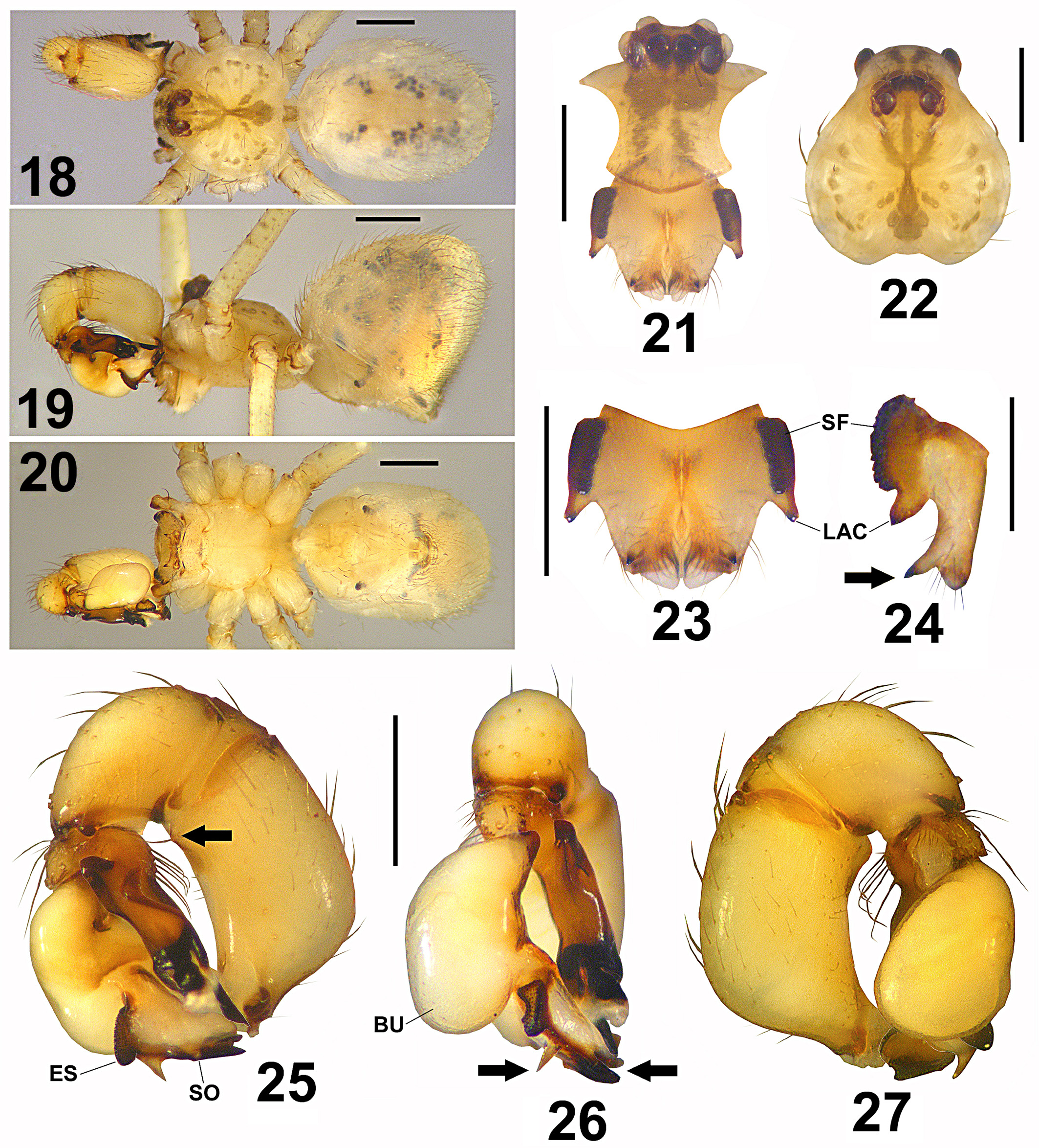
Figures 18–33 View FIGURES 18 – 27 View FIGURES 28 – 33
P. paredesi Valdez-Mondragón, 2010 View in CoL ; page 76, figs. 204–207 (description ♂).
Type data. MEXICO: Oaxaca: Municipio Asunción Ixtaltepec: river near to Nizanda (N 16.6575°, W 95.0105°): 1 male holotype (CNAN-T0420), 19 August 2002, R. Paredes, E. Cabrera cols., diurnal collecting on vegetation.
Material examined. MEXICO: Oaxaca: km 278 road Tehuantepec-Juchitán de Zaragoza (N 16.3682°, W 95.1675°; 27 m): 1 male, 1 female ( CNAN), 27 March 2010, A. Valdez, O. Francke, C. Santibáñez, J. Cruz cols. Municipio Santa María Mixtequilla: Santa María Mixtequilla (N 16.3759°, W 95.2421°; 55 m): 1 male ( CNAN), 27 March 2010, A. Valdez, O. Francke, C. Santibáñez, J. Cruz cols.
Diagnosis. Resembles P. gertschi ( Valdez-Mondragón, 2010; figs. 140–143), male distinguished by lateral apophyses of chelicerae more rounded basally in lateral view ( Fig. 24 View FIGURES 18 – 27 ); subdistal frontal apophyses on chelicerae (arrow, Fig. 24 View FIGURES 18 – 27 ); procursus slightly curved dorsally and curved just in the base ventrally ( Fig. 25 View FIGURES 18 – 27 ), without the small distal notch ( Fig. 25 View FIGURES 18 – 27 ); the distal spine on the procursus longer and wide distally ( Fig. 25 View FIGURES 18 – 27 ). Females distinguished from P. gertschi ( Valdez-Mondragón, 2010; figs. 144–146) by epigynum with paired apophyses smaller, more widely separated from each other than on P. g er t s c hi ( Fig. 31 View FIGURES 28 – 33 ); without paired and curved concavities; and by pore plates oval and short ( Fig. 33 View FIGURES 28 – 33 ).
Redescription. Male (holotype): [Note: The next male redescription is a translation from Spanish to English from the original description made by Valdez-Mondragón (2010), useful and convenient to future taxonomic work].
Prosoma: Carapace with pale gray irregular pattern, which is around the fovea, extended to posterior part of ocular region, trident-shaped on posterior part of ocular region ( Fig. 22 View FIGURES 18 – 27 ). Clypeus with gray pattern extending to three quarters of its total length, on distal fourth the pattern is bifurcated ( Fig. 21 View FIGURES 18 – 27 ). Chelicerae without sclerotized cones, with a large concavity formed by both chelicerae ( Figs 21, 23 View FIGURES 18 – 27 ). In frontal view, the lateral apophyses of chelicerae are thin and conical ( Fig. 23 View FIGURES 18 – 27 ); in lateral view are curved in proximal part, and with a wide tip distally ( Fig. 24 View FIGURES 18 – 27 ). Sternum uniformly pale orange ( Fig. 20 View FIGURES 18 – 27 ). Endites longer than wide, labium wider than long ( Fig. 20 View FIGURES 18 – 27 ). Legs: Coxae and trochanters uniformly pale yellow ( Fig. 20 View FIGURES 18 – 27 ). Femora with numerous pale gray spots ( Fig. 19 View FIGURES 18 – 27 ). Tibiae with numerous pale gray spots smaller than on femora. Tibiae with one basal gray ring and another one distally. Metatarsi and tarsi pale orange. Tarsi with paired claws, curved. Tibiae and metatarsi without curved setae. Opisthosoma: Globular, longer than high ( Fig. 19 View FIGURES 18 – 27 ), gray, with numerous small gray and white irregular spots laterally ( Figs 18, 19 View FIGURES 18 – 27 ), midline unspotted. Plate of genital gonopore pale orange, square ( Fig. 20 View FIGURES 18 – 27 ). Spinnerets pale gray ( Fig. 20 View FIGURES 18 – 27 ). Palp: Femur wide, slightly curved ventrally; with distal apophysis ventrally, which is rounded, inconspicuous, with one long seta apically (arrow, Fig. 25 View FIGURES 18 – 27 ). Procursus brownish, paler proximally, black distally, with dorsal apophysis long and wide, and the ventral notch notably deep ( Fig. 25 View FIGURES 18 – 27 ). Procursus ventrally with several long setae ( Fig. 25 View FIGURES 18 – 27 ), with brush of pseudotrichia next to the distal spine ( Fig. 25 View FIGURES 18 – 27 ). Distal spine of procursus long and wide ( Fig. 25 View FIGURES 18 – 27 ). Bulb wide, with a long and wide trapezoidal embolus ( Fig. 25 View FIGURES 18 – 27 ). Embolic sclerites placed on dorsal part of embolus ( Figs 25, 26 View FIGURES 18 – 27 ). Embolus pale yellow retrolaterally, dark sclerotized only in the margins ( Fig. 25 View FIGURES 18 – 27 ), with a conspicuous apical spine (left arrow, Fig. 26 View FIGURES 18 – 27 ). Spermatic operculum subdistal ( Fig. 25 View FIGURES 18 – 27 ); in dorsal view, the embolus is bifurcated apically (right arrow, Fig. 26 View FIGURES 18 – 27 ). Measurements: Total length 2.4. Carapace 1.1 long, 1.2 wide. Clypeus 0.5 long. Diameter AME 0.12, ALE 0.14, PME 0.11, PLE 0.13. Distance AME-PME 0.07, ALE- PME 0.06, PME-PME 0.16. Leg I: 21.9 (5.9+0.4+6.1+8.5+1.0). Tibia II: 3.9, tibia III: 52.6, tibia IV: 4.3; tibia I L/ d 43.
Female. Similar to the male, differences: Prosoma: Carapace slightly darker orange than on male ( Fig. 28 View FIGURES 28 – 33 ). Dorsal pattern of coloration of carapace more marked than on the male, with three groups of gray spots on each side ( Fig. 28 View FIGURES 28 – 33 ). The trident-shaped pattern on posterior part of ocular region more marked than the male ( Fig. 28 View FIGURES 28 – 33 ). Clypeus with a wide, longitudinal gray region, which has a white spot medially. Chelicerae pale gray, basally pale yellow. Sternum, endites and labium yellow ( Fig. 30 View FIGURES 28 – 33 ). Legs: Shorter and more robust than on male, coloration pattern as on male, with numerous gray spots bigger and more marked than on the male ( Fig. 30 View FIGURES 28 – 33 ). Coxae and trochanters pale orange ( Fig. 29 View FIGURES 28 – 33 ). Opisthosoma: More voluminous than the male ( Fig. 29 View FIGURES 28 – 33 ). Epigynum: Wider than long ( Fig. 31 View FIGURES 28 – 33 ). Ventral apophyses of epigynum paired and small ( Fig. 31 View FIGURES 28 – 33 ), located on median part ( Figs 31, 32 View FIGURES 28 – 33 ).
Pore plates oval ( Fig. 33 View FIGURES 28 – 33 ). Measurements: Total length 3.2. Carapace 1.1 long, 1.0 wide. Clypeus 0.4 long. Diameter AME 0.10, ALE 0.11, PME 0.09, PLE 0.12. Distance AME-PME 0.03, ALE-PME 0.05, PME-PME 0.10. Leg I: 13.7 (3.8+0.4+3.9+4.7+0.9). Tibia II: 2.5, tibia III: 1.8, tibia IV: 2.9; tibia I L/d 26.
Natural history. The male specimen from Santa María Mixtequilla was collected in a dry tropical forest, among boulders on the ground and rock walls.
Distribution. Asunción Ixtaltepec, Oaxaca, Mexico, type locality. Santa María Mixtequilla, Oaxaca, Mexico, additional material ( Fig. 34 View FIGURE 34 ).


No known copyright restrictions apply. See Agosti, D., Egloff, W., 2009. Taxonomic information exchange and copyright: the Plazi approach. BMC Research Notes 2009, 2:53 for further explanation.
|
Kingdom |
|
|
Phylum |
|
|
Class |
|
|
Order |
|
|
Family |
|
|
Genus |
Physocyclus paredesi Valdez-Mondragón, 2010
| Valdez-Mondragón, Alejandro 2014 |
P. paredesi Valdez-Mondragón, 2010
| Valdez-Mondragon 2010 |