
Holopothrips striatus, Jorge, Nina Castro, Cavalleri, Adriano, Bedetti, Cibele Souza & Isaias, Rosy Mary Dos Santos, 2016
|
publication ID |
https://doi.org/ 10.11646/zootaxa.4200.1.8 |
|
publication LSID |
lsid:zoobank.org:pub:4839B23B-3818-41C5-8F55-75D5707006B6 |
|
DOI |
https://doi.org/10.5281/zenodo.5673013 |
|
persistent identifier |
https://treatment.plazi.org/id/244087A3-FF83-FFE3-ED97-FA44578CF9BE |
|
treatment provided by |
Plazi |
|
scientific name |
Holopothrips striatus |
| status |
sp. nov. |
Holopothrips striatus View in CoL sp. n.
( Figs 1, 3–9 View FIGURES 1 – 5 View FIGURES 6 – 9 )
Macropterous female. Body uniformly dark brown including legs, fore tarsi somewhat paler; antennal segment I concolorous with head, II brownish at basal 2/3 and yellowish at tip, III–IV largely yellow but lightly shaded in apical third; V–VI with apical half brown; VII and VIII uniformly brown; fore wings pale but scale brown; without median dark line; tube dark brown at basal third and paler at apex.
Head about 1.4 times as long as greatest width ( Fig. 4 View FIGURES 1 – 5 ), cheeks straight and with faint lines of transverse reticulation; eyes about 0.4 times as long as head and not holoptic; po long and dilated at tip; maxillary stylets not retracted as far as po and about 1/3 of head width apart; antennae slender and 8-segmented, III and IV with three simple sense cones each; segment VI and VII with a narrow basal neck, segment VIII not broadly joined to VII ( Fig. 5 View FIGURES 1 – 5 ). Mouth-cone pointed and reaching ferna ( Fig. 3 View FIGURES 1 – 5 ).
Pronotum transverse and smooth; four pairs of long and capitate setae, only one pair of elongate setae on epimera, am pointed and short, usually not longer than discal setae ( Fig. 6 View FIGURES 6 – 9 ); prosternum with well-developed ferna and basantra sometimes faintly sclerotized ( Fig. 3 View FIGURES 1 – 5 ); epimeral sutures incomplete; mesonotum with elongate transverse reticles; metanotum closely striate and with few internal markings ( Fig. 8 View FIGURES 6 – 9 ); fore wings parallel sided, each with three long and capitate setae arising basally and about 15 duplicated cilia. Pelta reticulate and triangular in shape, but with distinct lateral lobes extending posteriorly ( Fig. 7 View FIGURES 6 – 9 ); paired campaniform sensilla present; abdominal tergites II–VII each with three pairs of wing-retaining setae; tergite IX setae S1 and S2 long and acute; length of tergite X about 0.65 of head length. Spermatheca not enlarged.
Measurements of female (holotype), in microns: Length about 2,875; head length 325, greatest width across cheeks 225, po length 80, eye dorsal length 135, interval between eyes (dorsal) 72; median length of pronotum 160, width 300, am length 20, aa length 48, ml length 58, ep length 107, pa length 105; tergite IX setae S1 262, S2 277; tergite X length 212, basal width 100, apical width 50; length (width) of antennal segments II–VIII 62 (30), 100(35), 88(35), 85(32), 75(27), 70(22), 37(12), respectively.
Macropterous male. Similar to female in structure and coloration, though smaller and with fore tibiae and tibiae paler ( Fig. 1 View FIGURES 1 – 5 ); abdominal sternites VII and VIII each with one transverse pore plate extending across the posterior region (interrupted medially on VII) and two oval pore plates placed anterolaterally ( Fig. 9 View FIGURES 6 – 9 ); S2 setae on abdominal tergite IX acute and not shortened.
Measurements of male (paratype): Length about 2,625; head length 293, greatest width across cheeks 210, po length 77; tergite IX setae S1 225, S2 247; length (width) of antennal segments II–VIII 60 (30), 97(30), 80(32), 75(30), 70(22), 62(22), 32(12), respectively.
Larva II. Body color largely white but antenna and abdominal segments IX–X brown, transverse bands of red internal pigmentation present in thorax and abdomen ( Fig. 2 View FIGURES 1 – 5 ).
Material examined. Holotype female, Brazil, Rio Grande do Sul, São Francisco de Paula , 17.ii.2014, on Myrcia retorta galls (A. Cavalleri) ( UFRGS 3451 View Materials ) . Paratypes: 2 females, 3 males collected with holotype.
Remarks. Holopothrips striatus might be related to H. orites from Peru, which also exhibits a long head and a closely striate metanotum. This type of metanotal sculpture is found only in few species of Holopothrips , with most species bearing elongate or equiangular reticles medially on the metanotum. The Brazilian H. affinis , H. erianthi and H. omercooperi also have a striate metanotum but the head shape and maxillary stylet position are distinct from H. striatus ( Table 1 View TABLE 1 ). The male sternites of these species also bear pore plates of similar shape to H. striatus , but in H. affinis and H. erianthi these structures are present on VI–VIII, whereas H. omercooperi has pore plates only on VIII. Moreover, the pelta of H. striatus is unusual in having lateral lobes.
The recognition of Holopothrips species is usually problematic due to extensive morphological variation within and between populations ( Mound & Marullo 1996; Cavalleri & Kaminski 2007), and the only available key to species ( Mound & Marullo 1996) does not include the species described in the last 20 years (see ThripsWiki 2016). Moreover, approximately 50% of the Brazilian species were described without any host-plant information or observation on their way of life. Information regarding the biology of these thrips certainly can be useful for delimiting species and understanding the origin of the galling behaviour in this group. Many Holopothrips species have been considered as gall-inducers, but no attempt has been made previously to investigate the tissue alterations induced by this lineage of Neotropical thrips.
Holopothrips striatus induces green galls with brown spots, on the upper surface of Myrcia retorta leaves, provoking a fold/roll movement of leaf lamina upwards, along both sides of the mid rib ( Figs 10–11 View FIGURES 10 – 12 ). The galling insects develop inside a chamber formed by the whole leaf, which shelters an aggregation of H. striatus in all stages of development. The number of thrips per gall is variable, but more than 30 individuals (among immature and adults) were observed living in a single gall. The main feature of this gall is the alternation of sites with homogeneous cells and sites of HR along the altered leaf lamina. The cells of the homogeneous galled areas are alive, while the cells of the HR areas accumulate dense phenolic content ( Fig. 12 View FIGURES 10 – 12 ). The galls are permanently open, and allow the communication of the thrips with the outer environment.
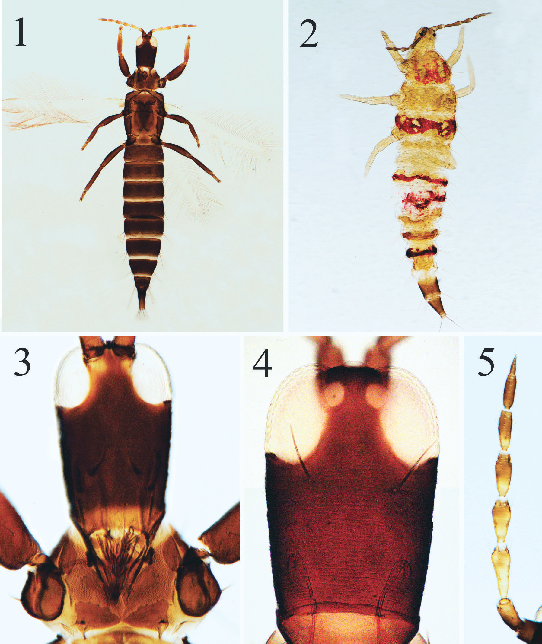
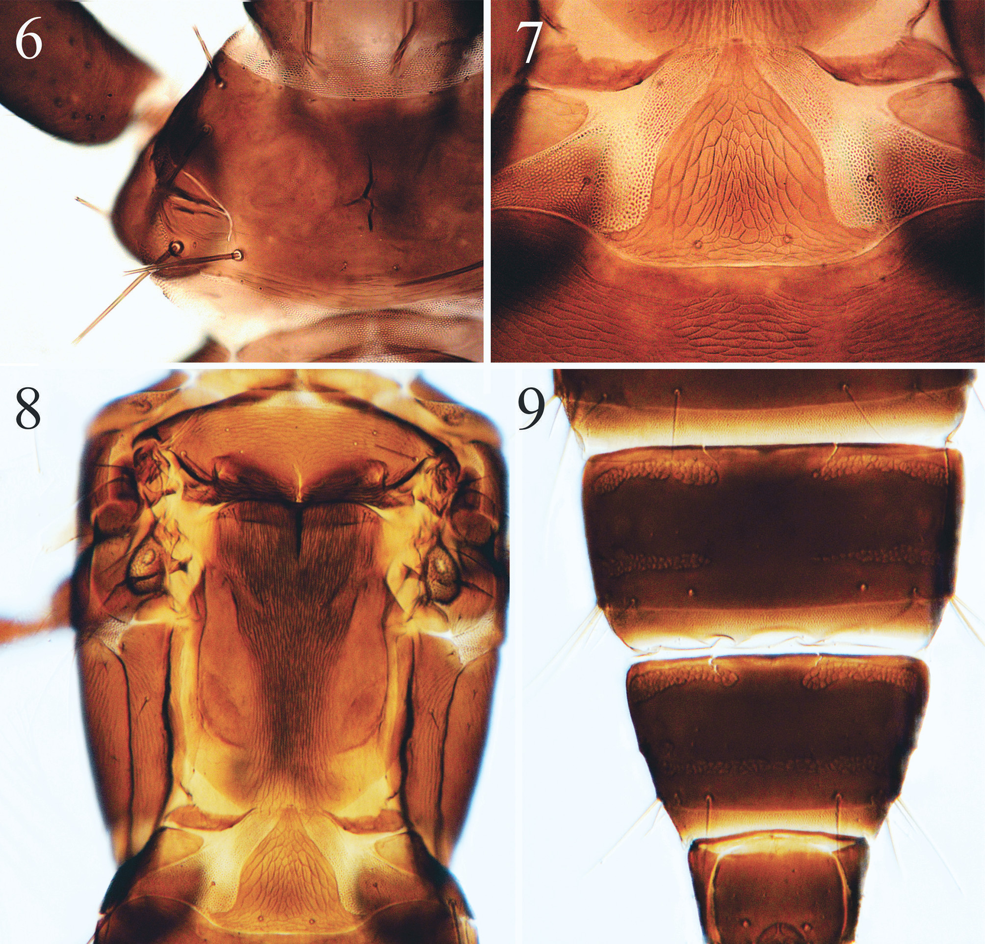

TABLE 1. Morphological comparison of the Holopothrips species with longitudinal striate sculpture on metanotum. L: W = Length / width ratio; am = pronotal anteromarginal setae.
| Species | Head L:W | Maxillary stylets AM setae | Male sternal pore plates |
|---|---|---|---|
| H. affinis (Bagnall) | 1.1 | wide apart and not retracted as very short, rarely larger than far as PO setae discal setae | VI – VIII |
| H. erianthi (Hood) | 1.3 | retracted as far as PO and well-developed close together medially | VI – VIII |
| H. omercooperi (Bagnall) | 1.2 | wide apart and not retracted as well-developed far as PO setae | VIII only |
| H. orites Hood | 1.6 | close together medially and well-developed retracted as far as PO setae | unknown |
| H. striatus sp. n. | 1.4 | wide apart and retracted to very short, rarely larger than basal third of head length discal setae | VI – VIII |
| Myrcia retorta galls |
| UFRGS |
Universidade Federale do Rio Grande do Sul |
No known copyright restrictions apply. See Agosti, D., Egloff, W., 2009. Taxonomic information exchange and copyright: the Plazi approach. BMC Research Notes 2009, 2:53 for further explanation.