
Diascorhynchus caligatus Ax, 1959
|
publication ID |
https://doi.org/ 10.5852/ejt.2020.595 |
|
publication LSID |
lsid:zoobank.org:pub:F81A7282-A44B-4E70-9A44-FE8F67E5C1EA |
|
DOI |
https://doi.org/10.5281/zenodo.3664221 |
|
persistent identifier |
https://treatment.plazi.org/id/245C87ED-4B1A-C83C-FDE4-FB416C3AEA06 |
|
treatment provided by |
Plazi |
|
scientific name |
Diascorhynchus caligatus Ax, 1959 |
| status |
|
Diascorhynchus caligatus Ax, 1959 View in CoL
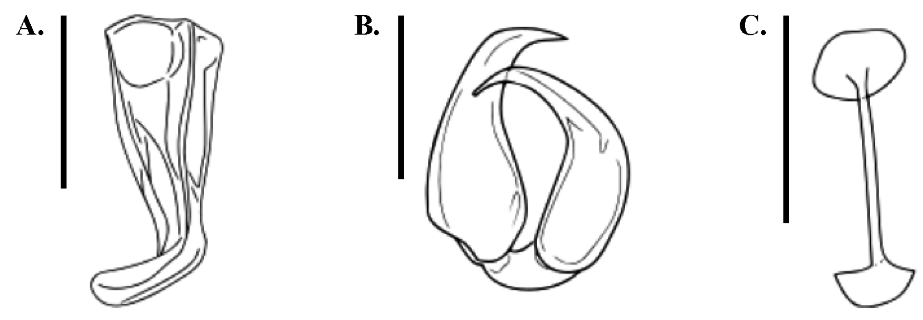
Fig. 4 View Fig
Material examined
PORTUGAL • 1 whole-mount (photographs of live specimen available); Algarve region , Olhos de Água; 37°05′20″ N, 08°11′17″ W; 15 May 2013; B. Tessens leg.; fine, clean sand from intertidal at open beach, west of rocky outcrop at depth of 0.5 m; HU X.2.06 GoogleMaps .
SPAIN • 1 whole-mount; Andalusia, Doñana National Park ; 6 Apr. 2008; B. Tessens and N. Van Steenkiste leg.; beach at 5 km from river mouth, sandy sediment from the lower tidal mark; HU X.2.07 .
Previously known distribution
Sile, Black Sea, Turkey ( Ax 1959).
Remarks
The overall habitus and internal organisation of the collected specimens correspond well to the description by Ax (1959). The body length measures between 1.6 and 1.7 mm, which is at the lower end of the size range of 1.6–2.0 mm reported by Ax (1959). The split proboscis bears a pair of large, sclerotised hooks ( Fig. 4B View Fig ). The dorsal hook is more robust and measures 30–32 μm in length, while the ventral hook measures 31–34 μm. Two elongate lateral gland sacs connect to the proximal end of the proboscis. The rosulate pharynx is situated slightly posterior to the body centre. Six globular testes lie in a single, medial row anterior to the pharynx.
The copulatory bulb varies in length between 61 μm and 86 μm and holds a sclerotised stylet ( Fig. 4A View Fig ). The stylet measures 42 to 48 μm. These measurements are in line with the descriptions of Ax (1959).
A single ovary and a bursa occur near the caudal body end. In the specimen from Doñana, we observed a sclerotised, spermatic duct, connecting the bursa to the ovary. Similar to what was described by Ax (1959), this duct is a straight tube, measuring around 26 μm in length and terminating at both ends in a small, sclerotised disk ( Fig. 4C View Fig ). The spermatic duct was not observed in the Portuguese specimen. Vitellaria are paired and occur laterally, between the ovaries and the brain.
No known copyright restrictions apply. See Agosti, D., Egloff, W., 2009. Taxonomic information exchange and copyright: the Plazi approach. BMC Research Notes 2009, 2:53 for further explanation.
|
Kingdom |
|
|
Phylum |
|
|
Class |
|
|
SubClass |
Trepaxonemata |
|
Order |
|
|
SubOrder |
Kalyptorhynchia |
|
InfraOrder |
Schizorhynchia |
|
Family |
|
|
Genus |