
Coronamyia, Bravo & Lopes & Araújo, 2023
|
publication ID |
https://doi.org/10.11646/zootaxa.5351.1.6 |
|
publication LSID |
lsid:zoobank.org:pub:E43B08B1-4DC0-4327-B239-4F47366A4D67 |
|
DOI |
https://doi.org/10.5281/zenodo.8391522 |
|
persistent identifier |
https://treatment.plazi.org/id/2565122B-0174-FFCF-03B8-FE59FE91D61B |
|
treatment provided by |
Plazi |
|
scientific name |
Coronamyia |
| status |
gen. nov. |
Coronamyia gen. nov.
Type species: Coronamyia paschoali sp. nov., by present designation.
Etymology. From Latin, Corona, meaning crown, and Greek, Myia, meaning fly, referring to the setous ring around the aedeagus. Feminine gender.
Diagnosis. Wing with R 5 ending in a pointed wing apex. Thoracic sensory area with microtrichia in the dorsal area of anepimeron; small circular thoracic gland near the thoracic sensory area with microtrichia. Aedeagus with a lightly sclerotized plate with long, thin setae surrounding it. Gonostyli glabrous, sclerotized, bifurcated, shorter than the gonocoxites, internal arm curved, external arm with basal lobes of different shapes. The hypopod is elongated and lacks an apical tenaculum. It has two groups of accessory tenacula dorsally: a group of short accessories tenacula at the base of the hypopod and one or two strips of long accessory geniculate tenacula in the remaining part of the hypopod, each inserted in a larger alveoli.
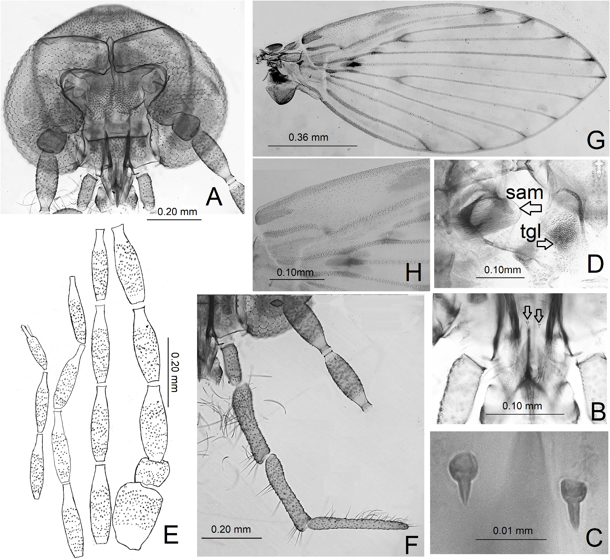
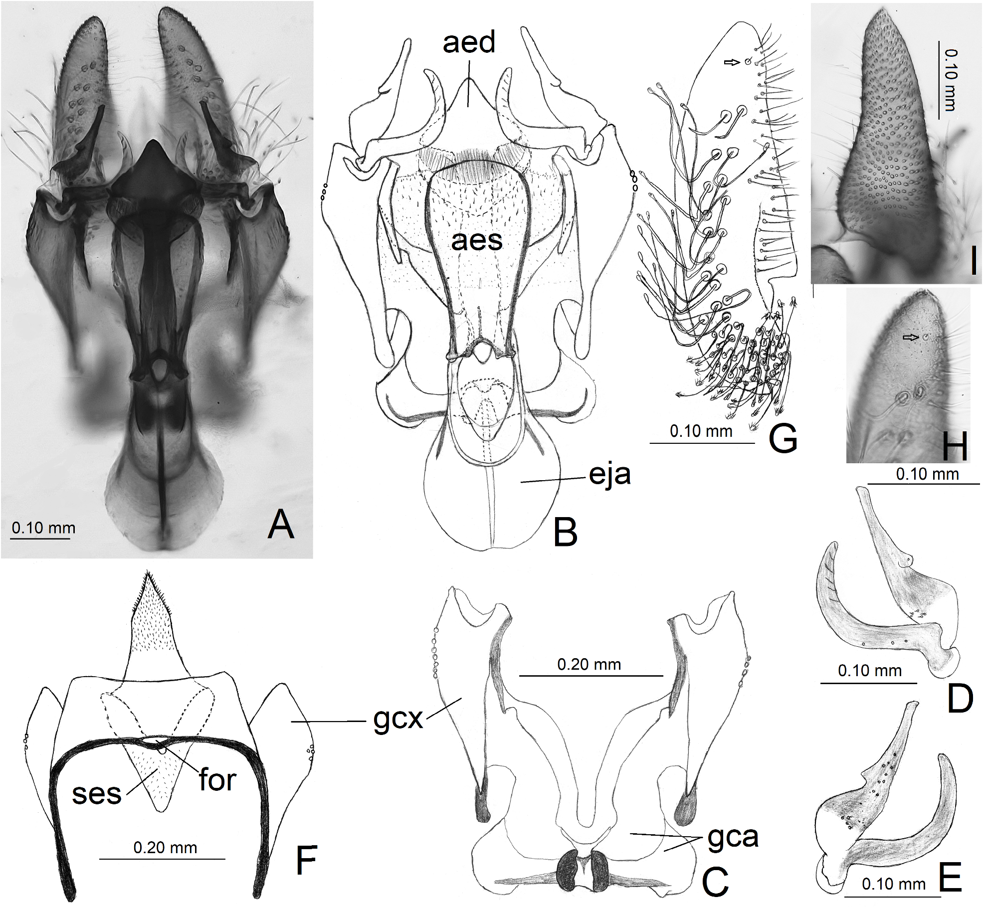
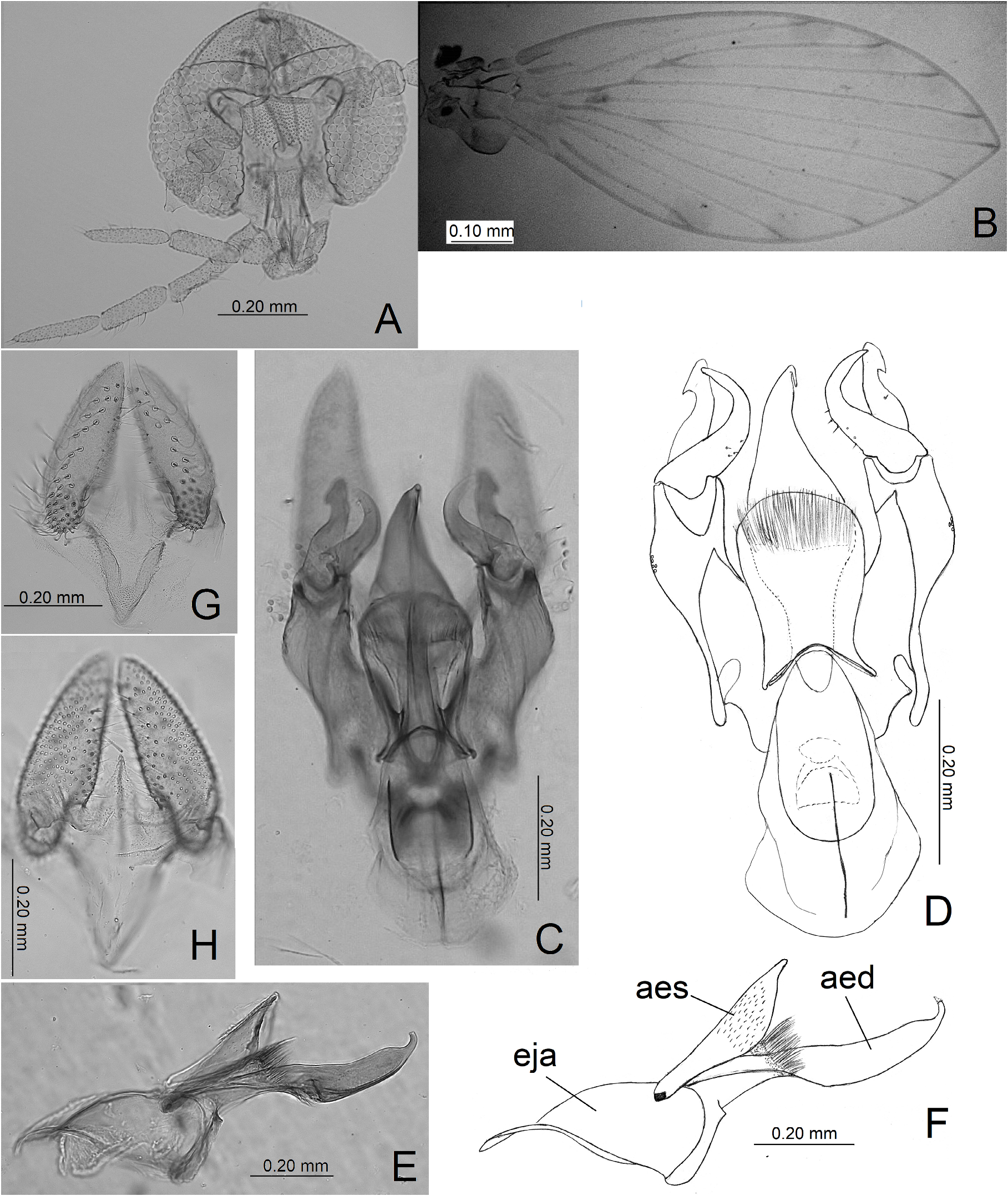
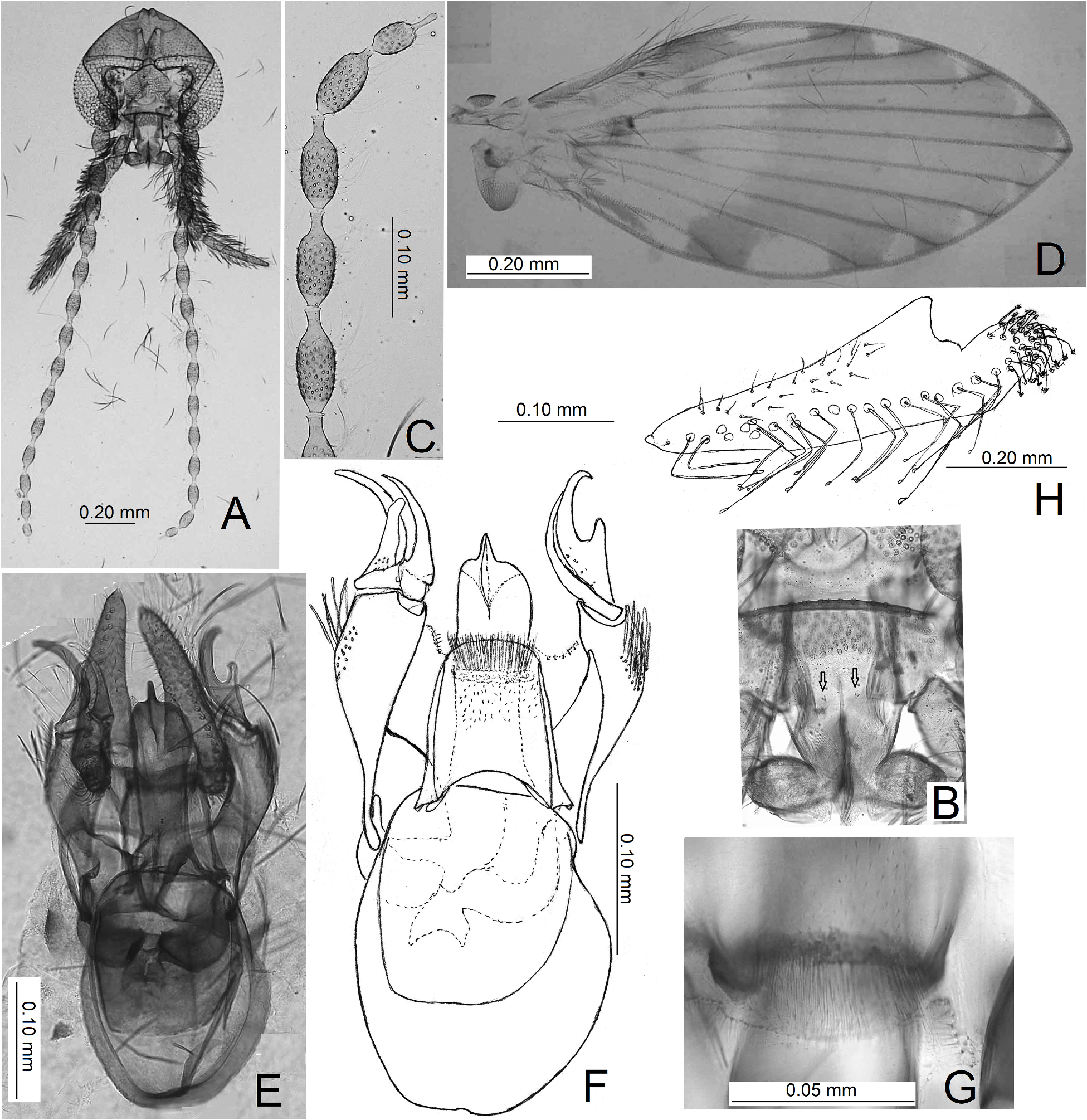
Description. male. Head: semicircular eye bridge with three or four rows of facets; eyes separate by 0.2 to 3.0 facet diameter; interocular suture as an inverted V-shaped; frontal patch of alveoli with anterior margin straight and posterior margin bilobed ( Figs. 1A View FIGURE 1 , 3B View FIGURE 3 , 6A View FIGURE 6 , 7A View FIGURE 7 , 8A View FIGURE 8 ); labrum subtriangular, dorsally with pointed apex ( Figs. 3B, C View FIGURE 3 ); labella bulbous with many setae; epipharynx with two spiniform setae dorsal to labial furca and at least 10 small spiniform setae in the middle of its surface ( Figs. 1B, C View FIGURE 1 , 3D, E View FIGURE 3 , 7B View FIGURE 7 ). Antenna with scape longer than pedicel, subcylindrical; pedicel almost spherical; flagellum with 14 fusiform flagellomeres decreasing in size from base to apex; apiculus cylindrical, approximately 0.5 the length of the last flagellomere; ascoids not visible ( Figs. 1E View FIGURE 1 , 3F View FIGURE 3 , 7A, C View FIGURE 7 , 8A View FIGURE 8 ). Palpus approximately 1/3 the length of flagellum with four segments, the last segment not annulate ( Figs. 1F View FIGURE 1 , 3G View FIGURE 3 , 6A View FIGURE 6 , 7A View FIGURE 7 ). Thorax: anepisternum and laterotergite with patch of alveoli, other pleuron sclerites glabrous ( Fig. 4A View FIGURE 4 ); coxa II with mesal anterior protuberance covered with pores. Wing ( Figs. 1G View FIGURE 1 , 4C View FIGURE 4 , 6B View FIGURE 6 , 7D View FIGURE 7 , 8B View FIGURE 8 ): base of C vein, after last break, wider than remaining vein; base of costal area with microsetae near C ( Figs. 1H View FIGURE 1 , 4D View FIGURE 4 ); R 2+3 not reaching R 4; R 5 ending at pointed apex of wing, not reaching R 4; R 2+3 fork Approximately at the same level of M 1+2 fork, or M 1+2 basal to R 1+2; small circular thoracic gland near the thoracic sensory area with microtrichia (tgl, Figs. 1D View FIGURE 1 , 4A View FIGURE 4 ); thoracic sensory area with microtrichia in the dorsal area of anepimeron (sam, Figs. 1D View FIGURE 1 , 4A View FIGURE 4 ). Terminalia: gonocoxites horn-like, wider distally than basally, with subapical group of setae in external margin ( Figs. 1B, C, F View FIGURE 1 , 2B, D View FIGURE 2 , 6D View FIGURE 6 , 7F View FIGURE 7 , 8E View FIGURE 8 ); anterior condyles of gonocoxites plate-like, fused at midline forming a gonocoxal bridge, with medium elevation directed to the ejaculatory apodeme ( Figs. 2C View FIGURE 2 , 5E View FIGURE 5 ). Gonostyli glabrous, sclerotized, bifurcated, shorter than gonocoxites, internal arm turned outward, external arm with basal or medial short to long lobe ( Figs. 2A, B, D, E View FIGURE 2 , 5A, B, C, D, F, G View FIGURE 5 , 6 C, D View FIGURE 6 , 7E, F View FIGURE 7 , 8C, E View FIGURE 8 ). Aedeagus symmetrical. Plate with tuft of long setae around the aedeagus ( Figs. 1A, B View FIGURE 1 , 5B, C, D View FIGURE 5 , 6D, E, F View FIGURE 6 , 7F View FIGURE 7 , 8C, D View FIGURE 8 ). Aedeagal sheath lightly sclerotized with apical microtrichia ( Figs. 1B, D View FIGURE 1 , 6D View FIGURE 6 , 7F View FIGURE 7 ). Ejaculatory apodeme wider than aedeagus, usually much more so ( Figs. 2A, B View FIGURE 2 , 5A, B View FIGURE 5 , 6C, D View FIGURE 6 , 7E, F View FIGURE 7 ). Epandrium bare, subrectangular, wider than long, with one basal foramen and anterior sclerotized band ( Figs. 2F View FIGURE 2 , 5H View FIGURE 5 ). Hypopod elongated, base wider than apex, without apical tenaculum, dorsally with one or sometimes two rows of big alveoli bearing accessory geniculate tenacula and a group of short accessories tenacula at base ( Figs. 1G, H View FIGURE 1 , 5H View FIGURE 5 , 6G View FIGURE 6 , 8G View FIGURE 8 ), ventrally covered by small alveoli bearing setae ( Figs. 1I View FIGURE 1 , 5I View FIGURE 5 , 6H View FIGURE 6 ). Epiproct not observed, probably reduced. Hypoproct subtriangular in dorsal view, with apical microtrichia ( Figs. 2F View FIGURE 2 , 5H View FIGURE 5 )
Female. Unknown.
Distribution. Brazilian Atlantic Rain Forest: Bahia, Espirito Santo and S„o Paulo.
Included species. Coronamyia biapicalis ( Bravo, Lago and Castro, 2004) comb. nov., Coronamyia paschoali sp. nov., Coronamyia santosi sp. nov., Coronamyia triangularis sp. nov., and Coronamyia trilobata sp. nov.
Remarks. The new genus Coronamyia falls within the tribe Maruinini , as delimited by Kvifte (2018), with fusiform flagellomeres, Rs not pectinate, and radial fork at the same level as the medial fork (in the diagnosis of the tribe “radial fork basal to medial fork”). The other characters are as the diagnosis of the tribe.
The new genus Coronamyia is morphologically similar to Alepia . This is due to the presence of an eye bridge with 3 or 4 facet rows (contiguous or separated); flagellomeres fusiform; pleuron with alveoli only on anepistemum and lalerotergite; the wings generally with infuscated patterns; R 5 ending at the wing apex; gonostyli often bifurcated; hypopods without apical tenaculum and with accessory tenacula (characters obtained from Quate 1996; Quate & Brown 2004; Ježek et al. 2011).
However, this new genus can be differentiated from the other genera of Maruinini by the above mentioned diagnostic characters, mainly the presence of a circular plate with a tuft of long setae around the aedeagus (observed only in the species included in Coronamyia new genus), a bifurcated gonostylus with an arched internal arm and lobed external arm; elongated, subovoid hypopods without apical tenacula; and accessory tenacula of two kinds, the basal type with umbellate tips, with the rest of dorsal area having accessory tenacula with clavate tips.
The hypopods of Alepia clara Bravo, Lago & Castro, 2004 and Alepia martinicana Wagner, 1993 are morphologically similar to those of the described species of Coronamyia , although the gonostyli of the first two species lack internal arched and external lobated arms. The gonostylus of Alepia viatrix Jaume-Schinkel, Kvifte, van der Weele & Mengual, 2022 is similar to the species of the new genus, although the shape of the hypopods, the presence of apical tenacula, and the absence of two kinds of tenacula differentiate it from the other species of the new genus.


No known copyright restrictions apply. See Agosti, D., Egloff, W., 2009. Taxonomic information exchange and copyright: the Plazi approach. BMC Research Notes 2009, 2:53 for further explanation.
|
Kingdom |
|
|
Phylum |
|
|
Class |
|
|
Order |
|
|
Family |
|
|
Tribe |
Maruinini |