
Litargosuchus leptorhynchus, Clark & Sues, 2002
|
publication ID |
https://doi.org/ 10.1046/j.1096-3642.2002.00026.x |
|
DOI |
https://doi.org/10.5281/zenodo.5106327 |
|
persistent identifier |
https://treatment.plazi.org/id/26418E48-FF8A-FFB8-FCD9-FD6CFACC11A7 |
|
treatment provided by |
Carolina |
|
scientific name |
Litargosuchus leptorhynchus |
| status |
sp. nov. |
LITARGOSUCHUS LEPTORHYNCHUS SP. NOV.
1988 Pedeticosaurus sp. Gow & Kitching, p. 518. Etymology. From Greek leptos, thin, delicate, and Greek rhynchos, snout, muzzle.
Diagnosis. Parietals partially fused and devoid of sagittal cresting. Squamosal broad transversely and without dorsolateral crest. Dentaries forming extensive symphysis, with little or no splenial involvement.
Holotype. BP/1/5237, skull, mandible and much of the postcranial skeleton, first identified as Pedeticosaurus sp. by Gow & Kitching (1988). The postcranium is represented by the mostly articulated presacral vertebral column and scattered parts of caudal series, both scapulae and coracoids, both humeri, left radius and ulna, part of the left ilium, both ischia, left hindlimb (with the exception of the proximal portion of the femur and most of the pes), and distal portion of right femur.
In view of its small size and gracile build, BP/1/5237 possibly represents a juvenile. Closure of the neurocentral sutures along the vertebral column provides a particularly useful osteological criterion for ontogenetic assessment ( Brochu, 1996). Open neurocentral sutures are apparent on the cervical vertebrae, but preservation and current state of preparation do not permit identification of this feature in the dorsal column.
Type horizon and locality. Upper Elliot Formation (Stormberg Group), 2 m below the contact with the Clarens Formation, on the farm Eagles Crag, Barkley East, South Africa. Age: Early Jurassic.
SKULL
Gow & Kitching (1988) provided only a brief account of the structure of the skull, and examination of the specimen revealed much additional detail.
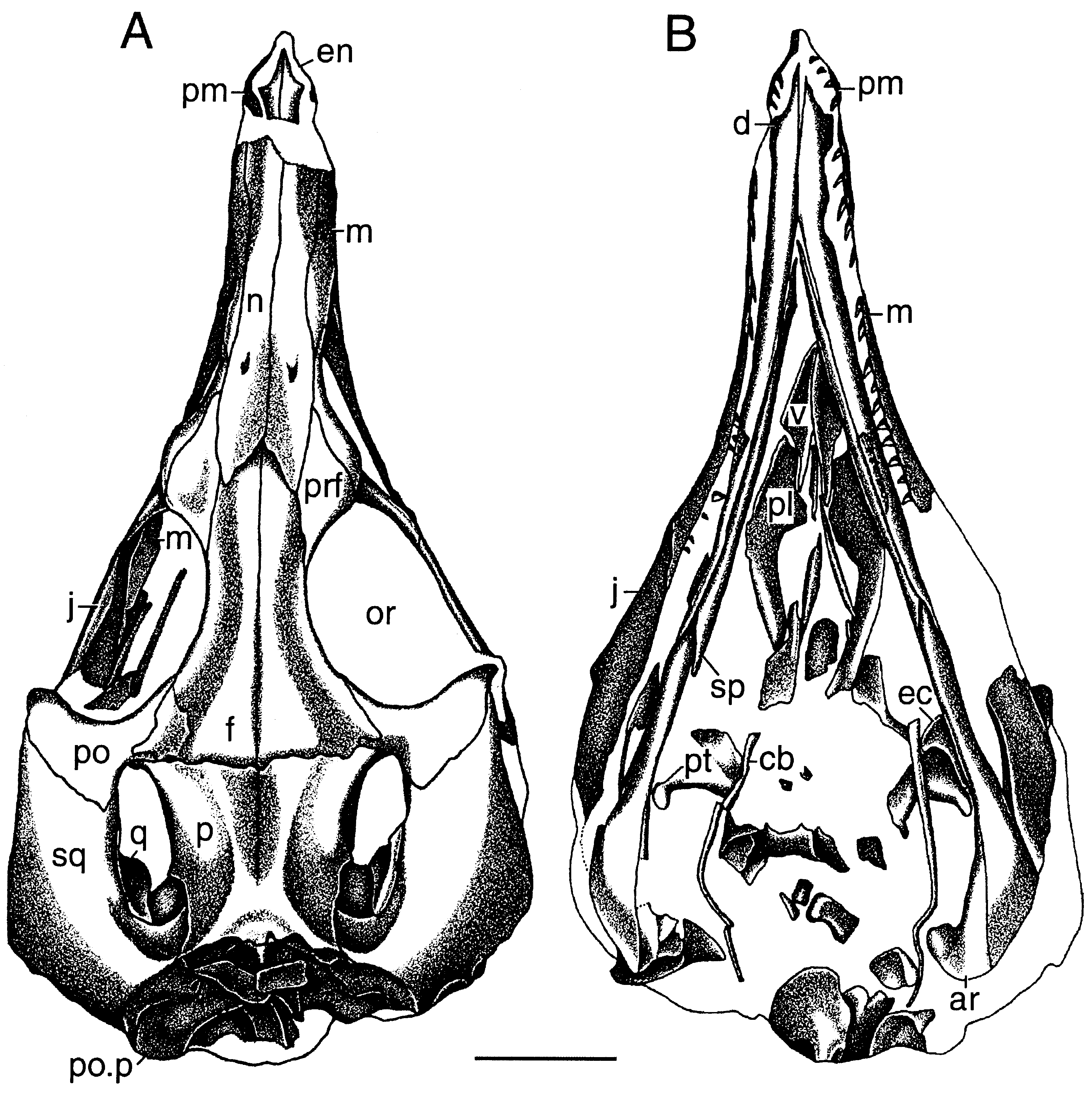
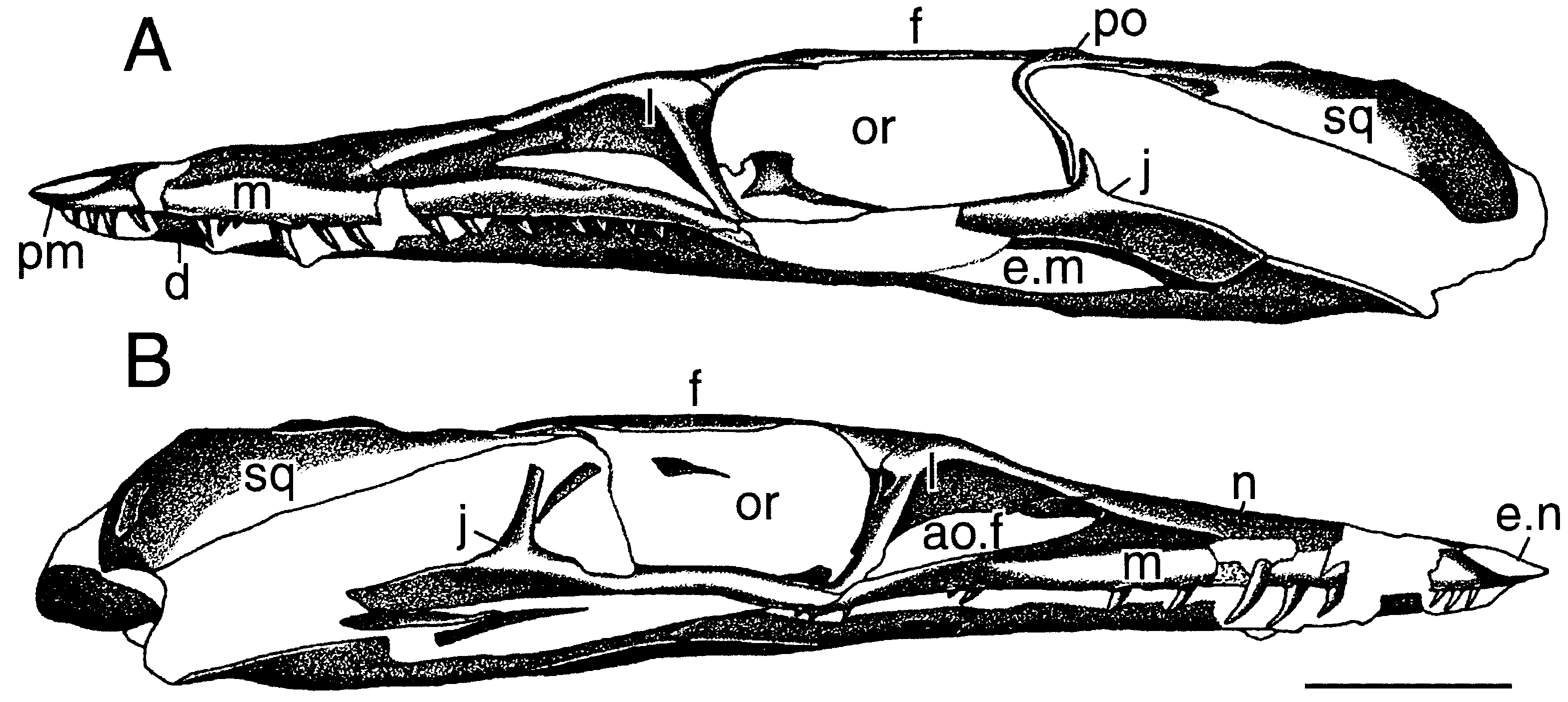
The skull ( Figs 1 View Figure 1 , 2 View Figure 2 ) was dorsoventrally flattened during fossilization. As a result of this crushing, the mandibular rami are now tightly appressed to the skull. The individual bones, with the exception of those constituting the palate and ventral portion of the braincase, are well-preserved, but some are traversed by fractures. They are delicate, smooth and devoid of sculpturing. The specimen was preserved in a hard, haematite-rich matrix, and removal of the encrusting haematite layer by mechanical means resulted in local loss of surficial bone. The occipital region is partially obscured by unidentified pieces of bone.
In dorsal view, the skull has a narrow snout and a transversely broad temporal region (although its width was accentuated by dorsoventral compression). It is 63 mm long (measured along the midline of the skull roof from the anterior tip of the snout to the anteromedial end of the occipital embayment). The orbit is large, with an anteroposterior diameter of about 16 mm. The supratemporal fossae are delimited medially and posteriorly by faint ridges on the parietal and squamosal. The supratemporal fenestrae are much longer than wide, resembling those of Terrestrisuchus ( Crush, 1984) .
On the better preserved left side, the recurved posterolateral process of the small premaxilla overlaps the nasal and maxilla on the side of the snout, excluding the latter element from participation in the posterior margin of the laterally directed external naris. The anterior ends of the premaxillae appear to be drawn out into a point. The left premaxilla holds four teeth, the first of which is the smallest; three teeth are preserved in the more incomplete right premaxilla. Contra Gow & Kitching (1988), the presence of a lateral notch between the premaxilla and maxilla cannot be ascertained due to poor preservation of this region on either side of the snout.
The low but long maxilla forms most of the rostral portion of the skull. In lateral view, its alveolar margin is almost straight. The facial portion of the maxilla is vertical. Its ascending process projects posteriorly as well as slightly dorsally and contacts the anterior ramus of the lacrimal half way along the dorsal rim of the antorbital fenestra, excluding the nasal from participation in the dorsal margin of the antorbital fossa. The ventral margin of the large, subtriangular antorbital fossa is deeply recessed relative to the remainder of the lateral surface of the maxilla. The antorbital fenestra is situated in the posteroventral corner of the antorbital fossa. It is long anteroposteriorly but low dorsoventrally. The more completely preserved left maxillary tooth row comprises at least 18 teeth (16 of which are completely or partially preserved) and terminates posteriorly just behind the anterior margin of the orbit.
The nasal extends back almost to the level of the anterior margin of the orbit. Its lateral sutural contacts with the maxilla, lacrimal and prefrontal are nearly straight. Its tapered anterior portion forms most of the dorsal margin of the narial fenestra. The dorsal surface of each nasal bears several neurovascular foramina, especially a large one in the posterior quarter. The contact between the ascending process of the maxilla and the anterior process of the lacrimal excludes the nasal from the dorsal margin of the antorbital fossa.
The lacrimal has the shape of an inverted L and is inclined forward in lateral view. It forms the preorbital bar and contributes a broad medial lamina to the medial wall of the antorbital fossa. The lateral surface of its vertical portion bears a thin but distinct ridge. Posteriorly, the lacrimal forms an extensive lateral contact with the prefrontal along the preorbital bar. Dorsally, it is narrowly exposed on the skull roof.
The prefrontal forms the anteromedial portion of the dorsal rim of the orbit. It is overlapped by the frontal posterolaterally. The dorsal surface of the prefrontal is subtriangular in outline.
The posterior process of the jugal is deep dorsoventrally and faces ventrolaterally (although this may be due to dorsoventral compression of the skull). Its dorsal process for contact with the descending process of the postorbital is short and delicate. The anterior process of the jugal forms the ventral margin of the orbit but does not extend to the posteroventral corner of the antorbital fenestra and fossa anteriorly.
The dorsoventral crushing of the skull has pushed the quadrates up through the supratemporal fenestrae. Few details are visible.
The frontal is much longer than wide and forms most of the skull roof as well as the slightly raised dorsal rim of the orbit. Its dorsal surface is concave transversely. Although the frontals are somewhat thicker along their median sutural contact they do not form a distinct ridge as in Hesperosuchus ( Clark et al., 2001) and Sphenosuchus ( Walker, 1990) . Anteriorly, the frontals extend forward for a short distance between the posterior ends of the nasals along the midline of the skull roof. Posteriorly, they contact the parietals along a transverse suture. The frontal does not participate in the anterior margin of the supratemporal fenestra.
The interparietal suture is present only on the anterodorsal portion of the skull roof and cannot be traced more posteriorly. The parietals lack extensive posterolateral wings and form a weakly concave embayment in the occipital margin of the skull roof. Their dorsal surface is very slightly concave transversely between the medial margins of the supratemporal fossae. The sutural contact betwen the parietal and squamosal posterior to the supratemporal fenestra is short.
The postorbital is triradiate in lateral view. It forms the anterior half of the dorsal margin of the supratemporal bar and overlaps the squamosal posteriorly. Anteromedially, the postorbital contacts the posterolateral end of the frontal. Its ventral process for contact with the ascending process of the jugal is delicate.
No palpebral bones are preserved.
The rather broad squamosal forms the posterolateral corner of the skull roof and resembles the homologous element in Crocodyliformes. It is thin and ventrally somewhat concave. The lateral edge of the squamosal is deflected and overhangs the infratemporal region and suspensorium laterally. Anteriorly, the squamosal extends ventral to the postorbital to participate in the formation of the postorbital bar. The dorsal surface of the squamosal is broad and gently convex transversely. It lacks the posterolateral crest bordering the supratemporal fossa found in most basal crocodylomorph archosaurs except Terrestrisuchus ( Crush, 1984) .
The distal end of the paroccipital process formed by the otoccipital (fused exoccipital and opisthotic) is expanded dorsoventrally. The post-temporal foramen is located at the sutural contact between the squamosal, otoccipital, and supraoccipital and is very small.
The palate is poorly preserved; it is represented by the posterior portions of the vomers, the palatines and fragments of the pterygoids. The vomers contact each other along the midline. The long medial portion of the ectopterygoid extends posteriorly along the anterolateral edge of the transverse flange of the pterygoid.
Long, rod-like bones preserved on both sides of the palate probably represent the ceratobranchialia I of the hyoid apparatus, as in extant crocodylians. They have been somewhat warped during fossilization.
MANDIBLE
The mandibular rami are long and very slender. The external mandibular fenestra is long and low. The number of dentary teeth cannot be determined due to the tight contact between the mandibular rami and the skull.
The dentaries form a rather long symphysis, which extends back to the level of the third maxillary tooth. The splenial either did not enter into the symphysis or contributed only minimally to its formation. The articular forms a distinct, dorsomedially directed process.
DENTITION
All teeth have labiolingually flattened crowns with anterior (mesial) and posterior (distal) carinae. Preservation of individual teeth is poor, and it cannot be determined whether the carinae were serrated or smooth; the carinae are serrated in all other known basal crocodylomorphs. The premaxillary and anterior maxillary teeth have slender and distinctly recurved crowns. In the left maxilla, the teeth in positions 4–6 appear to have the tallest crowns. The more posterior teeth have shorter and less recurved crowns.
No known copyright restrictions apply. See Agosti, D., Egloff, W., 2009. Taxonomic information exchange and copyright: the Plazi approach. BMC Research Notes 2009, 2:53 for further explanation.
|
Kingdom |
|
|
Phylum |
|
|
Class |
|
|
Order |
|
|
Family |
|
|
Genus |
Litargosuchus leptorhynchus
| Clark, James M. & Sues, Hans-Dieter 2002 |
Pedeticosaurus
| van Hoepen 1915 |