
Strongylophthalmyia flavimarginata, Zhou & Evenhuis & Yang, 2024
|
publication ID |
https://doi.org/ 10.11646/zootaxa.5403.5.2 |
|
publication LSID |
lsid:zoobank.org:pub:6BC79A0E-45A6-4685-AAD0-5B3133F098B4 |
|
DOI |
https://doi.org/10.5281/zenodo.10562319 |
|
persistent identifier |
https://treatment.plazi.org/id/266487E3-FFAA-4A03-FF2A-1928FDAAB768 |
|
treatment provided by |
Plazi |
|
scientific name |
Strongylophthalmyia flavimarginata |
| status |
sp. nov. |
Strongylophthalmyia flavimarginata sp. nov.
( Figs 9–14 View FIGURES 9–10 View FIGURES 11–14 , 27 View FIGURES 26–29 , 31 View FIGURES 30–33 )
Type material. Holotype. CHINA: ♂, Yunnan, Jinping, Wutaishan , 1097 m, 31 Mar. 2019, Liang Wang leg. ( CAU) . Paratypes. CHINA: 1♀, same collection data as for holotype ( CAU) .
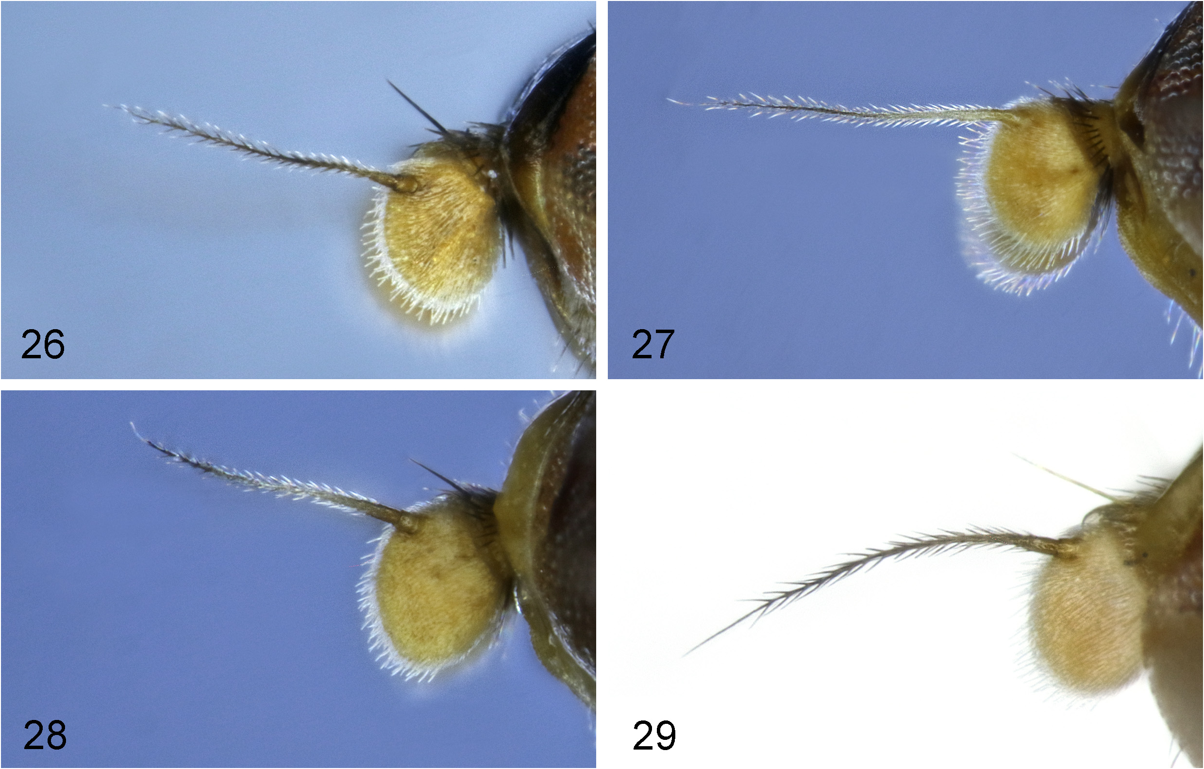
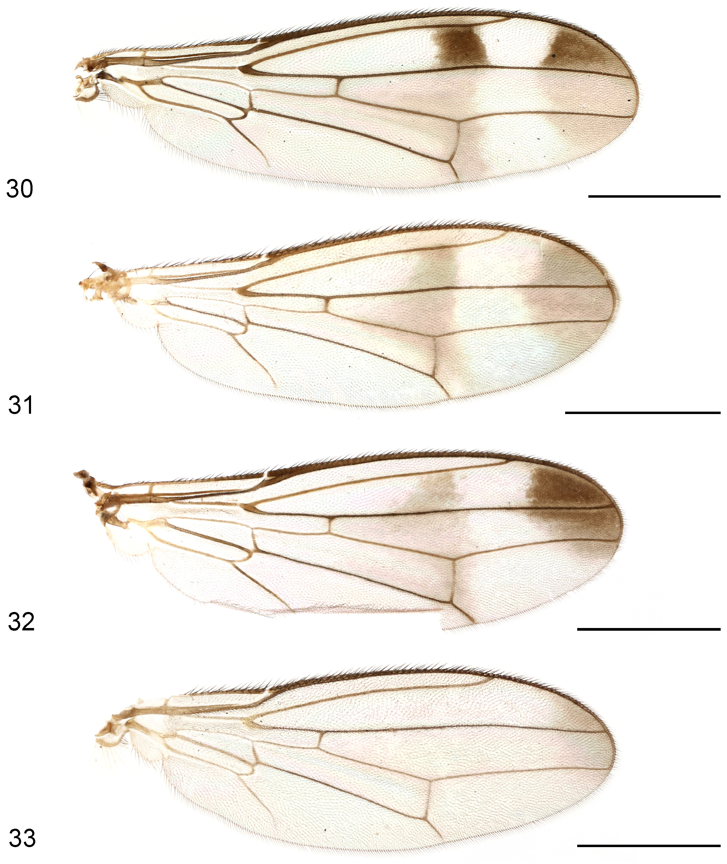
Diagnosis. Generally shining blackish brown, with lateral body surface mostly yellow to yellowish brown ( Figs 9, 10 View FIGURES 9–10 ); anterior third to whole of frons yellowish brown; face, parafacial and gena yellow; antennal arista bicolorous, with yellow basal half and dark brown apical half ( Fig. 27 View FIGURES 26–29 ); wing slightly infumate, with large dark suffusion at apex and vague broad median transverse band interrupted along cell r 1 ( Fig. 31 View FIGURES 30–33 ); legs mostly yellow, with mid and hind femora narrowly dark brown subapically, mid and hind tibiae largely dark brown; phallapodeme curved inwardly in apical half ( Fig. 14 View FIGURES 11–14 ); distiphallus about 1.52 times as long as phallapodeme, with hook-shaped sclerotized apical “glans”, membrane microtrichose only in basal half ( Figs 13, 14 View FIGURES 11–14 ).
Description. Male and female ( Figs 9, 10 View FIGURES 9–10 ). Body length 4.1–5.3 mm, wing length 3.5–4.1 mm.
Generally shining blackish brown with extensive paler markings ( Figs 9, 10 View FIGURES 9–10 ). Frons (in male only anterior third) yellowish brown; face, parafacial and gena yellow. Antenna yellow with apical half of arista dark brown ( Fig. 27 View FIGURES 26–29 ). Clypeus, proboscis and palpus yellow. Postpronotum, propleuron, basisternum and anterior third of anepisternum yellow. Mesonotum blackish brown with lateral quarter yellowish brown. Scutellum yellowish brown along apical margin. Wing slightly infumate, with large dark suffusion at apex and vague broad median transverse band at level of dm-m; median transverse band more or less interrupted along cell r 1 ( Fig. 31 View FIGURES 30–33 ); wing veins brown to dark brown. Halter white, slightly darkened basally. Legs mostly yellow; mid and hind femora narrowly dark brown subapically; mid and hind tibiae largely dark brown. Abdomen mostly yellow except tergites 1–6 in male or 2–5 in female with large dark brown median patch; tergite 7 blackish brown ( Figs 9, 10 View FIGURES 9–10 ).
Head with parafacial with sparse short whitish setulae; gena with silvery tomentose stripe along eye margin; postgena bulging, with several long black setulae. Head chaetotaxy: 1 inner vertical seta, 1 outer vertical seta, 2 fronto-orbital setae, 1 ocellar seta, 1 postocellar seta. Clypeus band-like in male, thick and bulbous in female; palpus elongate, with dense short dark setulae. Antennal scape with scattered marginal setae and 1 dominant dorsal seta; pedicel with single strong seta dorsally; first flagellomere ovate, wider than long, densely covered with long white setulae ( Fig. 27 View FIGURES 26–29 ).
Thorax with mesonotum covered with scattered short golden setulae, in dorsal view with distinct transverse suture.Anepisternum with short setulae along notopleural suture. Scutellum subtriangular, slightly inflated. Thoracic chaetotaxy: 1 presutural intra-alar seta, 1 anepisternal seta, 2 notopleural setae, 1 dorsocentral seta, 1 posterior supra-alar seta, 1 scutellar seta. Wing ( Fig. 31 View FIGURES 30–33 ) with R 4+5 and M 1+2 almost parallel apically; apical section of M 1+2 nearly straight; M 4 reaching wing margin; CuA+CuP closely approaching wing margin; r-m located about basal 0.43 of cell dm; apical section of M 4 shorter than dm-m; alula relatively large; anal lobe well-developed. Legs with dense dark setulae; fore coxa with several long white setulae anterodorsally.
Abdomen covered with dense long dark setae. Tergite 1 weakly sclerotized. Pregenital sclerites relatively weakly sclerotized.
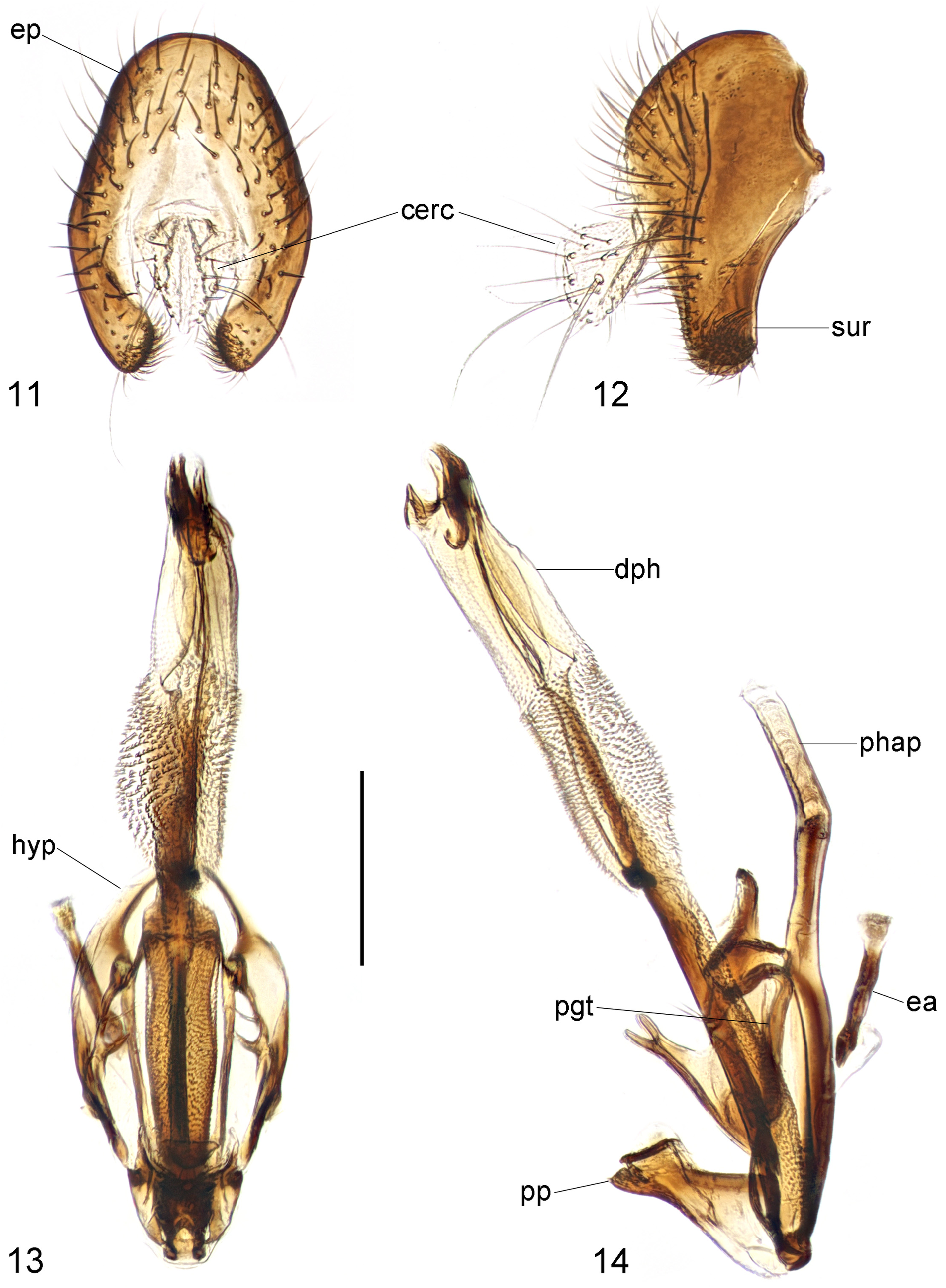
Male genitalia: Epandrium ( Figs 11, 12 View FIGURES 11–14 ) relatively short and broad, with dense long setae. Surstylus ( Figs 11, 12 View FIGURES 11–14 ) nearly parallel-sided in lateral view, with short stout setae on inner distal surface. Cerci ( Figs 11, 12 View FIGURES 11–14 ) distinctly expanded apically, with one rather long subapical seta and several shorter ones. Hypandrium ( Figs 13, 14 View FIGURES 11–14 ) strongly expanded laterally, with one pair of bifid anterior lobes. Phallapodeme ( Fig. 14 View FIGURES 11–14 ) long, curved inwardly in apical half. Pregonite ( Figs 13, 14 View FIGURES 11–14 ) long, narrow, band-like, basally fused to inner surface of hypandrium. Phallic plate ( Fig. 14 View FIGURES 11–14 ) thickened, divided into two articulating sclerites. Distiphallus ( Figs 13, 14 View FIGURES 11–14 ) long, about 1.52 times as long as phallapodeme, with hook-shaped sclerotized apical “glans”, membrane microtrichose only in basal half. Ejaculatory apodeme ( Figs 13, 14 View FIGURES 11–14 ) long, slightly expanded apically.
Etymology. The specific epithet is derived from Latin flavi- (meaning yellow) and -marginata (meaning marginated), referring to the yellow to yellowish brown patterns on the lateral body surface of the new species.
Distribution. China (Yunnan).
Comparative notes. Strongylophthalmyia flavimarginata sp. nov. superficially resembles S. elegantissima Frey, 1956 ( Myanmar). The latter species, which belongs to the S. fascipennis group, has the bare antennal arista and therefore can be easily separated from the new species. Among the S. stulate group, this new species is most similar to S. bifasciata (see above) but can be readily distinguished from the latter species by the very different color patterns on the head, thorax, and abdomen.

| CAU |
China Agricultural University |
No known copyright restrictions apply. See Agosti, D., Egloff, W., 2009. Taxonomic information exchange and copyright: the Plazi approach. BMC Research Notes 2009, 2:53 for further explanation.